985392
Description
Flashcards by rachel-chads, updated more than 1 year ago
|
|
Created by rachel-chads
over 11 years ago
|
|
| Question | Answer |
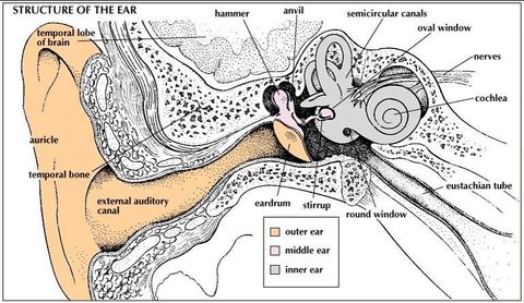
| Human auditory system (diagram) | |
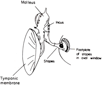
| Tympanic membrane (ear drum) and bones of ear | |
| Ears | All parts can be replaced Ear canal is special shape Ear drums is more efficient at the human voice range -> focuses the sound Middle ear is usually air filled in healthy people Fluid can be drained Bones can be replaced |
| Inner ear | Cochlea -> coiled tube Vestibular system -> 3 semi-circular canals, for balance All connected All use sensory hair cells |
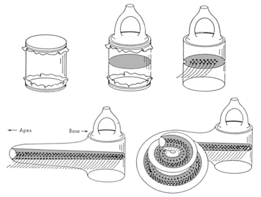
| Cochlea - development (diagram) |
Image:
cochlea.PNG (image/PNG)
|
| Tonotopic Map | Frequency sensitivity due to mechanism of basilar membrane, different portions of the membrane are maximally deformed by the sound of different frequencies. When auditory axons in the auditory-vestibular nerve synapse in the cochlear nuclei, they do so in an organised pattern based on characteristic frequency. |
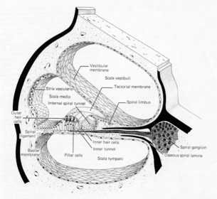
| Cochlear cross section (diagram) | |
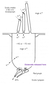
| Perilymph | Found in scala vestibuli and scala tympani Ionic content similar to cerebrospinal fluid Low K+ -> 7mM High Na+ -> 140 mM |
| Endolymph | Found in Scala media Completely different to the other fluids 140 mmol K+ +80mV |
| Stria vascularis | Difference in ion content of endolymph in scala media caused by active transport taking place in the stria vascularis Reabsorbs Ca2+, secretes K+ - against their concentration gradients Sets up endocochlear potential Enhances auditory transduction |
| Hair cell (diagram) | |
| Hair cell | Top is in endolymph, bottom is in perilymph Sound is caused by the movement of the hairs going backwards and forwards (more laterally) |
| Ménière's disease | Affects both auditory and vestibular system Deafness, Tinnitus Vertigo, Balance problems Due to fluid imbalance Other diseases due to the 'battery' loss Less K+ in the endolymph |
| Organ of Corti (diagram) | |
| Organ of Corti | Specialised sensory epithelium Direct mechanical stimulation (not like visual system) Outer hair cells -> amplification Inner hair cells -> 3,500 cells, primary |
| Organ of Corti - Movement of basilar membrane (diagram) | |
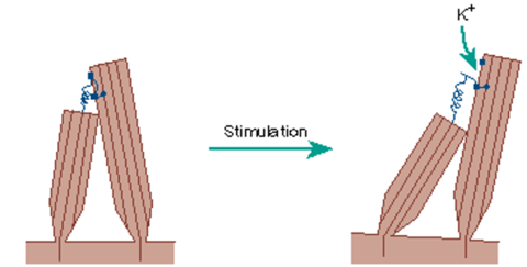
| Mechano-Electrical Transduction (Sensory) | Actin filaments - make the stereocilia rigid rods that only bend at the base Cross-link filaments link stereocilia so they move as a unit -> joint by tiplinks (cadherin) Stereocilia - 3 micrometres in diameter |
| Stereocilia (diagram) |
Image:
Stereocilia (image/png)
|
| Mechano-Electrical Transduction | It is the electrical gradient that is driving the positive ions in - not the concentration gradient Depolarisation of the cell Outward pump of K+ to restore/reset the membrane |
| Mechano-Electrical Transduction Hair cell (diagram) | |
| Electro-Mechanical Transduction (Amplification) | Outer hair cell changes length based on membrane potential Selectively processed Allows hearing in newborns (through having no hair cells) to be treated -> Cochlear inplant |
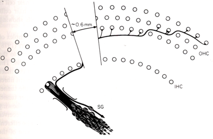
| Innervation - outer and inner hair cells (diagram) | |
| Innervation - outer and inner hair cells | 3 outer hair cells for every inner hair cell 95% of the spiral ganglion neurones communicate with the inner hair cellls IHC doesn't share spiral ganglion with other hair cells - wouldn't be able to tell the frequency 95% of the efferent neurones modulate our hair cells -> to protect the ears (temporary threshold shift) |
| Analysis of sound: Freuquency (pitch) | Encoded in nerves by location along the basilar membrane |
| Analysis of sound: Intensity (loudness) | Encoded bin nerves by numbers responding and by firing rate |
| Analysis of sound: Sound transduction | Inner hair cells (and OHCs) |
| Analysis of sound: Amplification | Outer Hair Cells |
| Auditory Tract | Primary Auditory Cortex ^ Inferior Colliculus (IC) ^ Lateral Lemniscus Superior Olivary Complex (S) ^ Cochlear Nuclei - AVCN, PVCN, DCN ^ Auditory Nerve Sprial Ganglion |
| Cochlear Nuclei | Anterior ventral (AVCN), Posterior ventral (PVCN) and Dorsal (DCN) regions each have a tonotopic map Individual cells have specific functions eg. Primary responses in spherical & bushy cells (VCN) Onset responses in octopus cells (CN) Pauser responses in fusiform cells AVCN contains relay cells to Medial Superior Olive - Calyx of Held, a giant fast synapse |
| Superior Olive (SO) | Sound localisation Medial SO - low frequency analysis, interaural time differences, received faithful inputs for calyx of Held on both sides Lateral SO - high frequency analysis, interaural intensity differences |
| Inferior Colliculus (IC) | Site of convergence of projections with complex frequency responses Monoaural input from DCN - binaural from SO Tonotopic maps Responsible for attention reflexes, startle response, learned reflexes |
| Cortex | Tonotopic map retained Neurones with sensitivity to features in complex sounds Auditory space maps, Selective attention, Inhibition of inappropriate motor responses, Recognition of stimuli, Discrimination of temporal patterns and short term auditory memory |
| Vestibular system | |
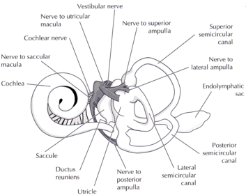
| Vestibular system | 5 different epithelia Go via 8th nerve to the brain |
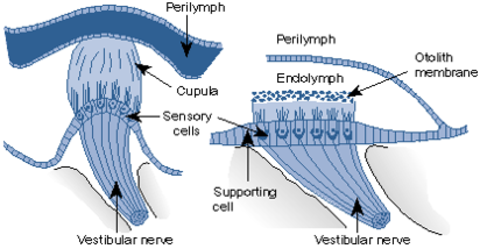
| Vestibular system cross section (diagram) | |
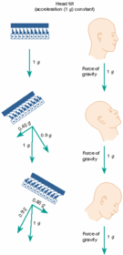
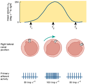
| Hair movement with head tilt (diagram) | |
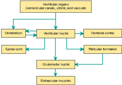
| Vestibular system (diagram) | |
| Links between systems |
Want to create your own Flashcards for free with GoConqr? Learn more.