
Mind map
Histology
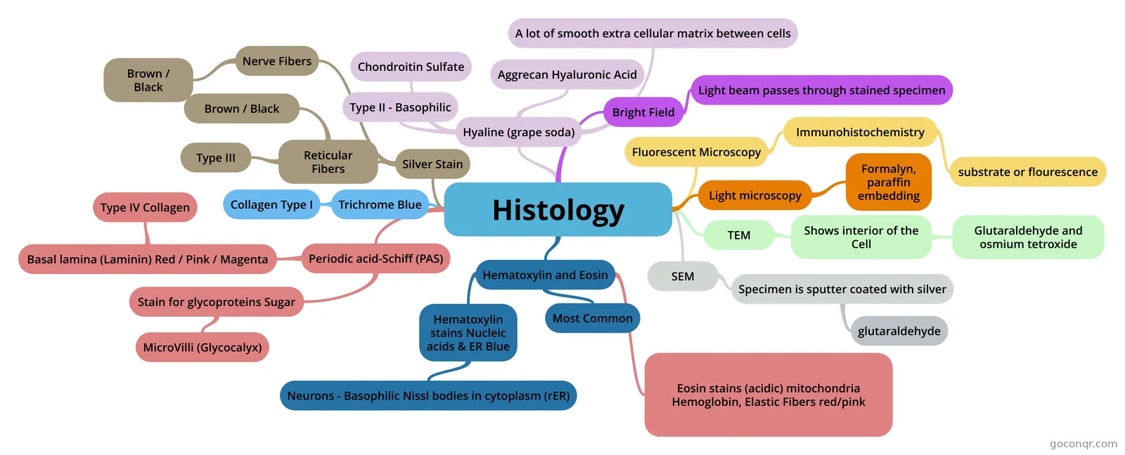
Mind map: Histology microscopy and stains
Map content
Histology
Light microscopy
- Formalyn, paraffin embedding
TEM
- Shows interior of the Cell
- Glutaraldehyde and osmium tetroxide
- Shows interior of the Cell
SEM
- Specimen is sputter coated with silver
- glutaraldehyde
- Specimen is sputter coated with silver
Fluorescent Microscopy
- Immunohistochemistry
- substrate or flourescence
- Immunohistochemistry
Hematoxylin and Eosin
- Most Common
- Hematoxylin stains Nucleic acids & ER Blue
- Neurons - Basophilic Nissl bodies in cytoplasm (rER)
- Eosin stains (acidic) mitochondria Hemoglobin, Elastic Fibers red/pink
Periodic acid-Schiff (PAS)
- Stain for glycoproteins Sugar
- MicroVilli (Glycocalyx)
- Basal lamina (Laminin) Red / Pink / Magenta
- Type IV Collagen
- Stain for glycoproteins Sugar
Trichrome Blue
- Collagen Type I
Silver Stain
- Reticular Fibers
- Type III
- Brown / Black
- Nerve Fibers
- Brown / Black
- Reticular Fibers
Bright Field
- Light beam passes through stained specimen
Hyaline (grape soda)
- Type II - Basophilic
- Aggrecan Hyaluronic Acid
- Chondroitin Sulfate
- A lot of smooth extra cellular matrix between cells
Information
- Description
- Histology Stains , Microscopes, Collagen etc
- Created by
- DrABC
- Views
- 152
- Tags
- Rating