3171466
Description
Mind Map by daniellekperry17, updated more than 1 year ago
|
|
Created by daniellekperry17
over 10 years ago
|
|
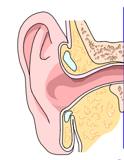
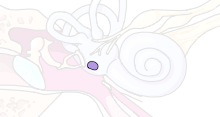
The Human Ear
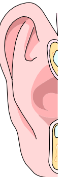
- The Outer Ear
- Pinna is a Convoluted funnel of cartilage that is attached to the side of the head. Its function is to help
collect sound vibrations near the opening of the ear; directs sound waves into the external auditory
canal.
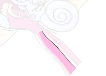
- External Auditory Canal: an air filled, s-shaped, 1 inch long passageway connected to the tympanic
membrane. Its function is for sound waves to be funneled down the canal and to be amplified; its
natural acidity helps to protect against infections; secretes cerumen to protect canal from drying out.
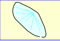
- tympanic membrane: a thin, tough flexible, fibrous membrane, approximately 1/3” in diameter
attached to the external auditory canal. Grey in color and cone-shaped resting on90-degree angle into
the middle ear cavity. Its function is sound waves from the external auditory canal hit this membrane
and cause it to vibrate; reproduces frequency and form of sound wave.
- Pinna is a Convoluted funnel of cartilage that is attached to the side of the head. Its function is to help
collect sound vibrations near the opening of the ear; directs sound waves into the external auditory
canal.
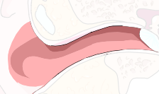
- The Middle Ear
- Eustachian Tube: 1 ¾ inch-long tube that connects the middle ear with the back of the nasal cavity;
normally closed except when swallowing or yawning. Its function is that it equalizes air pressure insde
and outside the tympanic membrance and allows the drainage of normal and diseased middle ear
secretions.
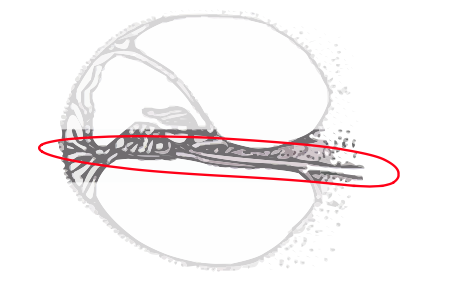
- Ossicles; Function of Auditory ossicles: form the mechanical links between the tympanic membrane
and the inner ear; deliver sound vibration to inner ear fluids; amplify airborne sound by
approximately 30dB.
- Malleus: a small bone (hammer shaped); its handle attaches to the tympanic membrane at a place
called the umbo. Its other end has a round head.
- Incus: a small bone (anvil shaped) in which the malleus rests and then connects to the stapes.
- Stapes: a small bone (stirrup-shaped) which is connected to the incus, and with its oval footplate
connects to the oval window of the ear.
- Malleus: a small bone (hammer shaped); its handle attaches to the tympanic membrane at a place
called the umbo. Its other end has a round head.
- stapedius muscle
- tensor tympani muscle
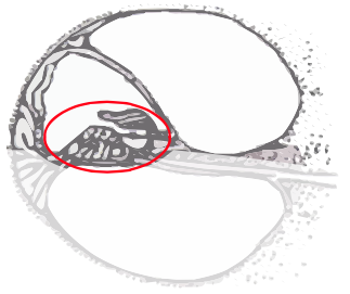
- Oval window: is a membrane that connects the middle ear with the upper half of the cochlea. Its
function is that vibrations from the ossicles are transferred to the cochlea via the action of the stapedial
footplate in the oval window.
- Round window: membrane that connects the middle ear with the lower half oof the cochlea;
simultaneous outward motion of the round window called the round window reflex. Its function is the
aid in fluid motion within the cochlea and serves to equalize the hydraulic pressure.
- Eustachian Tube: 1 ¾ inch-long tube that connects the middle ear with the back of the nasal cavity;
normally closed except when swallowing or yawning. Its function is that it equalizes air pressure insde
and outside the tympanic membrance and allows the drainage of normal and diseased middle ear
secretions.
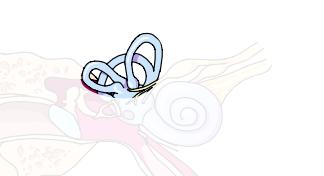
- The Inner ear
- semicircular canals (anterior, posterior, lateral);embedded in temporal bone; three separate loops;
contain clear, watery liquid. Its function is that hair cells within canals perceive sense of balance and
position in space; fluid flows in certain directions when you move your head; different movements
affect different canals; aids in maintaining balance and has nothing to do with hearing.
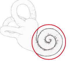
- Cochlea: a boney, spiral-shaped cavity coiled 2 ½ times; filled with fluid and divided into three sections;
pea-sized; houses organ of Corti. Its function is that it converts stimulus from outside environment into
nerve impluses for transmission to the brain.
- Lower Channel
- Middle Channel
- Upper channel
- Organ of Corti: center of cochlea; contains 15000 to 20000 microscopic hairs which, when displaced by
fluid movement, are the hydromechancal energy.
- Basilar membrane: divides cochlea lengthwise; final destination for vibrations that enter the ear, vibrate
in same pattern as sound waves
- semicircular canals (anterior, posterior, lateral);embedded in temporal bone; three separate loops;
contain clear, watery liquid. Its function is that hair cells within canals perceive sense of balance and
position in space; fluid flows in certain directions when you move your head; different movements
affect different canals; aids in maintaining balance and has nothing to do with hearing.
Media attachments
Want to create your own Mind Maps for free with GoConqr? Learn more.