551561
Blood
- Physical characteristics
and volume
- Blood is a sticky, opaque fluid with a metallic taste
Colour varies from scarlet (oxygen rich) to dark red
(oxygen poor) Temperature is 38 C slightly higher
than normal body temperature Blood accounts for
approximately 8% of body weight
- Blood is a sticky, opaque fluid with a metallic taste
Colour varies from scarlet (oxygen rich) to dark red
(oxygen poor) Temperature is 38 C slightly higher
than normal body temperature Blood accounts for
approximately 8% of body weight
- Circulating volume
- In a typical adult the circulating volume is
equivalent to the cardiac output per minute
- Cardiac Output = heart rate x stroke volume
- Cardiac Output = heart rate x stroke volume
- In a typical adult the circulating volume is
equivalent to the cardiac output per minute
- Normal blood pH
- 7.35-7.45
- 7.35-7.45
- Normal blood glucose
- 4-7mmol (FIVE)
- 4-7mmol (FIVE)
- Normal Blood Plasma Potassium
- 3.5-5.5 mEq/L (FIVE)
- 3.5-5.5 mEq/L (FIVE)
- Normal blood plasma sodium
- 135 to 145 (mEq/L)
- 135 to 145 (mEq/L)
- Normal carbon dioxide
- 4.7-6.0 Kpa (FIVE)
- 4.7-6.0 Kpa (FIVE)
- Full blood count
- Haemoglobin is the
coloured pigment inside
red blood cells that
carries oxygen round
the body.
Haemoglobin levels in
the blood are
measured in grammes
per 100 millilitres,
which is abbreviated to
g/dl. The normal
range of haemoglobin
for a man is 13.5 to
17.5 g/dl and for a
woman is 11.5 to 15.5
g/dl. Anything less
than these numbers is
called anaemia.
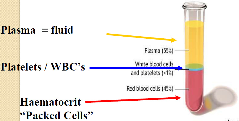
- Haematocrit
is the volume
percentage
(%) of red
blood cells in
blood.
Relative
volume of
blood
occupied by
erythrocytes
(normal 45%
for men 40
for women)
- Platelets
- A platelet count is a diagnostic test that determines the number of platelets in the patient's blood. Platelets, which are also
called thrombocytes, are small disk-shaped blood cells produced in the bone marrow and involved in the process of blood
clotting. There are normally between 150,000-450,000 platelets in each microliter of blood. Low platelet counts or abnormally
shaped platelets are associated with bleeding disorders. High platelet counts sometimes indicate disorders of the bone marrow.
- Prothrombin time
- The prothrombin time test belongs to a group of blood tests that assess the clotting ability of blood. The test is also known
as the pro time or PT test. The PT test is used to monitor patients taking certain medications (e.g. WARFARIN) as well as to
help diagnose clotting disorders. The PT test is used in combination with the partial thromboplastin time (PTT) test to screen
for hemophilia and other hereditary clotting disorders. The normal prothrombin time is 11-15 seconds
- Partial Thromboplastin time
- The partial thromboplastin time (PTT) test is a blood test that is done to investigate bleeding disorders and to
monitor patients taking an anticlotting drug (heparin). Liver disease decreases the production of clotting factors
increasing the PTT Heparin therapy increases the PTT Normal PTT results are between 35-45 seconds
- Haemoglobin is the
coloured pigment inside
red blood cells that
carries oxygen round
the body.
Haemoglobin levels in
the blood are
measured in grammes
per 100 millilitres,
which is abbreviated to
g/dl. The normal
range of haemoglobin
for a man is 13.5 to
17.5 g/dl and for a
woman is 11.5 to 15.5
g/dl. Anything less
than these numbers is
called anaemia.
- BLOOD CELLS
- Plasma
- Functions
- 1.Water: transport medium
- ◦carries heat ◦ nutrients,
wastes, gases, hormones
- ◦carries heat ◦ nutrients,
wastes, gases, hormones
- 2.Electrolytes
- ◦membrane excitability ◦ osmotic distribution of
fluid b/t ECF & ICF ◦ buffering of pH
- ◦membrane excitability ◦ osmotic distribution of
fluid b/t ECF & ICF ◦ buffering of pH
- 1.Water: transport medium
- Components
- electrolytes=
(Na+ & Cl-) 1%
- water= 90%
- Plasma Proteins 6-8 %
- Plasma Proteins: (albumins,
globulins, fibrinogen)
- 1. Maintaining colloid osmotic balance (albumins) 2.
Buffering pH changes 3. Transport of materials through blood
(such as water & hormones) 4. Antibodies (e.g. gamma
globulins, immunoglobulins) 5. Clotting factors (e.g.
fibrinogen)
- 1. Maintaining colloid osmotic balance (albumins) 2.
Buffering pH changes 3. Transport of materials through blood
(such as water & hormones) 4. Antibodies (e.g. gamma
globulins, immunoglobulins) 5. Clotting factors (e.g.
fibrinogen)
- Plasma Proteins: (albumins,
globulins, fibrinogen)
- Other components:
- • Nutrients (e.g. Glucose and amino
acids) • Hormones (e.g. Cortisol, thyroxine)
• Wastes (e.g. Urea) • Blood gases (e.g.
CO2, O2)
- • Nutrients (e.g. Glucose and amino
acids) • Hormones (e.g. Cortisol, thyroxine)
• Wastes (e.g. Urea) • Blood gases (e.g.
CO2, O2)
- electrolytes=
(Na+ & Cl-) 1%
- Functions
- 2. Leukocytes (White Blood Cells)
- Form the body’s defense system
against micro-organisms. Seek and
destroy functions: destroy invading
microorganisms destroy abnormal cells
(ie: cancer ). Clean up cellular debris
(phagocytosis) assist in injury repair
- Types of WBC’s
- Agranulocytes
- Monocytes
- • Exit blood (diapedesis) to become macrophages •2-6 % of the
WBC's •Phagocytic = defend against viruses and bacteria
- • Exit blood (diapedesis) to become macrophages •2-6 % of the
WBC's •Phagocytic = defend against viruses and bacteria
- Lymphocytes (B and T cells)
- * B-lymphocytes: produce antibodies * T-lymphocytes:
directly destroy micro- organisms * 25-33 % of the WBC's
- * B-lymphocytes: produce antibodies * T-lymphocytes:
directly destroy micro- organisms * 25-33 % of the WBC's
- Monocytes
- Granulocytes
- neutrophils= 50-70% of all leukocytes (most abundant of
WBC’s) Important in inflammatory responses Phagocytic
(engulfs and absorbs waste material, harmful
microorganisms, or other foreign bodies in the bloodstream
and tissues.)
- Eosinophils= * 1-4% of the WBC's * Attack
parasitic worms * Important in allergic reactions
- Basophils= * Release histamine * 0.5% of the
WBC's * Contribute to allergic reactions
- neutrophils= 50-70% of all leukocytes (most abundant of
WBC’s) Important in inflammatory responses Phagocytic
(engulfs and absorbs waste material, harmful
microorganisms, or other foreign bodies in the bloodstream
and tissues.)
- Agranulocytes
- Types of WBC’s
- Form the body’s defense system
against micro-organisms. Seek and
destroy functions: destroy invading
microorganisms destroy abnormal cells
(ie: cancer ). Clean up cellular debris
(phagocytosis) assist in injury repair
- 1. Erythrocytes (Red Blood Cells)
- Shape - a biconcave disc with large surface area
- No Nucleus
- Contains hemoglobin
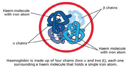
- Structure of haemoglobin
- 4 Haem molecules = carry gases.
Oxygenated haemoglobin : bright
Red (systemic) *Deoxygenated
Hemoglobin: Blue (venous
circulation)
- 4 Haem molecules = carry gases.
Oxygenated haemoglobin : bright
Red (systemic) *Deoxygenated
Hemoglobin: Blue (venous
circulation)
- Structure of haemoglobin
- Approximately 250 ml of oxygen are used every minute by a
conscious resting person (oxygen consumption) and therefore
about 25% of the arterial oxygen is used every minute.
- Primary Function = Transport oxygen from the lungs to
the cells of the body & assist with CO2 removal from
cells to lungs
- Short Life Span (~120 days) ◦Fragile - prone to rupture.
Ruptured RBCs: ◦are destroyed in spleen ◦phagocytic
WBC’s “clear the debris” up. Ruptured cells must be
replaced by new cells by: eythropoietin-from kidneys by
process called erythropoiesis in bone marrow
- Shape - a biconcave disc with large surface area
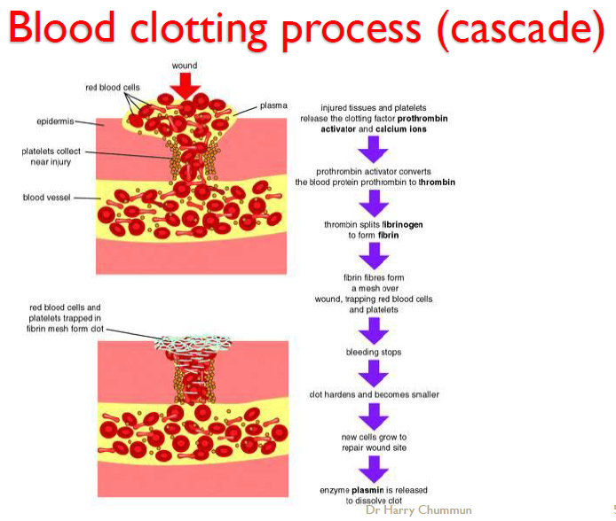
- 3. Thrombocytes (Platelets)
- Stop bleeding from a damaged vessel through:
◦Haemostasis by 3 steps: vascular Spasm formation of
a platelet plug blood coagulation (clotting)
- Stop bleeding from a damaged vessel through:
◦Haemostasis by 3 steps: vascular Spasm formation of
a platelet plug blood coagulation (clotting)
- Plasma
- Total Blood Volume
- 8 % of body weight. ◦2.75 / 5.5 liters of blood is
plasma ◦(remaining is the cellular portion)
- 8 % of body weight. ◦2.75 / 5.5 liters of blood is
plasma ◦(remaining is the cellular portion)
- FUNCTION OF BLOOD
- Defense: Foreign organisms
Injury/infection Clotting process
Body temperature
- Transports: Nutrients O2 & CO2 Waste Products
Hormones Electrolytes. Oxygen, Carbon Dioxide
Food Heat Waste Hormones Disease Clotting
- Both defense and transports
maintain Homeostasis
- Both defense and transports
maintain Homeostasis
- Transports: Nutrients O2 & CO2 Waste Products
Hormones Electrolytes. Oxygen, Carbon Dioxide
Food Heat Waste Hormones Disease Clotting
- Defense: Foreign organisms
Injury/infection Clotting process
Body temperature
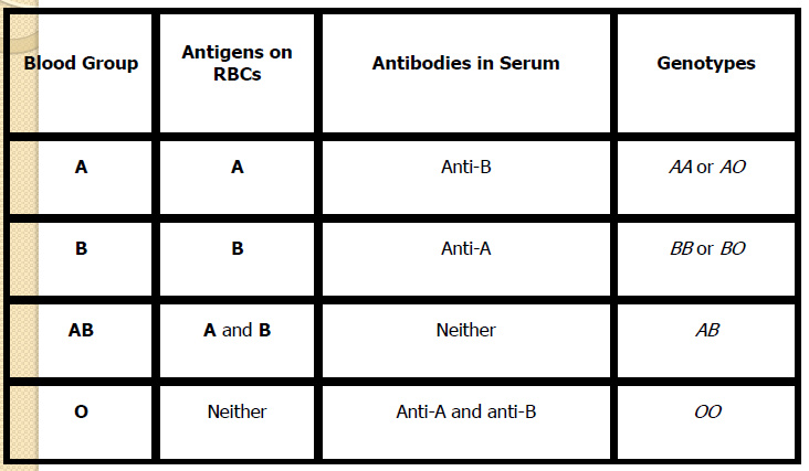
- The ABO Blood Groups System
- Discovered in 1901 by Dr. Karl Landsteiner 4
Main Phenotypes (A, B, AB, O) ABO gene
located on long arm of chromosome 9
- Antigens & Antibodies
- Universal Donor and Recipient
- Universal Donor Group O ◦Carries no A or B antigens
◦Packed and processed units have little antibody
- Universal Recipient Group AB ◦Patient has no anti-A or anti-B present
◦Cannot lyse any transfused cells ◦Beware: other ◦antibodies may be present
- Universal Donor Group O ◦Carries no A or B antigens
◦Packed and processed units have little antibody
- The Rh(D) Antigen
- Rh is the most complex system, with
over 45 antigens Discovered in 1940
after work on Rhesus monkeys
- Significance of Rh(D)
- 80% of Rh(D) –ve persons exposed to Rh(D) +ve
blood will develop anti-D Anti-D can also be
stimulated by pregnancy with an Rh(D) +ve baby
Rh(D) -ve women of childbearing potential should
never be given Rh(D) +ve blood products
- 80% of Rh(D) –ve persons exposed to Rh(D) +ve
blood will develop anti-D Anti-D can also be
stimulated by pregnancy with an Rh(D) +ve baby
Rh(D) -ve women of childbearing potential should
never be given Rh(D) +ve blood products
- Significance of Rh(D)
- Rh is the most complex system, with
over 45 antigens Discovered in 1940
after work on Rhesus monkeys
- Universal Donor and Recipient
- Discovered in 1901 by Dr. Karl Landsteiner 4
Main Phenotypes (A, B, AB, O) ABO gene
located on long arm of chromosome 9
Media attachments
Want to create your own Mind Maps for free with GoConqr? Learn more.