6180467
Description
Mind Map by Mason Hayward , updated more than 1 year ago
|
|
Created by Mason Hayward
over 9 years ago
|
|
Tissues
- Cells are organized in layers or groups to form tissues
- intercellular junctions are specialized connect cells.
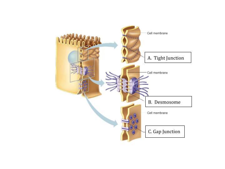
- Tight junction → Close space between cells
by fusing cell membranes
- Desmosomes → Bind cells by forming “spot welds”
between cell membranes
- Gap Junction → Form tubular channels
between cells that allow the exchange of
substances
- Tight junction → Close space between cells
by fusing cell membranes
- intercellular junctions are specialized connect cells.
- Four types of human tissue
- Epithelial
- Function → protection,secretion,absorption,excretion
Location → covers body surface, covers and lines internal
organs, compose glands. Characteristics → lack blood
vessels, cells readily divide, cells are tightly packed
together.
- Function → protection,secretion,absorption,excretion
Location → covers body surface, covers and lines internal
organs, compose glands. Characteristics → lack blood
vessels, cells readily divide, cells are tightly packed
together.
- Connective
- Function → bind,support,protect,fill spaces,store
fat,produce blood cells Location → widely distributed
throughout the body Characteristics → Mostly have a
good blood supply, cells are farther apart than epithelial
cells, extracellular matrix in betw
- Function → bind,support,protect,fill spaces,store
fat,produce blood cells Location → widely distributed
throughout the body Characteristics → Mostly have a
good blood supply, cells are farther apart than epithelial
cells, extracellular matrix in betw
- Muscle
- Function → movement Location → attached to
bones, in the walls of hollow internal organs, heart
Characteristics → Able to contract in response to
specific stimuli
- Function → movement Location → attached to
bones, in the walls of hollow internal organs, heart
Characteristics → Able to contract in response to
specific stimuli
- Nervous
- Function → conduct impulses Location → brain,
spinal cord, nerves Characteristics → cells
communicate with each other and other body parts
- Function → conduct impulses Location → brain,
spinal cord, nerves Characteristics → cells
communicate with each other and other body parts
- Epithelial Tissues → Epithelium
- Location Covers all free body surfaces Forms
the inner lining of body cavities Lines hollow
organs
- A basement membrane anchors epithelium
to connective tissue.
- Cancer cells secrete a substance that
dissolves the basement membrane,enabling
the cells to invade other tissue layers
(metastasis)
- Cancer cells also produce fewer adhesion proteins (help cells to
“stick” together) which allows them to spread into surrounding
tissues.
- Cancer cells also produce fewer adhesion proteins (help cells to
“stick” together) which allows them to spread into surrounding
tissues.
- Cancer cells secrete a substance that
dissolves the basement membrane,enabling
the cells to invade other tissue layers
(metastasis)
- Location Covers all free body surfaces Forms
the inner lining of body cavities Lines hollow
organs
- COVERING & LINING EPITHELIA
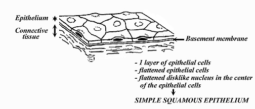
- Simple Squamous
- D: One thin layer of flat cells (easily
damaged) F: Diffusion,Filtration ,secretion
L:Avoli of the lungs,kidney glomeruli,lining
the heart
- D: One thin layer of flat cells (easily
damaged) F: Diffusion,Filtration ,secretion
L:Avoli of the lungs,kidney glomeruli,lining
the heart
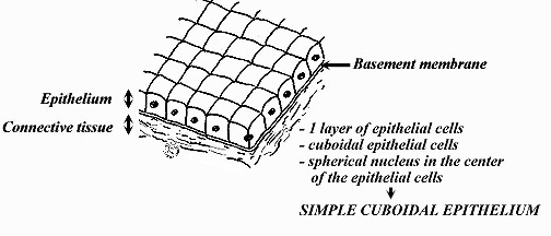
- Simple Cuboidal
- D: One layer of cube shaped cells
F:Secretion and absorption L:Small
glands,kidney tubules and ovary
surfaces
- D: One layer of cube shaped cells
F:Secretion and absorption L:Small
glands,kidney tubules and ovary
surfaces
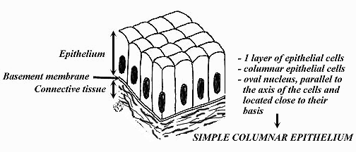
- Simple Columnar
- D: One layer of elongated cells. They can be ciliated or nonciliated.
(Ciliated = tiny hairs)(non ciliated= contains microvilli) F: Absorption
and Secretion L: non ciliated Found mainly in the lining of the
digestive tract. Ciliated Found in the small broncus tubes.
- D: One layer of elongated cells. They can be ciliated or nonciliated.
(Ciliated = tiny hairs)(non ciliated= contains microvilli) F: Absorption
and Secretion L: non ciliated Found mainly in the lining of the
digestive tract. Ciliated Found in the small broncus tubes.
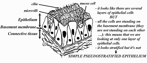
- Simple Pseudostratified
- D: One layer of cells that are attached
basement membrane, cells at different heights
F: Secretion and movement of mucus L: Ciliated
in respiratory tract
- D: One layer of cells that are attached
basement membrane, cells at different heights
F: Secretion and movement of mucus L: Ciliated
in respiratory tract
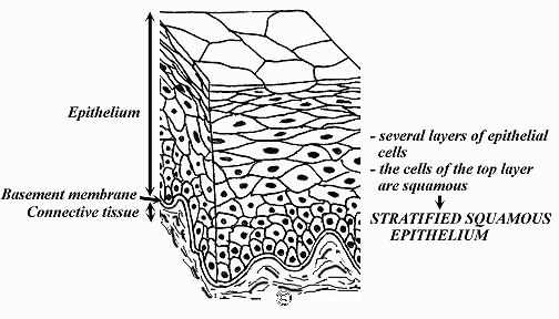
- Stratified Squamous
- D: Several layers of cells,top layers is flattened,thickest
layer of epithelial tissue F: Protection L: Keratinized
found on epidermis nonkeratinized found on mouth,
- D: Several layers of cells,top layers is flattened,thickest
layer of epithelial tissue F: Protection L: Keratinized
found on epidermis nonkeratinized found on mouth,
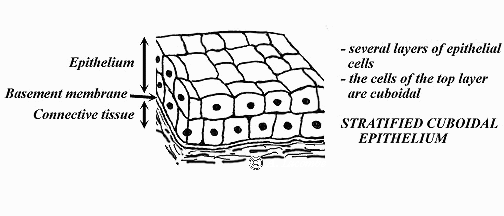
- Stratified Cuboidal
- D: Several cube shape layer F: protection
L: Found in largest ducts of sweat glands
- D: Several cube shape layer F: protection
L: Found in largest ducts of sweat glands
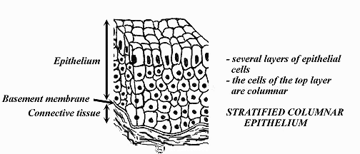
- Stratified Columnar
- D: Several layers,top layer contains elongated cells F:
Protection and secretion L:Very rare in the body,it lines part
of the urethra,large ducts of some glands
- D: Several layers,top layer contains elongated cells F:
Protection and secretion L:Very rare in the body,it lines part
of the urethra,large ducts of some glands
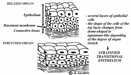
- Stratified Transitional
- D: Several layers of cells that can be stretched.
F:Allows for distention of urinary organs L: It is
found lining surfaces of organs subjected to
stretch, such as the bladder, the ureters and part
of the urethra.
- D: Several layers of cells that can be stretched.
F:Allows for distention of urinary organs L: It is
found lining surfaces of organs subjected to
stretch, such as the bladder, the ureters and part
of the urethra.
- Simple Squamous
- Epithelial
- Connective tissue
- Connective tissue connect, supports, protects, provides frameworks, fills spaces, stores fat, produces blood
cells, protects against infection, and helps repair damaged tissues. Connective tissue cells usually have
considerable extracellular matrix between them. This extracellular matrix consists of fibers and a ground
substance (gel-like material).
- Fibroblasts produce collagen and elastic fibers Macrophages are phagocytes (“eat cells”) Mast cells may release heparin and
histamine
- Major Cell Types
- Connective Tissue Fibers
- Collagen fibers have a great tensile strength. Elastic fibers are composed of elastin and are stretchy. Reticular
fibers are fine collagen fibers.
- Collagen fibers have a great tensile strength. Elastic fibers are composed of elastin and are stretchy. Reticular
fibers are fine collagen fibers.
- Loose Connective Tissue
- Areolar → forms thin membranes between organs and binds them together. Found beneath the skin and
surrounds organs Adipose → stores fat, cushions, and insulates. Found beneath the skin; in certain
abdominal membranes; and around the kidneys, heart, and various joints. Reticular → thin branched
reticular fibers. Supports the walls of the liver and spleen.
- Dense Connective Tissue
- Dense Regular → strong collagen fibers that bind structures as parts of tendons and ligaments. Dense
Irregular → thicker, randomly distributed collagen fibers and is found in the dermis.
- Dense Regular → strong collagen fibers that bind structures as parts of tendons and ligaments. Dense
Irregular → thicker, randomly distributed collagen fibers and is found in the dermis.
- Connective tissue connect, supports, protects, provides frameworks, fills spaces, stores fat, produces blood
cells, protects against infection, and helps repair damaged tissues. Connective tissue cells usually have
considerable extracellular matrix between them. This extracellular matrix consists of fibers and a ground
substance (gel-like material).
Media attachments
Want to create your own Mind Maps for free with GoConqr? Learn more.