6374593
Early Embryology
- Time Lines
- Zygote
Annotations:
- Between 12 - 24 hours
- Morula
Annotations:
- At the 16 cell stage, approximately 3 days after fertilization
- Inner Cell Mass
Annotations:
- Divides into the epiblast and hypoblast
- Epiblast
Annotations:
- Forms all tissues of embryo and lining of newly developed amniotic sac
- Notocord
Annotations:
- Specialized cells that migrate through the primitive node between the endo and ectoderm. They travel cranially and caudally along the midline to form tubular aggregates of cells. These provide structure to developing embryo until the vertebrae is formed and induces development of the CNS.
- Amnion
Annotations:
- The cavity formed above the epiblast and lined by a thin layer of epiblast cells. Fills rapidly with fluid and wraps around the embryo. It surrounds the umbilical vein as it passes from placenta to embryo
- - To Protect against mechanical shock - Allows for symmetrical growth - Prevents adherence of fetus to amnion - Maintains consistent temperature - Important for respiratory development - Barrier to infection - Homeostasis of fluid and electrolytes
- Hypoblast
Annotations:
- Will form embryonic yolk sac, which is essentially the former blastocyst cavity
- Yolk Sac
Annotations:
- Yolk sac is used to produce early red blood cells
- Outer Cell Mass
- Blastocyst
Annotations:
- When the morula fills with fluid. This is what enters the uterine cavity. Zona pellucida dissolves to make ready for implantation.
- Zygote
- Implantation
Annotations:
- Starts to occur around 5 - 6 days after implantation. Completed by the second week. Usually implants in the anterior or posterior wall of the uterus
- Trophoblasts
Annotations:
- Responsible for implantation. Mutlilayered, multinucleated structures from the outer cell mass. They erode the endometerium to implant deeper. Will form lacunae and produces hCG
- Placenta
Annotations:
- Developed from trophoblast cells. Their job is to secrete enzymes that digest the endometrial wall for implantation, and enlarges around the embryos. It erodes endometrial vessels to form trophoblastic lacuna to allow for early maternal-fetal exchange
- The placenta is the chorionic villi and the associated endometrium
- Function is placentral transfer and hormone production
- Placental
Transfer
Annotations:
- Gases, nutrients, waste products, hormones, electrolytes, maternal antibodies (IgG), drug metabolites and some infectious agents can cross
- Hormone Production
- hCG
Annotations:
- This is the hormone measured in pregnancy tests. It is usually detectable about 1 - 2 weeks after fertilization
- The job is to maintain the corpus luteum
- Progesterone
Annotations:
- Job is to maintain the endometrium
- Estrogen
- Human Placental Lactogen
- Chorionic Thyrotropin
- Corticotropin
- hCG
- Chorionic Villi
Annotations:
- Contain embryonic vessels. They are finger-like projections that will become the site of maternal-fetal exchange beside lacunae and branches many times to increase the surface area for exchange.
- Lacunae
Annotations:
- Fill with maternal blood and allow for exchange of oxygen, carbon dioxide, nutrients, waste products, hormones, maternal antibodies, electrolytes, some harmful agents
- Ectopic Pregnancy
Annotations:
- Any pregnancy in an abnormal location. Most common are in the uterine tube but can also occur in the abdominal cavity, cervix and ovary. The embryo will die and will result in severe bleeding and abdominal pain.
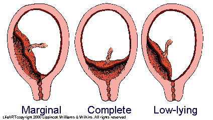
- Placenta Previa
Annotations:
- May result from implantation at the cervix. The placenta covers the cervical opening and is high risk for severe bleeding. Classified as either marginal, complete or low-lying.
- Gastrulation
Annotations:
- The formation of the 3 germ layers from epiblast into ecto, meso and endoderm. When cells from the epiblast migrate to the midline at the caudal end of the embryo, they form a thick band known as the "Primitive Streak" which elongates cranially. The cranial tip is known as the "Primitive Node"
- Ectoderm
Annotations:
- When cells from the primitive streak and node do not migrate. Derivatives: epidermis, brain, spinal cord, peripheral nervous system, autonomic nervous system and skull
- Mesoderm
Annotations:
- When cells from the primitive streak and node migrate downward and fill the space between the epiblast and hypoblast. Derivatives are: Skeletal muscle, heart and blood vessels, kidneys and gonads, skeleton, most connective tissue
- Paraxial Mesoderm
Annotations:
- Found along the midline
- Somites
Annotations:
- Starting around the 3rd week. First pair develops in the occipital region and continues cranio-caudal. By end of the 5th week there are 42 - 44 pair. Divided into 3 subunits
- Sclerotome
Annotations:
- Found next to the neural tube. Migrates around the neural tube and notocord to form vertebrae and ribs
- Myotome
Annotations:
- The part adjacent to neural tube forms epimeres. The lateral part forms hypomeres
- Epimeres
Annotations:
- Develops into the intrinsic muscles of the back
- Hypomeres
Annotations:
- Develops into muscles of the body wall and limbs, except the head and neck
- Dermatome
Annotations:
- Migrate to the dorsal region of the trunk to become the dermis of the back (hair follicles, glands, fatty tissue, blood vessels)
- Somatomeres
- Lateral Plate Mesoderm
Annotations:
- Extends laterally
- Somatic (Parietal) Layer
Annotations:
- With folding will end up along the body wall of the embryo under the ectoderm. Forms the many structures of the body wall and limbs
- Splanchnic (Visceral) Layer
Annotations:
- With folding will wrap around the part of the yolk sac that gets incorporated into the body cavity to form the serous layer around organs.
- Intermediate Mesoderm
Annotations:
- Between the paraxial and lateral mesoderms. Forms urogenital structures and gonads
- Endoderm
Annotations:
- When cells from the primitive streak and node migrate downward and displace the hypoblast. Derivatives: inner lining of gut tubes, respiratory tracts, bladder, some GI organs
- Maternal-Fetal Circulation
Annotations:
- 1. Endometrial Spiral Arteries 2. Intervillous Spaces / Lacunae 3. Chorionic Villi / Fetal Capillaries 4. Umbilical Vein 5. Fetus 6. Umbilical Arteries 7. Chorionic Villi / Fetal Capillaries 8. Intervillous Space / Lacunae 9. Endometrial Veins
Media attachments
Want to create your own Mind Maps for free with GoConqr? Learn more.