6847529
Description
Mind Map by Farah Mansour, updated more than 1 year ago
|
|
Created by Farah Mansour
over 7 years ago
|
|
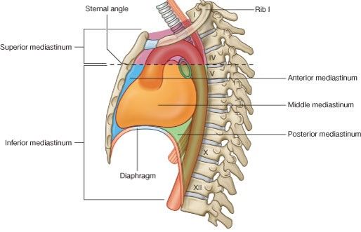
Inferior
Mediastinum
- Anterior
Mediastinum
- Behind the body and xiphoid process of the sternum
and in front of the middle mediastinum
- Boundaries
- Anterior : the body of the sternum.
- Posterior : middle mediastinum (the pericardium)
- On each side : the mediastinal layer of pleura.
- Anterior : the body of the sternum.
- Contents
- Superior and inferior sternopericardial ligaments.
- Part of the thymus gland
- Important component of the lymphatic system.
- Lies behind the manubrium sterni & may extend up
into the neck or down in the anterior mediastinum.
- It appears large at time of birth, gradually replaced
by fat in adult life.
- Important component of the lymphatic system.
- Internal mammary artery
- Two to three lymph nodes and lymphatics.
- Sternocostalis muscle. (part of transversus
thoracis muscles)
- Areolar tissue.
- Superior and inferior sternopericardial ligaments.
- Behind the body and xiphoid process of the sternum
and in front of the middle mediastinum
- Middle
Meiastinum
- Is the part of the inferior mediastinum that is
occupied by the pericardium and its contents.
- Boundaries
- Superiorly: the imaginary horizontal plane
- Inferiorly: diaphragm.
- Anteriorly: anterior mediastinum.
- Posteriorly: posterior mediastinum
- Superiorly: the imaginary horizontal plane
- Contents
- The pericardium and the heart.
- Ascending aorta
- The pulmonary trunk
- The lower half of the SVC
- The upper most part of the IVC
- The four pulmonary veins.
- Bifurcation of pulmonary trunk into
right and left pulmonary arteries.
- Bifurcation of the trachea.
- The right and left phrenic nerves: along the sides of the pericardium.
- Inferior tracheobronchial lymph nodes: below the bifurcation of the trachea.
- The pericardium and the heart.
- Is the part of the inferior mediastinum that is
occupied by the pericardium and its contents.
- Posterior
Mediastinum
- Between the pericardium and the vertebral column
- Boundaries
- Superiorly: the imaginary horizontal plane
- Inferiorly: diaphragm.
- Anteriorly: the pericardium above , and the diaphragm below.
- Posteriorly: the lower 8 thoracic vertebrae (from the 5th to the 12th).
- Superiorly: the imaginary horizontal plane
- Contents
- Descending thoracic aorta.
- (the aorta is ascending in the middle
mediastinum, arch in the superior, and
descending in the posterior)
- (the aorta is ascending in the middle
mediastinum, arch in the superior, and
descending in the posterior)
- Esophagus: on the right side of
the aorta, then crosses in front
of its lower part
- This is why the aortic opening of the diagram is the most
posterior (level of T12), while the one anterior to it is the
esophageal (level of T10) – vena cava opening is at T8)
- This is why the aortic opening of the diagram is the most
posterior (level of T12), while the one anterior to it is the
esophageal (level of T10) – vena cava opening is at T8)
- Azygos and hemiazygos veins.
- The two vagi nerves:
- The right vagus >> posterior gastric nerve
- the left vagus >> anterior gastric nerve (due to the rotation of
the stomach during the development of the GIT)
- The right vagus >> posterior gastric nerve
- The splanchnic nerves (greater, lesser and lowest splanchnic
nerves): they arise from the sympathetic trunk.
- The thoracic duct: ascends on the right side of the esophagus
till the level of the sternal angle where it becomes on the left
side of the esophagus.
- Posterior mediastinal lymph nodes.
- Descending thoracic aorta.
- Between the pericardium and the vertebral column
Media attachments
Want to create your own Mind Maps for free with GoConqr? Learn more.