8016586
Description
Mind Map by Connor Scott, updated more than 1 year ago
|
|
Created by Connor Scott
almost 9 years ago
|
|
The Spleen
- Gross Anatomy
- Surface anatomy
Annotations:
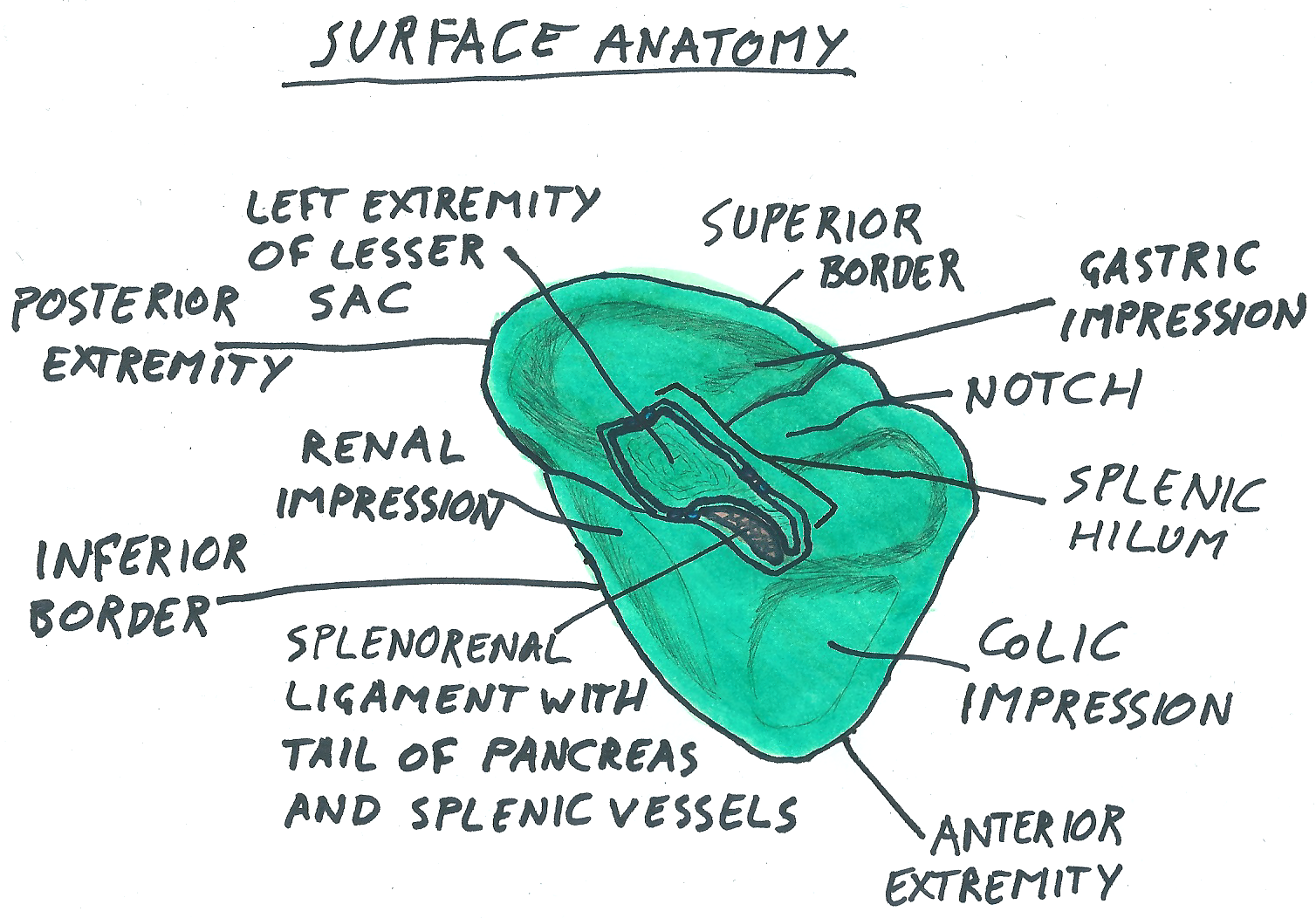
- Skandalakis et al. (2004) state that the spleen can be tetrahedral or triangular in shape, but is more commonly a wedge-shaped structure with a convex lateral surface, and a concave medial surface (pictured).
- The splenic hilum is located on the medial (or visceral) surface (pictured), which relates to the fundus of the stomach, the left kidney, the tail of the pancreas and the left colic flexure.
- The lateral surface is convex and fits into the concave space created by the diaphragm and ribs posterior to the spleen.
- The anterior and superior borders are notched whereas the posterior and inferior borders are rounded.
- Capsule
Annotations:
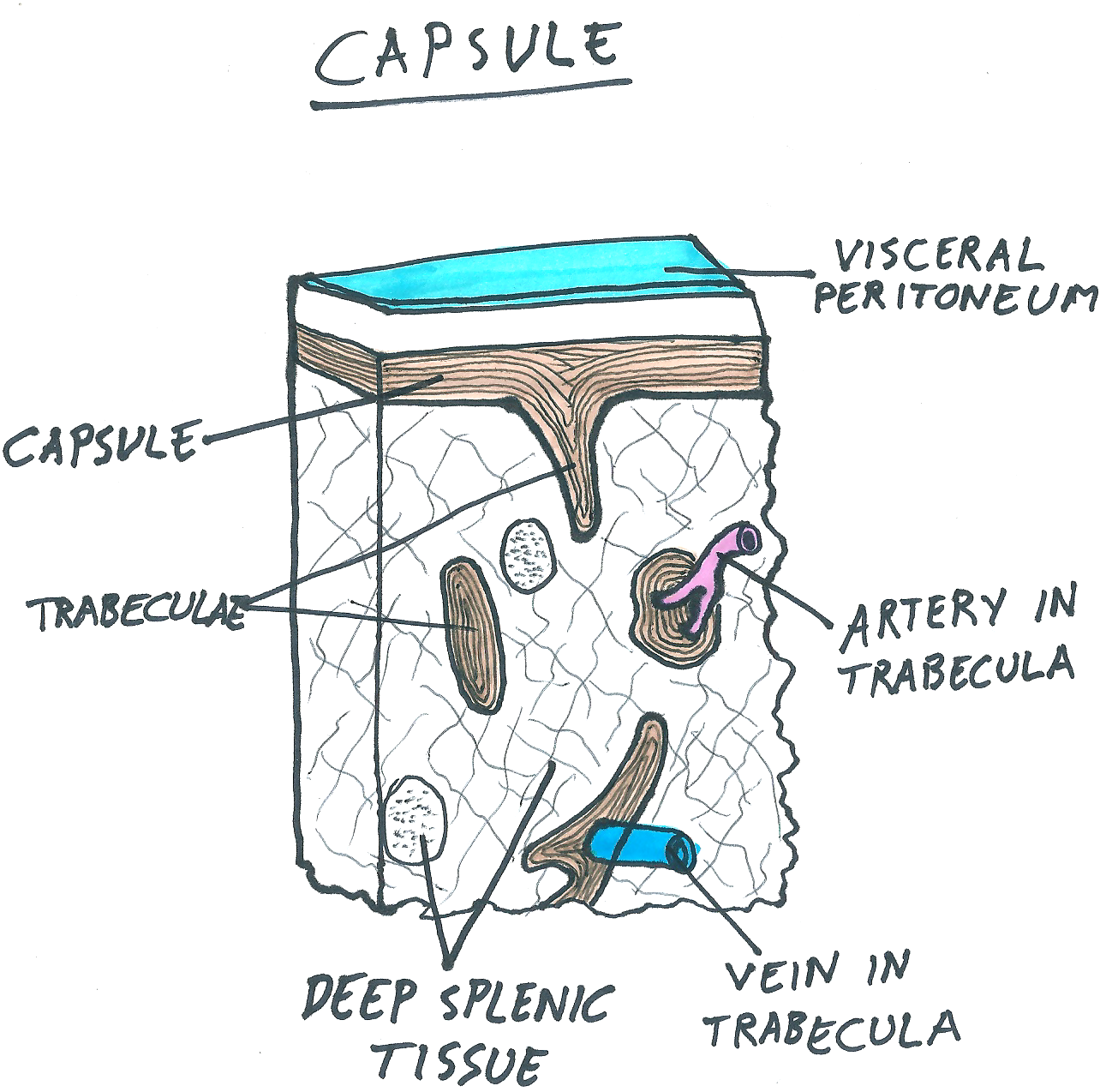
- A fibroelastic capsule composed of dense, irregular connective tissue surrounds the spleen and is thickened around the hilum. This layer is covered again by a layer of visceral peritoneum (pictured).
- Portions of the capsule project into the spleen and are called trabeculae. These projections carry blood vessels to and from the deep tissue of the spleen.
- The capsule and trabeculae contain smooth muscle, which periodically contract. This regulates the flow of blood into and out of the spleen.
- Innervation
Annotations:
- The nerves of the spleen come from the celiac plexus and branch out along the length of the splenic artery.
- These nerves provide sympathetic and parasympathetic innervation and carry out vasomotor functions.
- Surface anatomy
- Function
Annotations:
- The spleen has two main functions: - Immune system regulation and response and lymphocyte proliferation. - Recycling of old red blood cells.
- The immune function is carried out by the white pulp whilst red blood cell recycling is done by the red pulp.
- White Pulp
Annotations:
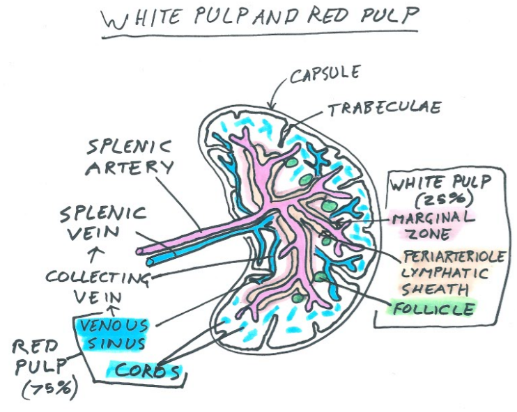
- The white pulp surrounds the arteries in the spleen and is composed of the periarteriole lymphatic sheath, marginal zone, and follicles (pictured). This tissue carries out immune responses and are important in producing antibodies.
- The periarteriole lymphatic sheath is a lymphoid tissue containing T cells, macrophages, and other immune cells.
- The marginal zone is another lymphoid tissue which surrounds the periarteriole lymphatic sheath. The primary cells present in the marginal zone are macrophages.
- Follicles are present around these other structures and contain B cells.
- Red pulp
Annotations:
- This system is associated with the venous system of the spleen which carries red blood cells out of the spleen. Red pulp is made up of venous sinuses and the cords of Billroth (pictured). This tissue is responsible for identifying and recycling old red blood cells.
- The venous sinuses transport the red blood cells to the collecting veins, which transport them to the splenic vein. The venous sinuses have slits that act as a selective barrier which cannot be traversed by old and deformed red blood cells.
- The cords sit between the ends of the arterioles and the venous sinuses. These are composed of macrophages mainly and act to break down old red blood cells.
- Ligaments
Annotations:
- The "ligaments" of the spleen are actually folds of peritoneum and a consensus has yet to be reached with regard to terminology associated with them.
- For example, Ellis (2013) and Mahadevan (2016) both agree on the terminology of the gastrosplenic ligament, but Ellis (2013) calls the splenorenal ligament (previously mentioned in the arterial supply section) the pancreatico-renal ligament. For this resource, this important ligament shall be referred to the splenorenal ligament.
- Other discrepancies exist regarding terminology but shall not be described in depth in this resource. The names of the ligaments are based on their attachements.
- The number of primary ligaments of the spleen also varies throughout the literature. Harris (2013) and Mahadevan (2016) only acknowledge two, whilst Dalpe and Cunningham (2003) describe seven major splenic ligaments. In this resource, four splenic ligaments described by Ostermann et al (1987) will be discussed.
- Gastrosplenic
Annotations:
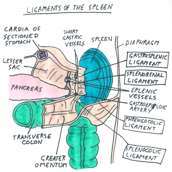
- This ligament attaches the greater curvature of the stomach to the spleen and grants passage to the stomach for the short gastric arteries and the left gastroepiploic artery. These are branches of the splenic artery.
- Splenorenal
Annotations:
- This ligament attaches the spleen to the left kidney. It is within this ligament that the splenic artery divides whilst on its way into the spleen. The splenic vein also passes within this ligament on exiting the spleen. The tail of the pancreas often penetrates this ligament (pictured).
- Phrenicocolic
Annotations:
- Attaching at the left colic flexure and passing below the spleen to the diaphragm at the level of the 10th and 11th ribs, the phrenicocolic ligament almost suspends the spleen.
- Splenocolic
Annotations:
- Continuous with the greater omentum, this ligament attaches the spleen to the transverse colon.
- Pathology
Annotations:
- The spleen is the most commonly injured organ in the abdomen, despite protection provided by ribs 9-12.
- Enlarged Spleen
Annotations:
- Pathology can result in swelling of the spleen, also known as splenomegaly. This is diagnosed by palpitation of the spleen which is not possible in the healthy individual. If the spleen can be felt inferior to the left costal margin, it is at least three times its normal size.
- Symptoms of splenomegaly are fatigue, jaundice and anemia. The risk of rupture is also increased.
- Treatment of the underlying cause of the enlarged spleen is normally the first approach. Failing this a splenectomy is performed.
- Wandering Spleen
Annotations:
- This is a rare condition caused by laxity or absence of the splenic ligaments. The condition results in an unusually mobile spleen which could cause torsion of the splenic vessels or rupture of the spleen. However, this is rare and around half of all cases are asymptomatic.
- Around 33% of cases are in children under the age of 10, thought to be congenital. In adults, women are more likely to get the condition than men, perhaps due to hormonal effects on the ligaments.
- In symptomatic and often dangerous cases, splenectomy is the treatment for wandering spleen.
- Rupture
Annotations:
- The close association between ribs 9-12 and the spleen can be problematic when these ribs fracture. The broken bone fragments can pierce the spleen and cause problems.
- Blunt force trauma to other areas of the abdomen can also be problematic as they can cause an increase in pressure in the abdomen. This may result in rupture of the splenic capsule and overlying peritoneum, as well as internal bleeding
- In rare cases, spontaneous splenic rupture may occur. This presents more insidiously than traumatic rupture, often resulting in poor prognosis. Other pathologies, such as infection or malignancy, are thought to be the cause of spontaneous splenic ruptures.
- The treatment for a ruptured spleen is usually removal of the organ, otherwise known as splenectomy. This can save the patient's life and prevent them from bleeding to death.
- Partial splenectomy is possible as the blood supply to specific areas is well defined (see "Arterial Supply" section). Patient's who have undergone total removal of the spleen can live normal lives as the splenic functions are also performed by the liver and bone marrow. However, susceptibility to certain bacterial infections is increased.
- How to use this resource
- Click the paper image at the top right of this bubble ^^
Annotations:
- This will open up notes on the topic in the bubble. Next, click the arrow at the top right of this note.
- This brings you to the next page of the notes on this topic. The number of pages are stated at the top when you first click the notepad.
- All bubbles with a note in the top right have information, so make sure you don't miss out on anything!
- To navigate around the mindmap, simply click anywhere on the screen and drag your mouse along in the direction you want to go.
- Zooming in and out
Annotations:
- The icon at the top of the resource next to the play icon can be used to zoom in and out as you navigate through the mindmap. This is especially useful when looking at images.
- Several images are located around the periphery of the mindmap. These relate to the topic within the closest proximity to it and are often used in the quizzes.
- Quizzes
Annotations:
- As well as notes attached to many of the bubbles, you may observe that each of the main bubbles coming off of the centre have paperclips in the top right corner.
- These paperclips contain short quizzes, relating to the information covered by the expansions of the bubble in which it resides. Simply click the paperclip, then press play to do the quiz. All of the answers are covered in this resource!
- Press Play
Annotations:
- At the top of the resource is an icon that allows you to see the creation of the mindmap bubble by bubble. You may alter the speed to suit yourself and it may give you a more global understanding of the whole resource.
- Click the paper image at the top right of this bubble ^^
- References
Annotations:
- Catalano OA, Soricelli A and Salvatore M. 2013. Abdominal imaging. Spleen anatomy, function and development. 2. 1479-1494.
- Dalpe C and Cunningham, M. 2003. Wandering spleen as an asymptomatic pelvic mass. Obstetrics and gynecology. 101(5 Pt 2), 1102-4.
- Ellis H. 2013. Anatomy of the pancreas and the spleen. Surgery (Oxford). 31(6), 263-266.
- Hasudungan A. 2017. Spleen Anatomy and Physiology. https://www.youtube.com/watch?v=RezL2xWFCe8&t=234s
- Kerr JB. 2010. Functional histology. 2nd ed. Sydney. Mosby
- Mahadevan V. 2016. Anatomy of the pancreas and spleen. Surgery (United Kingdom). 34(6), 261-265.
- Moore KL, Dalley AF amd Agur AMR. 2010. Clinically oriented anatomy. 6th ed. Philademphia. Lippincott Williams and Wilkins. Ostermann PA, Schreiber HW and Lierse W. 1987. The ligament system of the spleen and its significance for surgical interventions. Langenbecks Archiv für Chirurgie. 371(3), 207-16.
- Ostermann PA, Schreiber HW and Lierse W. 1987. The ligament system of the spleen and its significance for surgical interventions. Langenbecks Archiv für Chirurgie. 371(3), 207-16.
- Richman M, Hiyama DT and Wasson E. 2014. Wandering spleen. Surgery. 155(4), 728.
- Skandalakis PNS, Lee J, Kingsnorth AN, Colborn GL, Weidman TA, Hatch GF, Lauer RC, Skandalakis JE. Spleen. In: Skandalakis JE and Colborn GL, (eds). 2004. Skandalakis’ Surgical anatomy: the embryologic and anatomic basis of modern surgery. Athens: McGraw-Hill. 1230–87.
- Location and
Appearance
- Intraperitoneal
Organ
Annotations:
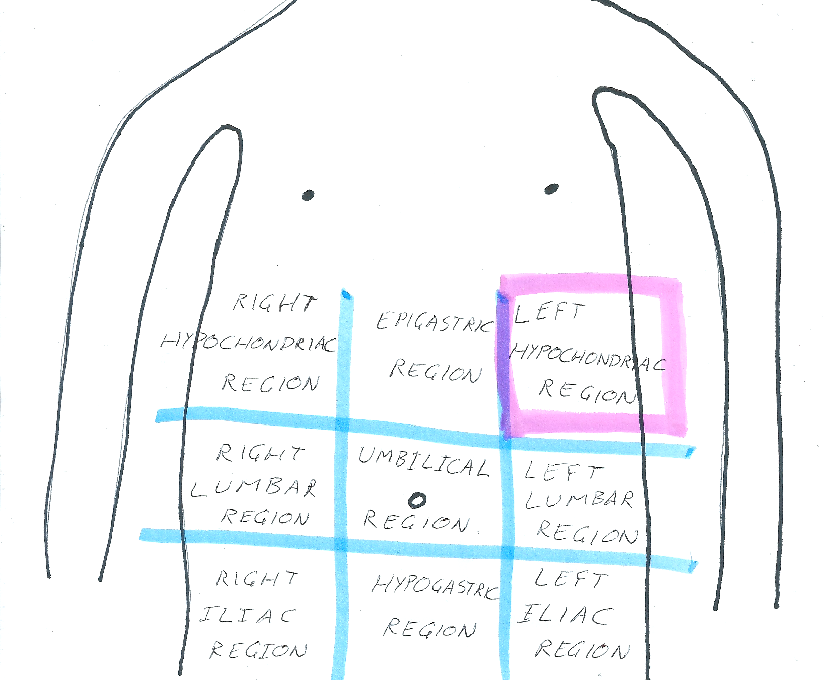
- The spleen is a soft, purple, ovoid organ that has been described as around the same size and shape as a fist.
- It is located in the upper left quadrant (hypochondrium) of the abdomen, protected by ribs 9-11.
- The spleen is the most vulnerable organ in the abdomen and the largest lymphoid organ in the body.
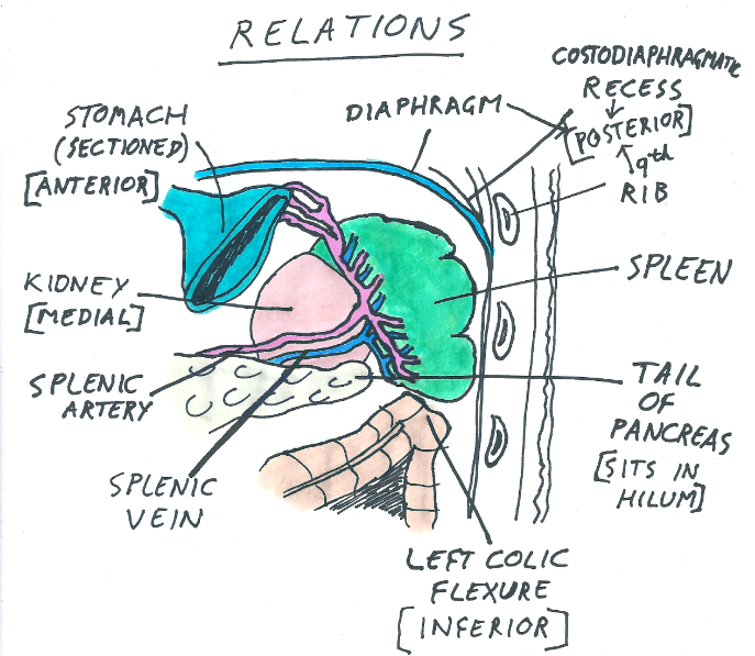
- Relations
Annotations:
- Anterior - Stomach. Posterior - Diaphragm, costodiaphragmatic recess and ribs 9-11. Inferior - Left colic flexure. Medial - Left kidney. Splenic hilum - In contact with the tail of the pancreas.
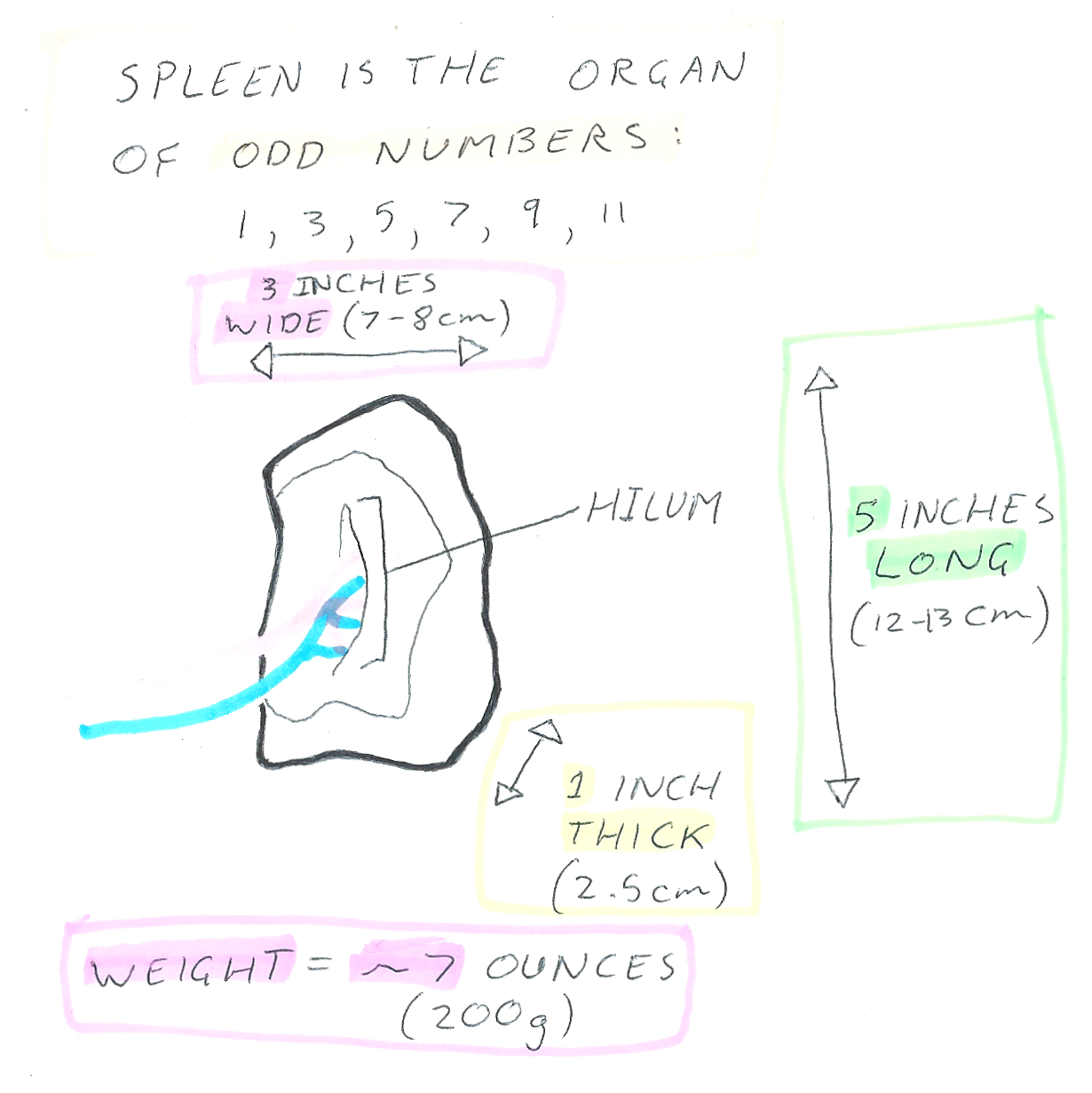
- Size
Annotations:
- The spleen is known as the organ of odd numbers because it is 1 inch thick, 3 inches wide, 5 inches long, 7 ounces in weight, and it is protected by ribs 9-11.
- Intraperitoneal
Organ
- Vasculature
- Venous Drainage
Annotations:
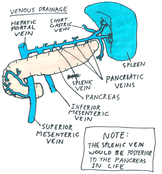
- The splenic vein drains the spleen and is formed from 3-6 branches which unite in the splenorenal ligament.
- The splenic vein is joined by the inferior mesenteric vein and 5-12 vessels that drain the pancreas. The vein runs posterior to the body and tail of the pancreas. The anastomosis between the splenic vein and the superior mesenteric vein, posterior to the neck of the pancreas, forms the hepatic portal vein.
- Arterial Supply
Annotations:
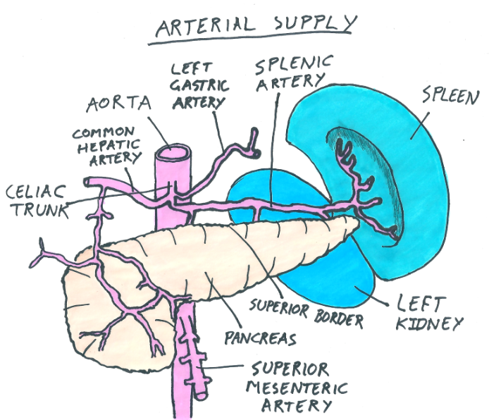
- The spleen gets its blood supply from the splenic artery which is the largest branch of the coeliac trunk. The splenic artery is also the primary supplier of blood to the pancreas as well as giving off the short gastric arteries and the left gastroepiploic artery to the stomach.
- The splenic artery runs posterior to the lesser sac of the abdomen and anterior to the left kidney. It takes a route in line with the superior border of the pancreas.
- As the artery passes between layers of the splenorenal ligament and into the hilum, it divides into five or more branches, which supply specific regions of the spleen. The lack of anastomoses within the spleen allows vascular regions to be defined, making partial spleen resection possible.
- Lymphatics
Annotations:
- Lymph from the spleen drains to nodes in the hilum before passing to the pancreaticosplenic lymph nodes. This group of nodes drain to the preaortic celiac lymph nodes.
- Venous Drainage
Media attachments
Want to create your own Mind Maps for free with GoConqr? Learn more.