Description
|
|
Created by Brittnee Gonzalez
over 6 years ago
|
|
Page 1
Bleeding Disorders
Failure of normal hemostatic mechanisms results in bleeding Can occur anywhere in the body Trauma Platelet factor abnormality Liver & spleen Coagulation factor abnormality Tiny vessels usually affected Deeper bleed Vascular abnormality Skin surface
General clinical manifestations Vary according to the type of defect Careful H & P Bleeding may be superficial or deeper within the body Petechiae With platelet factor abnormality Risk factors for bleeding (Chart 33-6) Severity/duration of thrombocytopenia Sepsis ( high platelet consumption) Increased intracranial pressure (rupture vessels; causes bleeding) Liver (coagulation factors) /renal dysfunction (erythropoietin) Dysproteinemia – low platelet function Alcohol abuse – low platelet production Splenomegaly – high platelet destruction Medications
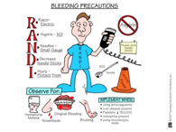
Medical management Treat underlying cause Anemia, blood loss Transfusions of blood products Hemostatic agents if fibrinolysis excessive (aminocaproic acid) Nursing Management Patient education Bleeding precautions, if Pt knows they are on bleeding precautions (keep body away from risk of falls, hurting, brushing teeth, if they bleed what will happen? It will not stop) Avoiding activities that increase the risk of bleeding Examination for bleeding Testing of all drainage and excreta
Thrombocytopenia
Low platelet level low production of platelets within bone marrow high destruction of platelets high consumption of platelets See table 33-3 for causes and treatments
Clinical manifestations < 50,000: possible bleeding < 20,000: Petechiae, nasal/gingival bleeding, excessive menstrual bleeding, excessive bleeding after surgery/dental extractions <5,000: spontaneous, fatal CNS bleeding or GI hemorrhage
Assessment & Diagnostic Bone marrow biopsy Genetic testing CBC Hepatitis screening
Medical Management Treat underlying cause Platelet transfusion Splenectomy Nursing Management Education Measures to minimize bleeding (see Chart 15-7 in Chapter 15)
Immune Thrombocytopenic Purpura (ITP)
Affects all ages but more common in children & young women Primary ITP occurs in isolation Secondary ITP occurs from autoimmune disease, viral infections, and medications
Pathophysiology Destruction of platelet by unknown stimulus Anti-platelet antibodies develop >>> antibodies bind to platelets >>> platelets ingested/destroyed >>> bone marrow increases platelet production to compensate >>> compensation becomes not as effective as thrombopoietin levels are not elevated in patients with ITP >>>> platelet production becomes diminished
Clinical Manifestations May have no symptoms Easy bruising Heavy menses Petechiae Dry purpura Mucosal bleeding (wet purpura) risk for intracranial bleeding
Assessment & Diagnostics History & physical HIV/Hepatitis C testing Bone marrow biopsy Helicobacter pylori
Medical Management Goal: “safe” platelet level Stop medication immediately if cause Pharmacological measures Immunosuppressive agents Corticosteroids Monoclonal antibodies Chemotherapeutic agents Thrombopoietin receptor agonist IVIG Splenectomy Nursing Management Determine risk of bleeding Medication history Sulfa-containing drugs STOP medication is PT has sulfa allergy present ASA based and NSAIDs Chart 33-9 Neurological assessments Avoid injections and rectal procedures Patient education
Platelet Defects
Qualitative defects; number of platelets normal but the platelets do not function normally Causes Aspirin and NSAIDs Use ESRD MDS Multiple myeloma Cardiopulmonary bypass Herbal therapy (Chart 33-9)
Acquired Coagulation Disorders
Liver Disease Diminished coagulation factors produced Prolonged PT Risk for bleeding Transfusion with FFP (in severe cases PRBCs and platelets)
Vitamin K Deficiency Synthesis of many coagulation factors depends on vitamin K Common in malnourished patients Prolonged use of antibiotics decreases intestinal flora that produce vitamin K à depleting vitamin K stores Vitamin k administration (PO or SQ) Adequate synthesis of coagulation factors is reflected by normalization of the PT Vitamin K is the antidote for warfarin toxicity Monitor INR
Anticoagulant Therapy Complications Most common in those taking warfarin INR monitoring Vitamin K antidote for warfarin toxicity Transfusion with fresh frozen plasma
Want to create your own Notes for free with GoConqr? Learn more.