Description
|
|
Created by eimearkelly3
over 10 years ago
|
|
Page 1
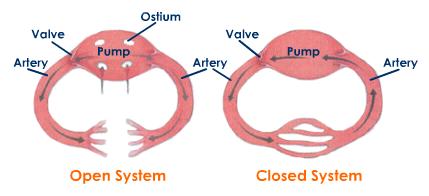
There are two forms of circulatory system :A) Open blood systemB) Closed blood systemOPEN CIRCULATORY SYSTEMThe heart pumps blood into open-ended blood vessels. The blood leaves these vessels and flows all around all of the cells of the animal's body.The blood flows back to the heart, entering it through openings in the heart walle.g. crabs, lobsters, insects, spiders, slugs, snailsCLOSED CIRCULATORY SYSTEMBlood remains in a continuous system of blood vessels, i.e. blood is always enclosed in blood vesselsExchange of material is possible through the thin capillary wallse.g. earthworms, humans
A closed system is more efficient for 2 reasons:A) The blood can be pumped around the body faster. This allows nutrients and, especially oxygen to be distributed faster to the cells. This in turn allows the animal to be more active (i.e. have a higher metabolic rate)B) A closed system allows the flow of blood to different organs to be increased or decreased, e.g. more blood can be supplied to the leg muscles if the animal is running.
The circulatory system is composed of:A) The BloodB) The HeartC) The Blood Vessels
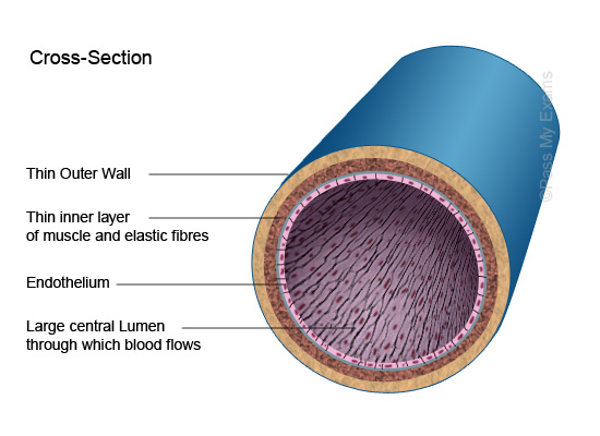
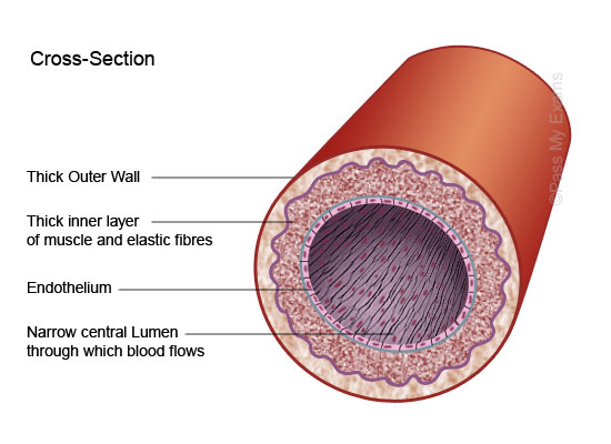
Blood vessels :The three main types of blood vessels are:-Arteries (carry blood away from the heart. Arteries divide into smaller vessels called arterioles --> oxygenated blood with the exception of the pulmonary artery)-Veins (carry blood to the heart. Small veins are called venules --> deoxygenated blood with the exception of the pulmonary vein)-Capillaries (tiny vessels that link arteries and veins)
ARTERIES AND VEINS Have an outer layer of tough, inelastic protein called collagen, which prevents the walls from over-expansion. A middle layer of muscle and elastic fibres. The muscle is involuntary and can alter the size of the vessel. e.g. during exercise, the arteries leading to muscles expand and allow up to 10 times more blood flow into the muscle. Also, when we are too hot, blood vessels in the face expand (or dilate) causing blushing. This allows more blood to enter the vessels. As a result, more heat is lost, allowing us to cool. The elastic fibres bring the vessel back to shape when the muscle relaxes. The recoil of the artery also helps to pump blood. An inner single layer of living cells called the endothelium, which surrounds the lumen.
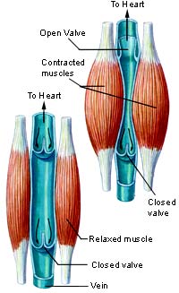
BLOOD PRESSURE AND VALVESBlood pressure is the force the blood exerts against the wall of a blood vessel (varies along the blood circuit).Blood pressure is highest in the arteries when the heart contracts. This pressure causes the artery to expand slightly. The expansion can be detected as a pulse.Pressure in veins is very low. Physical activity helps to push the blood in the veins back to the heart. This happens when the ordinary body muscles (skeletal muscles) contract. They squeeze the veins and help to return blood to the heart.People who are stationary often get weak because their muscles do not contract. Blood then collects in veins and the brain suffers a shortage of blood and oxygen.The pressure that forces the blood through the veins is low. In order to prevent backflow, and to ensure blood flows only towards the heart, veins have valves.* Valves prevent the backflow of blood.
Blood pressure in highest at the start of the artery and lowest at the entrance to the atrium. Blood pressure is measured at the start of the artery in the upper arm. The systolic (contraction) and diastolic (relaxation) pressures are measured. A normal adult should have a systolic pressure of 110-140 mm Hg and a diastolic pressure of 75-80 mm Hg.* Blood pressure is measured using a device known as a sphygmomanometer.
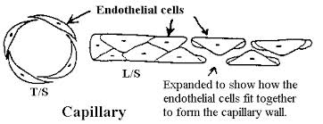
CAPILLARIESCapillaries are tiny, much-branched vessels. Their walls are made of a single layer of endothelium cells (one cell thick). Capillary walls are permeable so they allow for the exchange of materials between the blood and body tissues.-site of exchange between blood and tissue cells-one cell thick --> rapid diffusion-tissue cells are close to capillaries for sufficient exchange-blood flow in capillaries is slow for effective exchange.
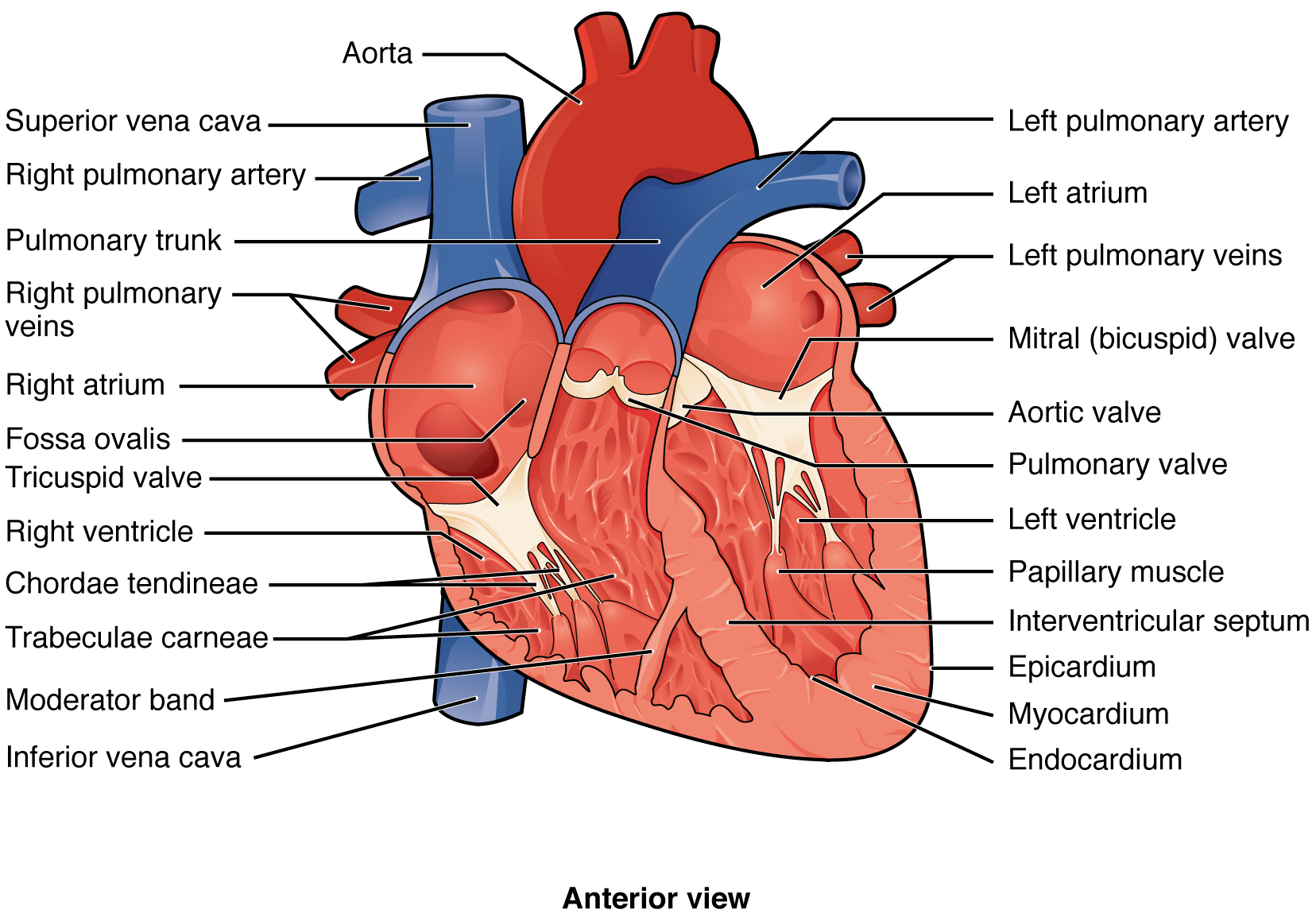
The heart is located between the two lungs (slightly to the left side of the chest, or thorax) and just above the diaphragm (thoracic cavity). It is made of cardiac muscle and surrounded by a double membrane called the pericardium. Pericardial fluid between these membranes helps to reduce friction when the heart beats.Cardiac muscle is a special type of muscle that is slow to fatigue.The heart is a double pump.The left and right side of the heart fill in unison. Each side pumps the same volume of blood.The wall of the left ventricle is 3x thicker than the right ventricle (due to the amount of cardiac muscle required to pump blood to the whole body)
Structure of the heart:The heart is divided into two sides by a wall called the septum. There are four chambers in the heart.-ATRIAThe two upper chambers are the atria (singular atrium). The atria pump blood to the lower chambers or ventricles. Because the distance is so short, the atria have thin walls.-VENTRICLESThe ventricles pump blood out of the heart. The right ventricle pumps blood to the lungs. The left ventricle pumps blood to the head and body. As this is a much longer circuit, the wall of the left ventricle is much thicker (and stronger) than the wall of the right ventricle.The atria and ventricles are seperated by valves. These are held in place by tough chords ('heart strings') called tendons. The tendons are attached to the wall of the heart by projections called papillary muscles.-VALVESThe valves ensure that blood can only flow from the atria to the ventricles (i.e. prevent the backflow of blood). The valve on the right hand side of the heart is the tricuspid valve (i.e. it has three flaps). The valve on the left is the bicuspid valve (i.e. it has two flaps).Semilunar valves (so called because their flaps are shaped like half-moons) allow blood to flow out of the heart into the two main arteries (i.e. the pulmonary artery and the aorta). They prevent blood returning to the heart.
BLOOD FLOW IN THE HEARTDeoxygenated bloodBlood that is low in oxygen (deoxygenated) enters the heart through the two venae cavae. The inferior vena cava carries blood from the lower part of the body. The superior vena cava carries blood from the head, arms, and chest.The blood enters the right atrium. This chamber contracts and forces blood down through the tricuspid valve. The venae cavae close to prevent blood flowing back out of the heart.The blood now enters the right ventricle. When this chamber contracts, the tricuspid valve closes. Blood is forced out of the heart to the lungs through the semilunar valve in the pulmonary artery.Oxygenated bloodOxygen-rich (oxygenated) blood returns to the heart from the lungs. It enters the left atrium through the pulmonary veins. It is pumped down, through the bicuspid valve, to the left ventricle. When this chamber contracts, the bicuspid valve closes and blood is pumped out to the body through the semilunar valve in the aorta.The semilunar valves allow blood to pass out of the heart. When the ventricles relax, these valves close to stop blood flowing back into the ventricles.
The oxygen content of the blood in the two sides of the heart can be recalled as follows:LORDLeft Oxygenated Right Deoxygenated
A portal system is a blood pathway that begins and ends in capillaries.The blood vessels in a portal system do not have a direct connection with the hearte.g. the hepatic portal vein connects the stomach and the intestines to the liver.
Blood supply to the heart: The muscle of the heart is supplied with blood by the coronary (or cardiac) arteries. These branch from the aorta at the point where it leaves the heart, just beyond the semi-lunar valve.The right coronary artery serves the right atria and right ventricle. The left coronary artery serves the left atria and the left ventricle (much bigger).There is an extensive capillary network throughout the cardiac muscle --> this allows for the exchange of materials e.g. oxygen, carbon dioxide, food etc.Blood flows through the heart tissue during diastole (i.e. when the cardiac muscle is relaxed) the coronary veins return deoxygenated blood to the right atrium.Blood is drained from the muscle of the heat by the coronary (or cardiac) veins. These return the blood directly to the right atrium (i.e. not to the venae cava)Blockage of the coronary arteries is a common cause of heart attack. Such attacks are often preceded by chest pains called angina.The cardiac ateries sub-divide to form numerous cardiac capillaries. These capillaries exchange materials with the muscular walls of the heart. The cardiac capillaries rejoin to form cardiac veins.
DOUBLE CIRCULATIONThe human heart is a double pump. The two sides are separated by the septum, this septum is imperative for the two circuit circulatory system, to ensure that oxygenated and deoxygenated blood do not mix.Pulmonary circuitThe right ventricle pumps blood around the pulmonary circuit, it is a short circuit that involves the blood being pumped to the lungs to gather oxygen and lose carbon dioxide. The oxygenated blood then returns to the left side of the heart. As it is such a short circuit, the walls of the right side of the heart have only a thin muscular layer.Systemic circuit (heart - body - heart)The left ventricle pumps oxygenated blood to the head, arms, trunk (torso) and legs. As this is a much longer route, a thicker, stronger layer of cardiac muscle is required in the left side of the heart.Double vs. single circulationThe double circulatory system prevents the mixing of oxygenated and deoxygenated blood.Also ensures blood pressure remains at a constant high to allow for the oxygenated blood to reach all parts of the body.Some animals have a single circulatory system, i.e. the blood is pumped from the heart, round the body and back to the heart again in one single circuit (e.g. earthworms and fish).A single circulatory system produces low blood pressure, thus restricting the metabolism of the animal.
Control of heartbeat.If a heart is removed from a body and kept in a nutritive oxygen-rich fluid it will continue to beat. This shows that a heart can beat independently of the brain.Heartbeat is controlled by the small bundle of specialised tissue called the pacemaker (lies close to the entry of the superior vena cava within the right atrium wall).The pacemaker sends out regular electrical impulses, which initially causes the atria to contract. These impulses then cause the ventricles to contract.These impulses can be increased or decreased by the brain.The heartbeat is controlled in the following way: The pacemaker (also called the SA or sino-atrial node) pulses and causes the atria to contract. The electrical impulse from the pacemaker stimulates the AV (atrio-ventricular) node. This is similar to the pacemaker but is located further down in the right atrium. The AV node sends an impulse down special muscle fibres in the septum. The impulse is passed out to the walls of the ventricles by thin fibres. The impulses from these fibres cause the ventricles to contract.
The pacemaker controls the rate of the heartbeat. However, nerves from the brain (along with hormones) can change the rate at which the pacemaker (and therefore the heart) operates.Factors that increase the rate of heartbeat include exercise, temperature, emotions, and shock. Factors such as relaxation, sleep and alcohol decrease the rate of heartbeat.*Electrodes placed on the chest can measure the electrical activity of the heart. A record of this activity is called an ECG (electrocardiogram)Two nerves from the medulla oblongata (brain) connect to the pace maker, influencing the rate of contraction.One nerve quickly accelerates the heart rate and the other quickly reduces it back to resting rate.
HEART ACTIONDeoxygenated blood from the superior vena cava flows into the right atrium and from here to the right ventricle.Oxygenated blood from the pulmonary veins (directly from the lungs) flow into the left atrium and from here into the left ventricle.The right and left ventricles fill with blood.The pacemaker (S.A. node) in the right atrium generates a nerve impulse causing the atria to contract.The contraction of the atria pushes further blood to the ventricles.The nerve impulse from the pacemaker then extends to the ventricles causing them to contract forcing the blood to the arteries.right side --> pulmonary arteryleft side --> aortaThe bicuspid valve on the left side of the heart prevents backflow of the blood to the left atria.The tricuspid valve on the right side prevents backflow of blood to the right atria.As the bicuspid and tricuspid valves close they create a 'lub' noise.The semi-lunar valves open allowing blood to flow out of the heart through the arteries.Once the ventricles empty, the semilunar close giving a 'dub' sound. This prevents the backflow of blood into the ventricles.The blood then flows from the arteries to that capillaries.
StagesBlood enters the heart- Diastole (filling phase, lasts approx. 0.4 seconds)- Relaxation (passive action)When the cardiac muscle is relaxed the heart fills with blood under low pressure from the veins.a. Blood flows from the veins into the atria.b. The cuspid valves open.c. The atria fill with blood.d. Blood flows into and fills the ventricles.e. The right side fills with deoxygenated blood and the left fills with oxygenated. 2. - Systole ( emptying phase approx. 0.4 seconds) - ContractionWhen the cardiac muscle contracts the chambers of the heart empty of blood.Two stages involved:Stage 1 (Atrial Systole approx. 0.1 seconds)a. Atria contract under stimulation from the sino-atrial node (pacemaker)b. Ventricles are topped up with extra blood from the atria.c. Delayed arrival of stimulus to the ventricles from the sino-atrial node.Stage 2 (Ventricular systole approx. 0.3 seconds)a. The impulse enters the ventricles at the A.V. node (atrio-ventricular node), lies at the bottom of the right atrium against the septum. The impulse travels along the septum and through the ventriculas walls, stimulating contraction..b. The cuspid calves close, the semi-lunar valves open.c. Cuspid valves close to prevent backflow to the atria.d. Once the blood enters the arteries, the semi-lunar valves close preventing backflow into the ventricles.*A heart murmur is any abnormal sound associated with the heartbeat. A heart murmur may indicate damage to one or more of the valves.
Pulse:- A pressure wave that passes along the walls of the artery.Caused by the rapid rise in pressure at the origins of the arteries as blood is forced from the ventricles. The alternate expansion and contraction of the arterial walls is referred to as the pulse. It is an indication of heart rate. The average pulse rate is 72 bpm (beats per minute)
The effects of smoking, diet, and exercise on the circulatory system...Smoking: Major cause of heart disease. Tobacco smoke contains nicotine and carbon monoxide (CO). Nicotine increases blood pressure and heart rate. CO interferes with the transport of oxygen to the body cells. High levels cause hardening of the arteries.Diet: Too much cholesterol from animal fats can build up on the inner walls of the arteries and reduce the rate of the flow of blood. A blockage in the cardiac artery prevents blood and oxygen getting to the cardiac muscle and will cause a heart attack. Eating fewer fatty meats, and fatty dairy products can reduce the risk of heart disease.Exercise: When we exercise, the heart beats faster -- makes the heart muscle stronger and more efficient at pumping blood. Improves the oxygen supply to the cardiac muscle and reduces blood pressure.
The circulatory system, blood vessels, and blood pressure
The Heart
Portal system
Heartbeat
Videos
Want to create your own Notes for free with GoConqr? Learn more.