Description
|
|
Created by Beckie Thorne
almost 11 years ago
|
|
Page 1
Functions of the cardiovascular SystemTransport and exchange of gas Carries oxygen for aerobic respiration from lungs to tissue Picks up CO2 from tissues and releases it in the lungs Transport nutrients from digestive system to cells Transport metabolic waste to excretory organs Transport hormones from glands to target cells Distrubution of metabolic heat and maintenance of body t
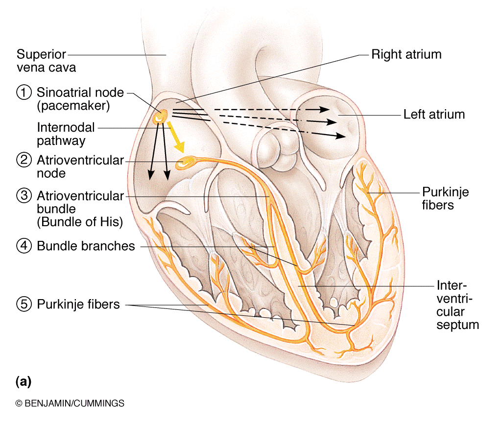
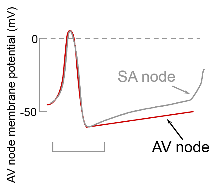
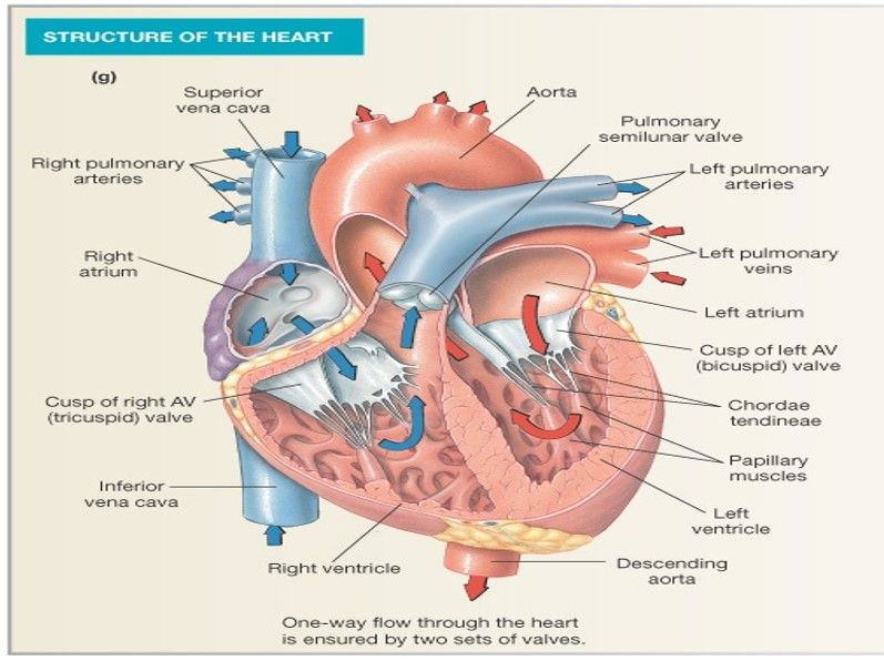
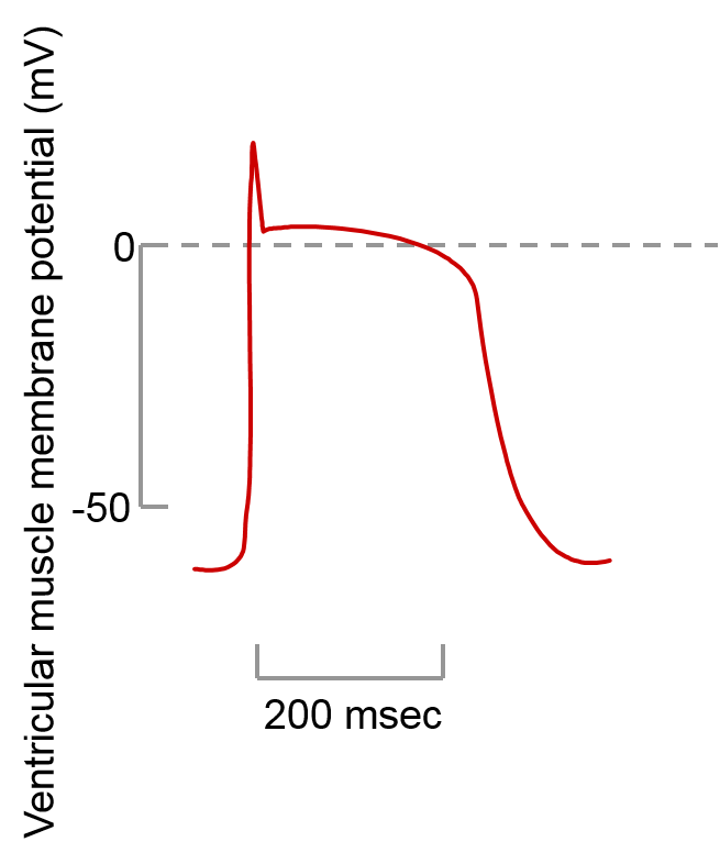
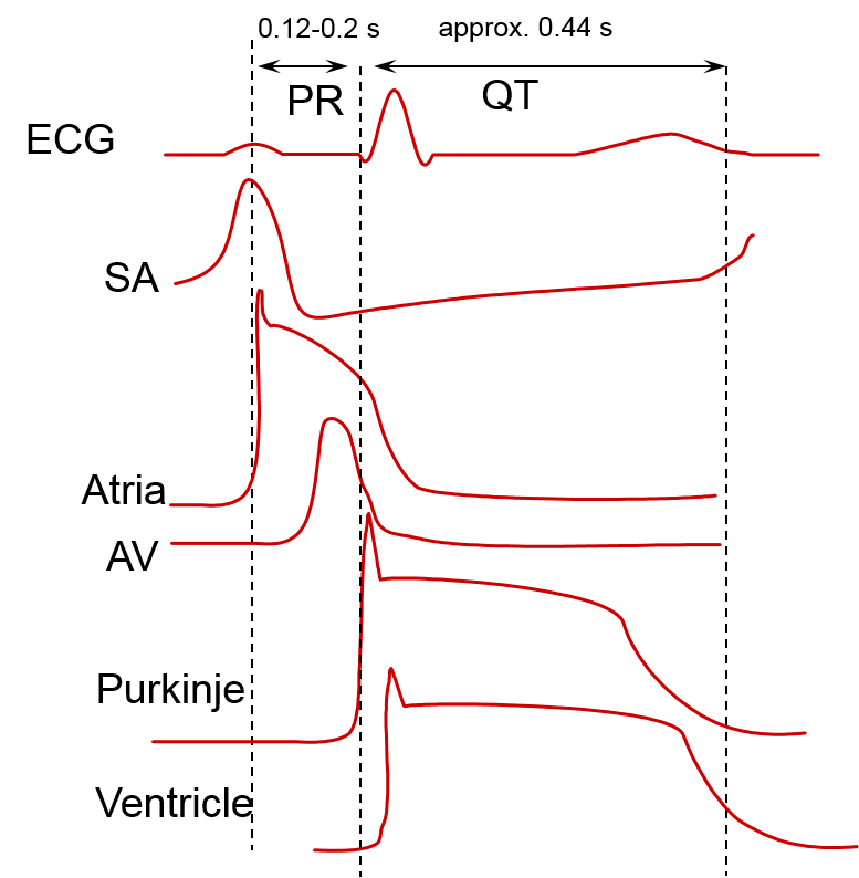
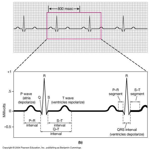
The HeartThe heart is a muscle(pump) made of chambers: left atruim, right atruim, left ventricle and right ventricle. It works by depolarization causing Ca2+ channels to open, Ca2+ influx from extracellular fluid and intracellular stores which causes a contraction. Cardiac cells are similar to neurons therefor the same principles of ion/charge movement apply. 99% of heart cell contract with every heart beat the other 1% have specialized function for normal heart excitationNormal heart beat is 60bpm but this changes depending on demand so it has a range of 40-200bpm.Anatomy:It is enclosed by the PERICARDIUM(fibrous sac) with the EPICARDIUM (fibrous layer) closely affixed to the heart. The space between the the two is filled by a fluid that allows movement of the heart within the sac. The MYOCARDIUM (wall of heart) is mainly cardiac muscle cells. ENDOTHELIAL CELLS line the cardiac chamber and blood vessels, INTERVENTRICAL SEPTUM separates ventricles and ATRIOVENTRICULAR VALVES are located between the atrium and ventricles. These valves are attached to papillary muscles which limit the valves movement to only allow blood to flow out of the atrium into the ventricles and open in response to pressure: When pressure in the atruim is greater than the ventricles they open When pressure in the ventricle is greater than the atrium they close This forces blood from the right ventricle into the pulmonary trunk and blood from the left ventricle into the aorta. These are controlled by semi lunar valves so that blood flow into them during contraction but prevents it moving back during relaxation. Small pressure differences produce large flows of blood due to the valves having little resistanceMyocardial Autoryhthmic CellsThe SA node, AV node and purkinje show INTRINSIC AUTOMATICITY which is the ability to generate a heart beat without nerve input. They receive their blood supply from the coronary arteries. The SA node is the PRIMARY PACEMAKER with a pacemaker potentional of ~80-100 bpm it drives the heart and supresses other pacemakers. The AV node has a rhythm of ~40-60 and the bundle of his ~15-30. Atrial and ventricular myoctes have no pacemaker potentialSinoatrial(SA) node located in the right atruim it is a specialized structure that send electrical impulses that cause atria and ventricles to contract. The electrical signals spread quickly due to gap juntions. Time 0: SA node activity and atrial contraction begins. Time 50msec: stimulus spreads across the atrial surfaces and reaches the AV node. Time 150msec: there is a 100msec delay at the AV node then the atrial contraction begins. Time 175msec: Impulse travels along interventricular septum within the AV bundle that branches to Purkinje fibres and via moderator band to papilary muscles of the right ventricle. Time 225msec: Purkinje fibres distribute and relay impulse through ventricular myocardium then ventricular contraction begins SA node action potential lasts 200msec and has no resting potential. Artificial pacemaker are able to regulate ventricular cells.The AV nodeThe SA node and AV node are similar exept the SA node reaches threshold potential quicker due to having a steeper pacemaker potential.Ventricular Muscle Action PotentialIt is not autorythmic as the signal comes from the AV node. It has a RMP of ~-80/-90mV. Its depolarisation is Na+ dependent with a Ca2+ plateu and K+ dependent repolarisation. Prolonged Ca2+ entry causes (muscle) cyocyte contraction, the long refractory period prevents muscle tetanus.Nervous System modulation of heart rate:Sympathetic: innervate the entire heart. Speeds up heart rate by increasing Ca2+ and If flow. Noradrenaline is released and detected mainlyby b-adrenergic receptors.Parasympathetic: mainly found on cells in the atria it slows heart rate down by increasing K+ efflux and decreasing Ca2+ influx. Ach is released and detected by muscarinic repeptors.The AP remains the same but the pacemaker potential gets quicker/ slowerElectrocardiogramIs a measure of current generated in extracellular fluid it detects electrical activity and is a combination of the SA node, atria, AV node, Purkinje and ventricles. It can be used to detect problems like atrial/ ventricular fibrilation (not contracting together) but can not detect if something is wrong with the hearts mechanical activity. P wave: corresponds to the current flow during atrial depolarization QRS complex: ventricular depolarization T wave: ventricular repolarization Atrial repolarization is not usually evident as it occurs at the same time as QRS complex. The ECG graph varies depending on placements of electrodes
Want to create your own Notes for free with GoConqr? Learn more.