
Flashcards
Anatomy sheets
Flashcards do conjunto (53)
- 1Describe the borders of the viscera in the gastrointestinal tract-Superior: thoracic diaphragm
-Inferior: pelvic floor
-Posterior: vertebral column/abdominal wall
-Lateral: psoas muscle, quadratus lumborum and transversus abdominus muscles - 2Adaptions of cavity where abdominal viscera is housed?-Largely flexible walls
-Allows for flexion of the trunk
-Accommodation of large volumes within the abdomino-pelvic cavity - 3Why is the inguinal canal of particular importance?-Association with inguinal herniae
-Parts of digestive tract and their peritoneal coverings herniate through weaknesses in the inguinal muscular walls - 4What is the general structure of the digestive tract?-Muscular tube lined with mucosa and submucosa surrounded by an outer connective tissue layer
-Epithelial cells line the stomach and pancreas, secreting enzymes. They are supplemented by secretions from accessory organs (salivary glands, pancreas and liver0 - 5What is the nervous supply like in the digestive system?-Muscles involved in mastication, swallowing and defaecation are under voluntary control
-Rest is autonomic (enteric) organised into 2 plexi. They are responsible for normal coordination of gut function and are influenced by the PNS/SNS
-Myenteric plexus is between 2 layers of muscle in the muscularis externa
-Submucosal plexus is in the submucosa - 6What is pain? Is pain from the viscera/visceral peritoneum poorly or well localised?An alerting, unpleasant sensation passing from tissue that is damaged or in the process of being damaged to the post-central gyrus of the brain where pain is perceived
Poorly localised - 7Describe the foregut Vasculature/Innervation?-Extends from terminal oesophagus to where the common bile duct enters the duodenum
-Vasculature: branches of celiac trunk of the aorta
-Sympathetic: celiac plexus and greater splanchnic nerve
-Parasym: vagus nerve - 8Describe the midgut Vasculature/Innervation?-Extends from duodenum to 2/3 of transverse colon
-Vasculature: branches of superior mesenteric arter
-Sympathetic: superior mesenteric plexus via lesser splanchnic nerve
-Parasym: vagus - 9Describe the hindgut Vasculature/Innervation?-Extends from distal 1/3 transverse colon to the rectum
-Vasculature: branches of the inferior mesenteric artery
-Sympathetic: inferior mesenteric plexus via the least splanchnic nerve
-Parasym: S2-S4 - 10What is the peritoneum? How can it be sub-divided?-A serous membrane lining the internal walls of the abdominal cavity/viscera.
-It is continuous
-Parietal peritoneum lines the abdominal walls
-Visceral peritoneum envelops the abdominal organs
-Layers are separated by a thin lubricating fluid secreted by the serous membrane allowing frictionless movement of the visceral organs - 11What are retroperitoneal organs?Organs that bulge into the peritoneal cavity slightly.
Include the kidneys, adrenals, ureters, colon, rectum - 12How do the lesser and greater sac communicate?Via the epiploic foramen of WInslow
- 13What stimuli initiate reflexes in the enteric NS?-Chemical
-Thermal
-Mechanical
-Osmotic - 14At what vertebral level is: -bifurcation of the aorta -coeliac artery -superior mesenteric artery -inferior mesenteric artery-L4
-T12
-L1
-L3 - 15Which vessels drain blood from the GIT? Where does it go?-Splenic vein
-Superior mesenteric vein
-Inferior mesenteric vein
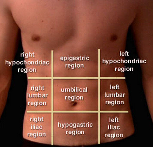
-All 3 drain into the portal vein to the liver - 16Describe how the abdomen can be divided into 9 regions How are the dividing lines formed?-2 vertical lines joining the mid clavicular point to the mid linguinal point
-Subcustal plane at the level of the 10th costal cartilage/L3
-Transtubercular plane joining 2 iliac crests - 17What is the cysterna chyli?-Dilated sac at the lower end of the thoracic duct
-Has fatty chyle from the intestines and lipid products of digestion - 18Describe the vasculature of the: foregut midgut hindgutForegut: celiac artery/gastric+splenic veins to the portal vein
Mindgut: Superior mesenteric artery/portal venous system
Hindgut: inferior mesenteric artery/portal venous system - 19Where is the site of sympathetic ganglion for the: foregut midgut hindgutForegut: celiac plexus
Midgut: superior mesenteric plexus
Hindgut: inferior mesenteric plexus - 20What is the parasympathetic supply to the: foregut midgut hindgutForegut: vagus nerve
Midgut: vagus nerve
Hindgut: pelvic splachnic nerve - 21Where is referred pain of the: foregut midgut hindgutForegut: epigastric region
Midgut: umbilical region
Hindgut: public region - 22Define mesentaryDouble layer of visceral peritoneum connecting an abdominal organ to the abdomen wall.
Also connects parts of the small intestine together - 23Define omentum What is the greater/lesser omentum?-Sheets of peritoneum that extend from the stomach and proximal part of duodenum to other abdominal organs
Greater: 4 layers of visceral peritoneum that descend from greater curvature of the stomach and fold back up to attach to the anterior surface of the transverse colon
Lesser: 2 layers of visceral peritoneum that attach from the lesser curvature of the stomach to the liver - 24Functions of mesenteries-Decreases friction between adjacent visceral surfaces
-Allows some movement of organs during digestion
-Allows communication between body wall/internal organs
-Holds/suspends organs in place to posterior abdominal wall - 25What are ligaments (in peritoneum)?Double fold of peritoneum connecting viscera together or connecting viscera to the abdominal wall
- 26What is an adhesion?-Internal scar tissue connecting tissues not normally connected
-Forms as a result of injury during surgery (stresses on organs)
-Internal organs can attach to surgical site/other organs
-Twisting/pulling of organs can result - 27What are peritoneal recesses?Spaces formed by peritoneum draping over viscera
- 28What is a haemoperitoenum? What is a pathological pneumoperitoneum?Blood fills up in peritoneum
Organs such as the bowls burst and leak contents - 29What is a hernia? Where is it likely to occur?When an internal body part pushes through weakness in muscle/tissue wall.
Hiatus (through diaphragm), incisional, linguinal and umbilical - 30The mature peritoneal cavity contains a few ml of fluid. To what volume can this space be distended by pathological conditions? Why might this be beneficialUp to 1.5 litres
Ascites (fluid collection in peritoneum) can occur. Common cause is cirrhosis of liver - 31Where does inflammatory exudate in the peritoneal cavity collect when: supine upright position?Supine: hepatorenal recess
Upright: rectrouterine recess/rectovesicle - 32Pain from the parietal peritoneum is severe and accurately localised. Why?Innervated by both somatic and visceral afferent fibres allowing accurate localisation of pain
- 33What is the vestible?-Part of the mouth which lies between the lips and cheeks and teeth
-It is very flexible - 34What is the oral cavity?-Part that contains the teeth and tongue
-Extends to hard and soft palate superiorly
-Extends to mylohyoid muscles and mandible inferiorly
-Opens into oropharynx posteriorly - 35What are the 3 salivary glands? Locations?
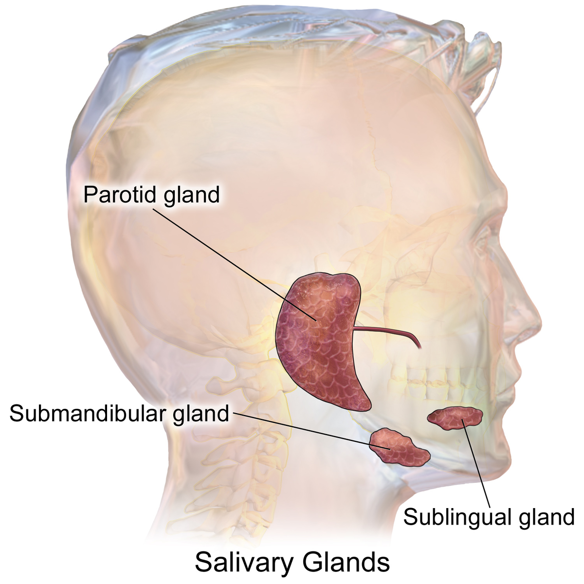
- 36Describe the parotid salivary gland-Produces watery serous saliva when stimulated by the paraNS component of the glossopharyngeal nerve
-Parotid saliva enters the mouth vestibule via the parotid duct that pierces the buccinator muscle (upper second molar) - 37Describe the submandibular salivary gland-Clasps free end of mylohyoid muscle
-Secretes via submandibular duct into the floor of the mouth
-Produces a mixed serous/mucous saliva
-Supplied by secretomotor fibres of the facial nerve - 38Describe the sublingual salivary gland-Produces mixed saliva that is thicker than submandibular saliva
-Requires many short small short ducts to deliver it to the oral cavity
-Supplie by chorda tympani branch of facial nerve - 39What is the tongue?-Mass of skeletal muscle
-Has distinctive perimysium covered in mucous membrane
-Has a thin midline fibrous septum running along its length separating the muscles - 40What are the intrinsic muscles of the tongue?-They run longitudinal, vertical and horizontally
-Concerned with shape of tongue - 41What are the extrinsic muscles of the tongue?-Originate on the styloid process (styloglossus), mandible (genioglossus), hyoid bone (hypoglossus) and soft palate (palatoglossus)
-Attach to the tongue muscle and fascia - 42Purpose of muscles of the tongue?-Manipulate food so that it meets the occlusal surface of the teeth
-Mixes food with saliva
-Forms food into a bolus for swallowing
-Pushes bolus towards the back of the tongue - 43What is the oesophagus?-Fibromuscular tube connecting the pharynx in the neck to the stomach in the abdomen above the dome of the diaphragm
-Cervical region: anteiror to C6-T1, posterior to trachea and has left/right laryngeal nerve + carotid sheaths next to it - 44What is the stomach?A distensible sac where food is churned, acidified, hydrated and where digestion is initiated
-Chyme flows to the small intestine which is controlled by the pyloric sphincter - 45Describe the innervation of the tongue
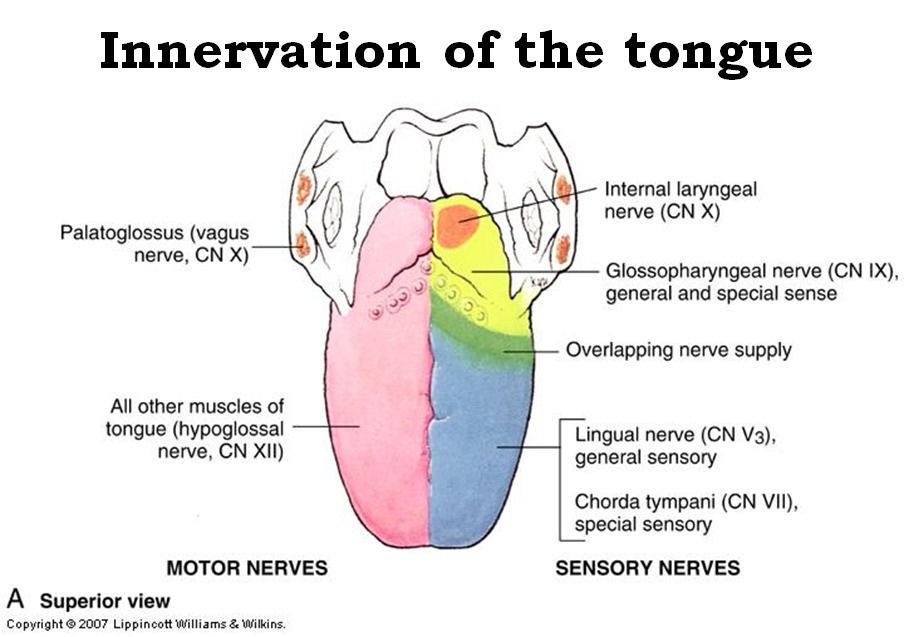
- 46Where is the oesophagus the narrowest?C6
- 47Describe the hiatuses of the diaphragm What goes through each and at what vertebral level?Caval hiatus (T8)- IVC
Oesophageal hiatus (T10)- oesophagus, Vagus nerve
Aortic hiatus (T12)- aorta, thoracic duct, azygous vein - 48What is the acute angle as the oesophagus enters the stomach? Significance?Angle of His
Prevents reflex - 49What vessels drain blood from the stomach?Left/right gastric veins into hepatic portal veins
Short gastric, left/right gastro-omental veins into superior mesenteric - 50If someone suffers a bilateral fracture to the body of the mandible, what measures are immediately necessary?-Ensure airways are clear, anaesthetise and suture muscles back
-Tongue may fall back into oropharynx blocking the airway
-Perfuse bleeding
-Intrinsic muscles are ruptured - 51A patient presents at your GP surgery complaining that immediately after a meal, particularly if it contains a bitter substance, a painful swelling appears under the left side of the body of his mandible which takes up to two hours to subside. What is the most likely cause of this symptom? How would you confirm your diagnosis?-Salivary duct obstruction due to stone formation in submandibular gland
-After eating, saliva should exit but doesn't
-It subsides as it is absorbed which can lead to infections of the gland
-Diagnosis can be confirmed with a sialography - 52What structures might be damaged during surgery to remove a malignant growth in the parotid gland?Facial nerve
- 53What is a hiatal hernia, and where will the pain be experienced?-Stomach goes through hiatus in the diaphragm into the thorax
-May be no symptoms
-Confirmed by chest X-ray
-GORD may occur- heartburn
-Pain in epigastric region
Informações
- Descrição
- Overview of the digestive tract and peritoneal coverings Oral cavity, salivary glands, oesophagus and stomach
- Criado por
- Dolu Falowo
- Visualizações
- 0
- Avaliação