
Flashcards
Msak

Flashcards do conjunto (53)
- 1—Leg Group posterior
Gastrocnemius
O: super Condylar line of the femur
I: posterior surface Calcaneus via calcaneal tendon
Action: Flex the Knee
, Plantar Flex the Ankle
Innervation: Tibial Nerve - 2—Leg Group Posterior
Soleus
O: Soleil line. Proximal posterior surface of the Tibia and posterior aspect of head of femur
I: Calcaneus tendon via calcaneal tendon
Action: Plantar flex the ankle
Innervation: Tibial Nerve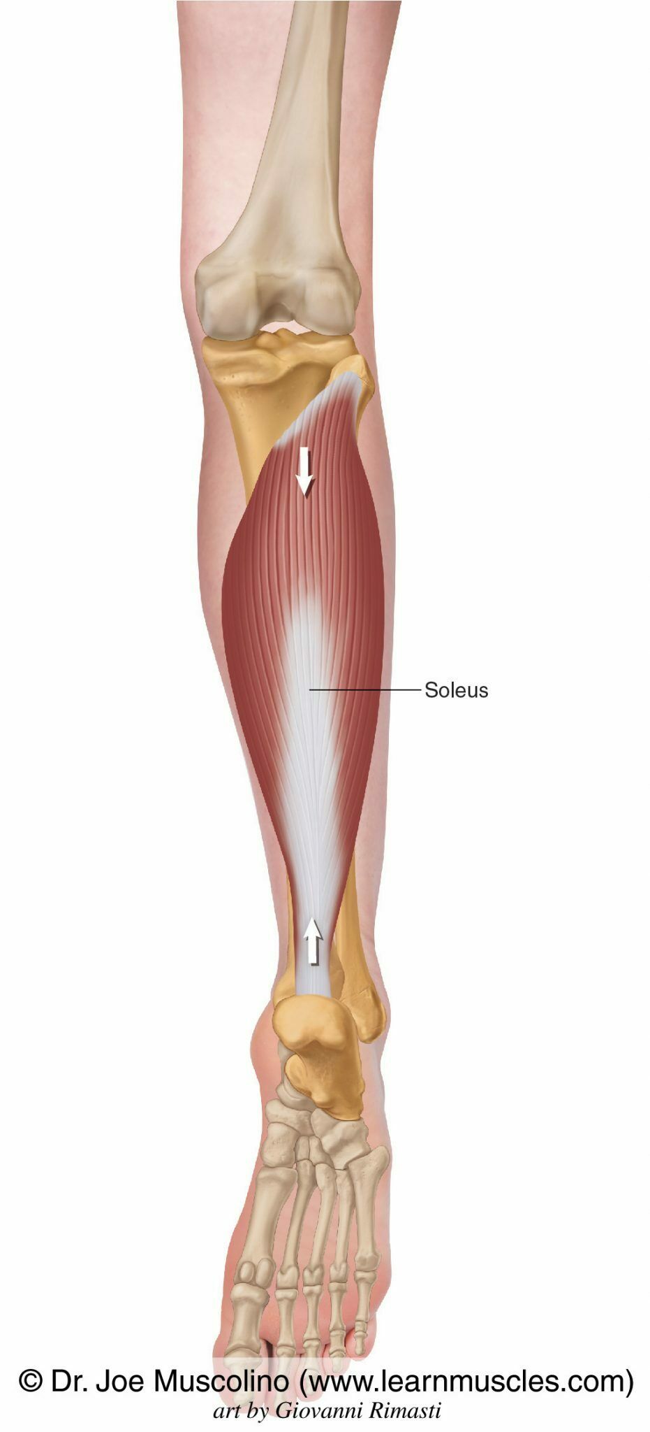
- 3—Leg Group Deep Posterior
Tibialis Posterior
O:Proximal posterior shaft of tibia and fibula, and interosseous membrane
I: All five tarsal bones and bases of second through fourth metatarsal
Action: Plantar flex the ankle invert the foot
Nervation; Tibial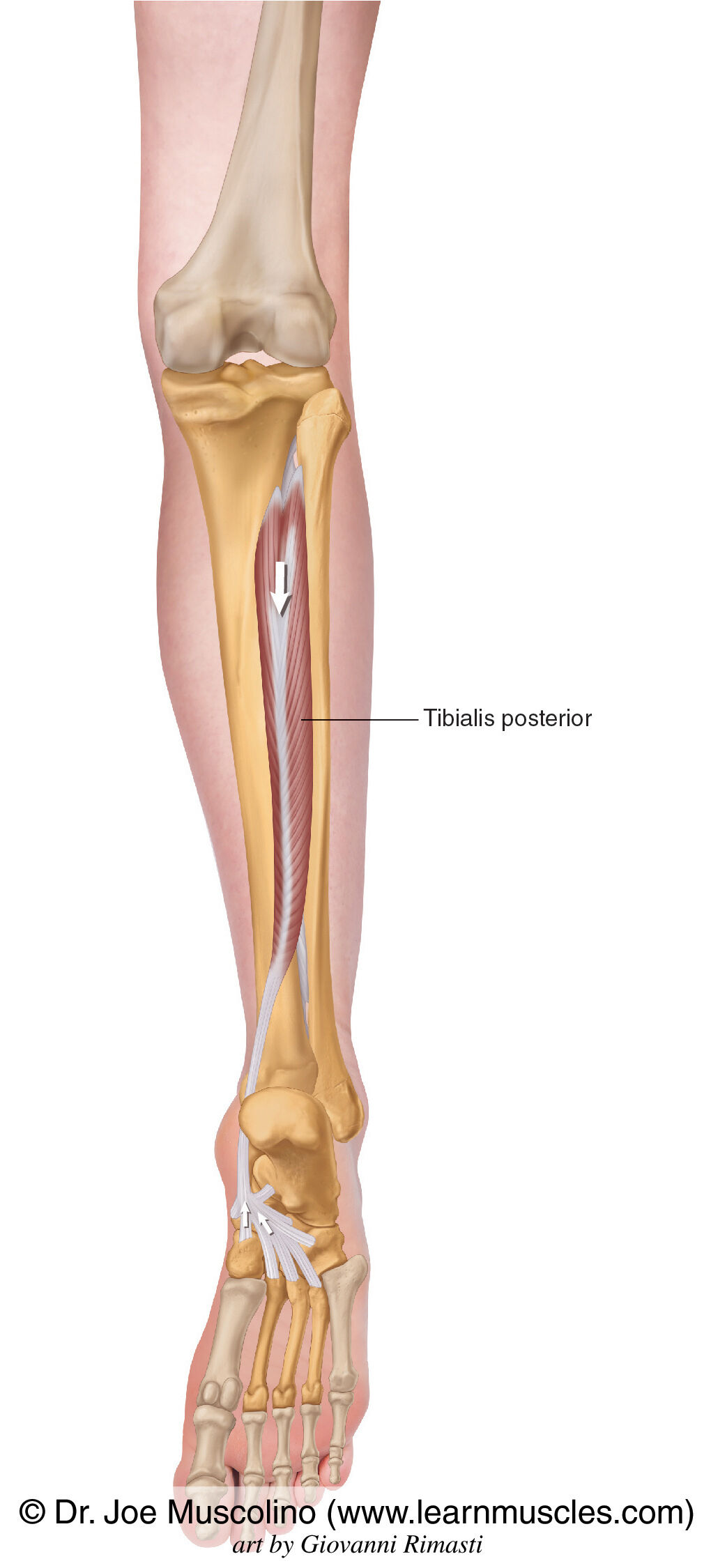
- 4—Leg Group Posterior
flex dIgItorum longues
O: Middle, posterior surface of tibia
I: Distal phalanges of second through fifth toes
Action: Flex the second through fifth toes. Weak plantar flexion of the ankle. Invert the foot
- 5—Leg Group Posterior
Flexor hallucis
Longus
O: Middle half of posterior fibula.
I: Distal phalanx of first toe
Action: Flex the first toe, weak plantar flexion of the ankle. Invert the foot.
Innervation: Tibial nerve
- 6—Anterior group
Tibialis Anterior
O: Lateral condyle of the tibia: Proximal lateral surface of the Tibia and interosseous membrane.
I: Medial cuneiform and base of the first metatarsal
Action: Invert the foot and dorsiflex the ankle.
Innervation: Deep fibular nerve.
- 7—Anterior group
Extensor Hallicus longues
O: Middle anterior surface of fibula and interosseous membrane
I: Distal phalanx of first toes
Action: Extend the first toe, Dorsiflex the ankle, Invert the foot.
Innervation: Deep fibular
- 8—Anterior group
Extensor digitorum Longus
O: Lateral condyle of tibia: proximal anterior shaft fibula and interosseous membrane.
I: Middle and distal phalanges of second through fifth toes
Action: Extend the second through fifth toes dorsiflex the ankle. Evert the foot.
Innervation: Deep fibula
- 9—Lateral compartment
Fibularis longus
O: Head of fibula and proximal head of fibula and two thirds of lateral fibula.
I: Base of first metatarsal and medial cuneiform
Action: Evert the foot, assist to plantar flex the ankle
Superficial fibular - 10—Lateral compartment of
Fibularis Brevis
O: Distal two thirds of lateral fibula
I: Tuberosity of fifth metatarsal
Action: Evert the foot, assist to plantar flex the ankle
Innervation: Superficial fibula - 11—Anterior compartment of leg
Fibularis Tertius
- 12—Dorsal surface of the foot
Extensor Digitorum Brevis
O: Dorsal aspect of calcaneus
I: Second through fourth toes
Action: Extend second to fifth toes
Innervation: Deep fibular nerve
- 13—Dorsal surface of the foot
Extensor hallucis Brevis
O: Dorsal aspect of calcaneus
I: Proximal phalanx of first toe
Action: Extend the first toe
Innervation: Deep fibular nerve
- 14—Superficial plantar surface of the foot
Abductor hallucis
O: Medial process of the calcaneus
I: Proximal phalanx of first toe
Abduct the fist toe assist to flex the first toe
Innervation: Medial plantar nerve
- 15—Superficial plantar surface of the foot
Abductor Digi Mini
O: Lateral process of the Calcaneus
I: Proximal phalanx of first toe
Action: Flex the fifth toe, abduct the fifth toe.
Innervation: Lateral plantar Nerve
- 16—Second layer of plantar surface of the foot
Quadratus Plantae
O:Medial and lateral sides of plantar surface of the calcaneus
I:Posterior and lateral aspect of the flexor digitorum longus tendon
Action: Assist flexor digitorum longus tendon to flex the second through fifth toes
Innervation: Tibial Nerve - 17—Second layer of plantar surface of the foot
Lumbercles pedis
O:Tendons of flexor digitorum longus
I: Bases of proximal phalanges of second through fifth toes
Actions: Flex the proximal phalanges of second through fifth toes
Extend the middle and distal phalanges of second through filth toes.
Tibial Nerve
- 18—Third layer of plantar surface of the foot
Flexor hallucis brevis
O: Plantar surface of the cuboid and lateral cuneiform
I: Medial and lateral surface of the base of proximal phalanx of first toe
Action: Flex the first toe
Innervation: Lateral plantar nerve
- 19—Third layer of plantar surface of the foot
Flexor digiti mini
O: Base of fifth metatarsal
I: Proximal phalanx of fifth toe
A: Flex the fifth toe
Nervation: Lateral plantar nerve
- 20—Third layer of plantar surface of the foot
Adductor Hallucis
O: Oblique head: bases of second through fourth metatarsals
I: Transverse head: Plantar ligament of third through Fifth metatarsophalangeal joints
Lateral surface of base of proximal phalanx of first toe
Action: Flex and adduct the first toe
Innervation: Medial plantar nerve - 21—Muscles of the shoulder
Muscles of the shoulder
Deltoid
Lateral ⅓ of clavicle, acromion and spine of scapula
Deltoid tuberosity of humorous
All fibers: abduct the shoulder
Anterior fibers: flex and medially rotate , horizontally adduct the shoulder
Posterior fibers: extend , lately rotate, horizontally abduct the shoulder
Axillary nerve
O: Lateral ⅓ of clavicle, acromion and spine of scapula
I: Deltoid tuberosity of humorous
Actions: All fibers: abduct the shoulder
Anterior fibers: flex and medially rotate , horizontally adduct the shoulder
Posterior fibers: extend , lately rotate, horizontally abduct the shoulder
Innervation: Axillary nerve - 22—Anterior trunk muscles
Pecrolis Major
O: Medial half of clavicle , sternum and cartilage of the first through sixth rib
I: Lateral crest of greater tubercle of humorous
Action: All fibers: adduct the shoulder. Assist to elevate the thorax
Upper fibers: Flex the shoulder, horizontally adduct the shoulder
Lower fibers: extend the shoulder
Innervation: Upper fibers : lateral pectoral nerve
Lower fibers: Lateral and medial pectoral nerve
- 23—Anterior trunk muscles
Pectoralis Minor
O:Third, fourth and fifth ribs.
I: Medial surface of the coracoid process of the scapula
Action: Depresses the scapula
Innervation: Pectoral nerve
- 24—Anterior trunk muscles
Subclavius
O:First rib and cartilage
I: Inferior surface of middle one third of the clavicle
Action: Elevate the first rib
Innervation: Subclavian nerve
- 25—Superficial back muscles
Trapezius
O: Spinous processes and fibers, external occipital protuberance(base of skull)
I: Lateral one third of clavicle, acromion, and spine of scapula
Action: Bilaterally; extend the neck and spine
Middle fibers adduct and stabilize the scapula
Lower fibers: depress the scapula
Innervation: Spinal portion of the cranial nerve - 26—Superficial back muscles
Rhomboids
Origin: Major: spinous processes of T2- T5 Minor: Spinous processes of C-7 and T1
Insertion: Major: Medial border of the scapula between the spine of the scapula and the inferior angle.
Minor: Upper portion of the Medial border of the scapula across from the spine of scapula
Action: Adduction and retraction of the scapula. Elevate the scapula. Downwardly rotate the scapula.
Innervation: Dorsal scapular nerve
- 27—Superficial back muscles
Latissimus Dorsi
O:Inferior angle of scapula, ,Spinous process of 6 thoracic vertebrae, Last four ribs of thoracolumbar fascia, and posterior iliac crest.
I: Intertubercular groove of the Humerus
Action: Extend the shoulder, Adduct the shoulder and medially rotate the shoulder
Innervation: Thoracodorsal nerve
- 28—Poster shoulder muscle
Teres Major
O:Inferior angle and lower one third of lateral border of scapula
I: Crest of lesser tubercle of humerus
A: Extend the shoulder, adduct the shoulder, and medially rotate the shoulder
Innervation: Lower subscapular nerve - 29—Rotator cuff
Supraspinatus
O: Supraspinous fossa of scapula
I: Greater tubercle of humerus
Action: Abduct the shoulder
Stabilize the head of humerus in the glenoid Cavity
Supra scapular Nerve
- 30—Rotator cuff
Infraspinatus
A: Infraspinous fossa of the scapula
O:The greater tubercle of humerus
Action: Stabilize the head of the humerus in the glenoid Cavity. Adduct the shoulder. Latterly rotate the shoulder
Innervation: Suprascapular Nerve - 31—Rotator cuff
Teres Minors
O:Upper two thirds of the Lateral border of scapula
O:Greater tubercle of humerus
Action: Stabilize the Head of the humerus is the glenoid cavity. Adduct the shoulder. Latterly rotate the shoulder
Innervation: Axillary nerve - 32—Rotator cuff
Subscapularis
O: Subscapular fossa of scapula
I: Lesser tubercle of humerus
Action: Medially rotate the arm, Stabilize the head of humerus in the the Joint Cavity
Innervation: Upper and lower Subscapular Nerve - 33—Anterior arm muscles
Biceps Brachii
O:Short head: coracoid process of scapula
Long head: supra glenoid tubercle of scapula
I: Tuberosity of the radius. Aponeurosis ( attaches to muscle not bone) of the Biceps Brachii.
Action: Flex the elbow, supinate the forearm, flex the shoulder and assist with adduction.
Innervation: Musculocutaneous Nerve
- 34—Anterior arm muscles
Coracobrachialis
O:Coracoid process of the scapula
I: Medial surface of Mid humeral shaft
Action: Flex the shoulder, adduct the shoulder
Innervation: Musculocutaneous Nerve - 35—Anterior arm muscles
Brachialis
O:Distal half of anterior surface of humerus (mid shaft )
I: Tuberosity, and coronoid process of ulna
Action: Flex the elbow
Innervation: Dual innervation: Musculocutaneous , small branch from radial - 36—Posterior arm muscles
Triceps Brachii
O: Long head: Infraglenoid tubercle of the scapula
Lateral head: Posterior surface of proximal half of humerus
Medial: Posterior surface of distal half of humerus
O: Olecranon process of ulna
Action: All heads: Extend the elbow
Long head: extends the shoulder, adducts the shoulder
Radial
- 37—Extensors of the wrist( Posterior muscles)
Extensor carpi radialis lounges
O: Distal one third of lateral supracondylar ridge of humerus
I: Lounges: base of second metatarsal
A: Extends and abducts the wrist, assist to extend the elbow
Radial N.
- 38—Extensors of the wrist extensor radialis carpi brevis
O: common extensor Tendon from lateral epicondyle of humerus
I :base of third metatarsal
A:Extends and abducts hand at wrist, assist to extend the elbow
Radial N. - 39—Extensors of the wrist Extensor carpi ulnaris
O: conmen extensor tendon from Lateral epicondyle of humerus
I: Base of 5th metacarpal
A: Extends and adducts wrist
Radial N.
- 40—Posterior muscles of the forearm (extensors of the wrist)
Extensor indicis
O:Posterior surface of distal shaft of ulna and interosseous membrane
I: Extensor expansion of 2nd digit
A: Extends 2nd digit and helps extend hand at wrist
Radial N.
- 41—Posterior muscles of the forearm
Extensor Digitorum ( muscles of the forearm extensors of the wrist)
O:Lateral epicondyle of humerus
I: Bases of middle and distal phalanges of second through fifth fingers
A:
Extends second through fifth digits, assists wrist extension
Radial N. - 42—Posterior muscles of the forearm
Posterior muscles of the forearm (extensor of the wrist )
Extensor Digiti Minimi
o: Lateral epicondyle of humerus
I: Extensor expansion of 5th digit
A:Extends 5th digit
Radial N.
- 43Muscles of the ElbowSupinator
Anconeus
Pronator Teres Pronator
Quadratus - 44—supinator
O: Lateral epicondyle of the humerus, radial collateral ligament , Annual ligament and supinator crest of ulna
I:Anterior, lateral surface of proximal one third of radial shaft
A: Supinate the forearm
N: Radial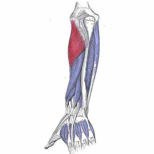
- 45—Anconeus
O: Lateral epicondyle of humerus
I: Olecranon prosses and posterior surface of ulna
A: extends the elbow
Radial Nerve
- 46—Pronator teres( muscles of posterior surface of the elbow)
O: Common flexor tendon from medial epicondyle of humerus & coronoid process of the ulna
I: Middle of lateral surface of the radialis s
A: Pronate the forearm/ assist to flex the elbow
N: Medial
- 47—Pronator Quadratus
O : Medial, anterior surface of distal ulna
I :Lateral, anterior surface of distal radials
A: Pronate the forearm
N: Median
- 48—Flexor carpi radialis ( flexors of the wrist)
O: Medial epicondyle of humerus
I: Base of 2nd and 3rd metacarpal
A: Flexes and abducts hand at wrist (radial deviation); assist flexion of elbow
N:Median N.
- 49—Palmaris longus ( flexor of the wrist)
O :Medial epicondyle of humerus
I:Distal half of flexor retinaculum and palmar aponeurosis
A:Flexes hand at wrist and tenses palmar aponeurosis
N:Median N.
- 50—Flexor carpi ulnaris( flexor of the wrist)
O: Superficial head: medial epicondyle of humerus
Deep head: olecranon and posterior border of ulna
I: Pisiform bone, hook of hamate bone, base of 5th metacarpal
A: Flexes and adducts hand at wrist
N: Ulnar nerve
- 51—Flexor digitorum superficialis ( flexor of the wrist)
O: Humeroulnar head: medial epicondyle of humerus, coronoid, process of ulna, ulnar collateral ligament
Radial head: superior half of anterior radius
I: Sites of middle phalanges of 2nd through 5th fingers
A: Flex the second thru 5th fingers, flex the wrist
N:Median N.
- 52—Flexor digitorum profundus ( flexors of the wrist )
O:Medial and anterior surface of proximal 3/4 of ulna and interosseous membrane
I:Bases of distal phalanges, palmar surface of second thru 5th fingers
A:Flex the second thru 5th fingers, flex the wrist
N: Ulnar and medial - 53—Brachial radials
O : Lateral condyle of humerus? prox 2/3 of the supracondylar ridge of humerus
I: styloid processes of radials
A: flex the elbow assist to pronate and supinate forearm when the movements are resisted
N: radial


Informações
- Descrição
- FlashCards sobre Msak, criado por Shanti Woolcock em 16-11-2023.
- Criado por
- Shanti Woolcock
- Visualizações
- 11
- Avaliação