
Flashcards
RCA Thorax 3: Mediastinum and Heart in situ
Flashcards do conjunto (15)
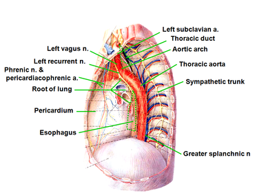
- 1List the structures on the left side of the mediastinum:1. Arch of the aorta
2. Ligamentum arteriosum with the left recurrent laryngeal nerve under it.
3. Left pulmonary artery
4. Left brachiocephalic vein
5. Hemiazygos veins
6. Left phrenic and vagus nerve
7. Descending thoracic aorta
8. Pericardium - 2List the structures on the right side of the mediastinum:1. Right brachiocephalic vein
2. Superior vena cava
3. Azygos vein
4. Brachiocephalic trunk
5. Right vagus nerve
6. Right phrenic nerve
7. Trachea and esophagus
8. Pericardium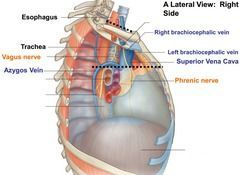
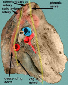
- 3Describe the relation of the phrenic and vagus nerve to the root of the lung:The phrenic nerve passes anterior to the root of the lung whereas the vagus nerve passes posterior to the root of the lung.
- 4Describe the superior, inferior, anterior and posterior borders of the anterior mediastinum: Describe the contents of the anterior mediastinum:Superior: Superior mediastinum above the level of the sternal plane (sternal angle, rib 2 to T4)
Inferior: Diaphragm
Anterior: Sternum
Posterior: Pericardium
Contents: Loose connective tissue, internal thoracic artery and vein branches, and parasternal lymph nodes.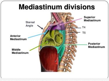
- 5Describe the general procedure shown: Describe the indication: Describe the two approaches:An emergency procedure called pericardiocentesis may be necessary if blood or fluid collects within the pericardial cavity outside of the heart e.g following a stab wound to the heart. The tough outer fibrous layer of pericardium cannot expand rapidly and so the blood or fluid is under pressure and compresses the pulmonary veins and atria of the heart restricting cardiac output- a life threatening situation called cardiac tamponade.
There are two main ways of inserting a needle into the pericardial cavity to aspirate blood or fluid in order to improve the patient’s condition until more definitive treatment is carried out:
Intercostal parasternal approach alongside the left border of the sternum (usually the left 5th intercostal space over the “bare area” of the heart where is no intervening lung).
Subxiphoid approach (the most popular)- the needle is inserted just inferior and to the left of the xiphoid process. Advance the needle into the pericardial cavity, aiming towards the tip of the left scapula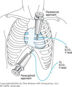
- 6Name the area highlighted in purple: Name the pericardiocentesis approach that enters the pericardial cavity this way:Bare area of the heart, where there is no intervening lung.
Intercostal parasternal approach.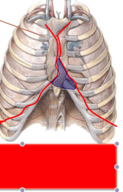
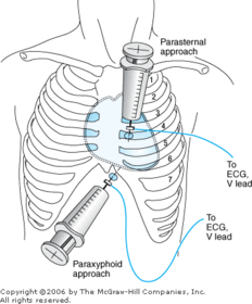
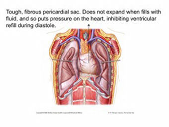
- 7What is characteristic of the outer layer of the pericardium and why is this important?The outer layer is composed of tough fibrous connective tissue lined by the thin parietal serous pericardium. It fuses with the adventitia of the great vessels. The pericardial cavity is the potential space between the parietal and visceral layers of serous pericardium. It normally contains a tiny amount of fluid that lubricates the movement of the heart. The tough outer fibrous pericardium can only expand a little and must do so gradually if the heart enlarges or if fluid or blood collects in the pericardial cavity; a sudden accumulation of fluid or blood in the pericardial cavity can cause life-threatening cardiac tamponade.
- 8Name the structure being demonstrated: What are the borders:Structure: Transverse pericardial sinus
Lies behind the pulmonary trunk, ascending aorta and superior vena cava.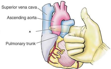
- 9Name the structure indicated by the arrow: Borders:Oblique sinus, between where the pulmonary veins enter the posterior wall of the left atrium.
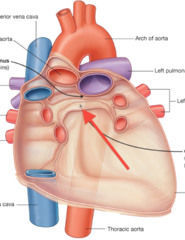
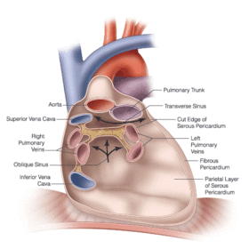
- 10Describe the surface projections of the margins of the heart:
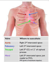
- 11—
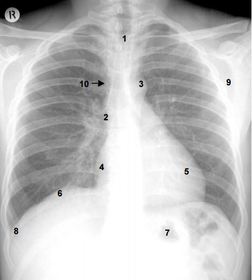
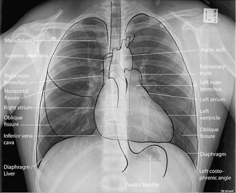
- 12Why is the right hemidiaphragm higher than the left?The traditional explanation is that it is pushed up by the underlying liver, although in reality it may be that the heart depresses the central tendon and left hemidiaphragm.
- 13On a chest radiograph, where is the approximate position of the left pulmonary artery?The left pulmonary artery is visible on a chest radiograph as a shadow just below the aortic knuckle but it is not always very distinct.
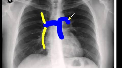
- 14—1. Right brachiocephalic vein.
2. Brachiocephalic trunk.
3. Trachea.
4. Left brachiocephalic vein.
5. Left common carotid artery.
6. Left subclavian artery.
7. Esophagus.
8. Intercostal muscles.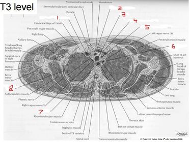
- 15—1. Internal thoracic artery and vein.
2. Pulmonary trunk.
3. Left main bronchus.
4. Descending aorta.
5. Esophagus.
6. Superior vena cava.
7. Ascending aorta.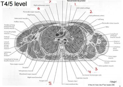
Informações
- Descrição
- RCA Thorax 3: Mediastinum and Heart in situ
- Criado por
- Ashutosh Kumar
- Visualizações
- 465
- Tags
- Avaliação