
Mapa mental
SKIN Pathology
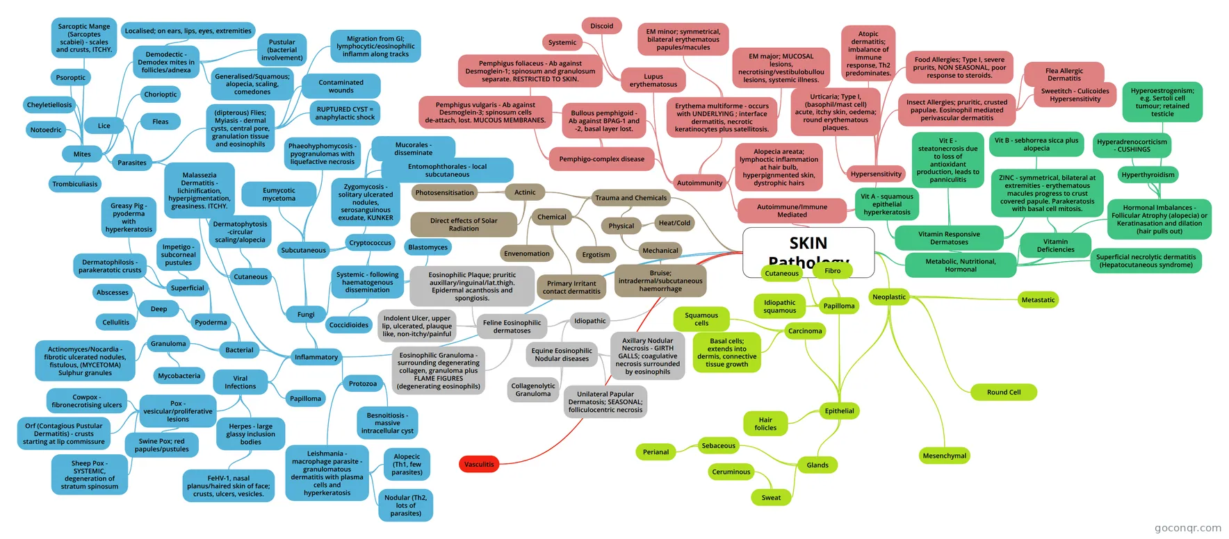
Mind map: SKIN Pathology
Conteúdo do mapa
SKIN Pathology
Vasculitis
Inflammatory
- Bacterial
- Pyoderma
- Superficial
- Impetigo - subcorneal pustules
- Greasy Pig - pyoderma with hyperkeratosis
- Dermatophilosis - parakeratotic crusts
- Deep
- Abscesses
- Cellulitis
- Superficial
- Granuloma
- Actinomyces/Nocardia - fibrotic ulcerated nodules, fistulous, (MYCETOMA) Sulphur granules
- Mycobacteria
- Pyoderma
- Viral Infections
- Pox - vesicular/proliferative lesions
- Cowpox - fibronecrotising ulcers
- Orf (Contagious Pustular Dermatitis) - crusts starting at lip commissure
- Sheep Pox - SYSTEMIC, degeneration of stratum spinosum
- Swine Pox; red papules/pustules
- Herpes - large glassy inclusion bodies
- FeHV-1, nasal planus/haired skin of face; crusts, ulcers, vesicles.
- Papilloma
- Pox - vesicular/proliferative lesions
- Fungi
- Cutaneous
- Malassezia Dermatitis - lichinification, hyperpigmentation, greasiness. ITCHY.
- Parakeratotic hyperkeratosis plus epidermal hyperplasia and spongiosis. Yeast found in aggregates in the corneum/crusts.
- Dermatophytosis -circular scaling/alopecia
- Fungi use keratin as a nutrient, so migrate to the hair follicle. Furunculosis my occur, leading to mycotic pyogranulomas (Kerion) that my look like tumours.
- Malassezia Dermatitis - lichinification, hyperpigmentation, greasiness. ITCHY.
- Subcutaneous
- Eumycotic mycetoma
- MYCETOMA - tumour-like lesion, with draining tracts and granules in the discharge. Papules/nodules form due to chronic suppuration/fibrosis. Granules are clumps of hyphae in Eumycotic mycetoma.
- Phaeohyphomycosis - pyogranulomas with liquefactive necrosis
- Zygomycosis - solitary ulcerated nodules, serosanguinous exudate, KUNKER
- Mucorales - disseminate
- Entomophthorales - local subcutaneous
- Eumycotic mycetoma
- Systemic - following haematogenous dissemination
- Cryptococcus
- Blastomyces
- Coccidioides
- Cutaneous
- Protozoa
- Leishmania - macrophage parasite - granulomatous dermatitis with plasma cells and hyperkeratosis
- Alopecic (Th1, few parasites)
- Nodular (Th2, lots of parasites)
- Besnoitiosis - massive intracellular cyst
- Definitive hosts are Cats, but the lifecycle involves an intermediate host; often cattle/horses.
- Leishmania - macrophage parasite - granulomatous dermatitis with plasma cells and hyperkeratosis
- Parasites
- (dipterous) Flies; Myiasis - dermal cysts, central pore, granulation tissue and eosinophils
- Contaminated wounds
- Migration from GI; lymphocytic/eosinophilic inflamm along tracks
- RUPTURED CYST = anaphylactic shock
- Lice
- Fleas
- Mites
- Sarcoptic Mange (Sarcoptes scabiei) - scales and crusts, ITCHY.
- Burrows into and below the stratum corneum, leading to hyperkeratosis; both ortho- and patchy para-, spongiosis, leukocyte exocytosis and formation of eosinophilic pustules.
- Demodectic - Demodex mites in follicles/adnexa
- Generalised/Squamous; alopecia, scaling, comedones
- Pustular (bacterial involvement)
- Localised; on ears, lips, eyes, extremities
- Chorioptic
- Psoroptic
- Notoedric
- Trombiculiasis
- Cheyletiellosis
- Sarcoptic Mange (Sarcoptes scabiei) - scales and crusts, ITCHY.
- (dipterous) Flies; Myiasis - dermal cysts, central pore, granulation tissue and eosinophils
- Bacterial
Trauma and Chemicals
- Physical
- Heat/Cold
- Mechanical
- Bruise; intradermal/subcutaneous haemorrhage
- Phagocytes and leukocytes recruited to remove RBCS. Heamoglobin catabolism responsible for colour change (biliverdin, bilirubin, heamosiderin).
- Bruise; intradermal/subcutaneous haemorrhage
- Chemical
- Primary Irritant contact dermatitis
- Ergotism
- Envenomation
- Actinic
- Direct effects of Solar Radiation
- Photosensitisation
- Physical
Autoimmune/Immune Mediated
- Autoimmunity
- Pemphigo-complex disease
- Pemphigus foliaceus - Ab against Desmoglein-1; spinosum and granulosum separate. RESTRICTED TO SKIN.
- Starts on the periocular/nasal region but spreads to the ears, neck and ventral abdomen. The lesions start as erythematous maculae, develop to pustules and eventually erosion occurs, with crust formation.
- Pemphigus vulgaris - Ab against Desmoglein-3; spinosum cells de-attach, lost. MUCOUS MEMBRANES.
- Desmoglein 3 found in mucous membranes/mucocutaneous junction, connecting the spinosum cells above the basal layer (suprabasilar) - spongiosis and vacuolation. The lesions (vesicles/bullae) start in the mouth.
- Pemphigus foliaceus - Ab against Desmoglein-1; spinosum and granulosum separate. RESTRICTED TO SKIN.
- Bullous pemphigoid - Ab against BPAG-1 and -2, basal layer lost.
- Lupus erythematosus
- Systemic
- Discoid
- Erythema multiforme - occurs with UNDERLYING ; interface dermatitis, necrotic keratinocytes plus satellitosis.
- Immune complexes deposited in the vasculature and basal membrane, following Type III and IV reactions.
- EM minor; symmetrical, bilateral erythematous papules/macules
- EM major; MUCOSAL lesions, necrotising/vestibulobullou lesions, systemic illness.
- Alopecia areata; lymphoctic inflammation at hair bulb, hyperpignmented skin, dystrophic hairs
- Pemphigo-complex disease
- Hypersensitivity
- Urticaria; Type I, (basophil/mast cell) acute, itchy skin, oedema; round erythematous plaques.
- Atopic dermatitis; imbalance of immune response, Th2 predominates.
- Excessive amounts of IgE are produced in response to an innocuous antigen. High Th2 means the cell mediated response is deficient; making the animal susceptible to local infections e.g. Malassezia.
- Food Allergies; Type I, severe prurits, NON SEASONAL, poor response to steroids.
- Lymphocytes are exposed and sensitised to the allergens in the GI tract, and then travel to the skin.
- Insect Allergies; pruritic, crusted papulae. Eosinophil mediated perivascular dermatitis
- Antigens include venom, faeces, saliva or the whole body of the insect. Type I or IV Hypersensitivity.
- Flea Allergic Dermatitis
- Sweetitch - Culicoides Hypersensitivity
- Autoimmunity
Metabolic, Nutritional, Hormonal
- Vitamin Responsive Dermatoses
- Vit A - squamous epithelial hyperkeratosis
- Vit E - steatonecrosis due to loss of antioxidant production, leads to panniculitis
- Steatonecrosis - fat necrosis Panniculitis - inflammation of fatty subcutaneous tissue
- Vit B - sebhorrea sicca plus alopecia
- Vitamin Deficiencies
- ZINC - symmetrical, bilateral at extremities - erythematous macules progress to crust covered papule. Parakeratosis with basal cell mitosis.
- Hormonal Imbalances - Follicular Atrophy (alopecia) or Keratinasation and dilation (hair pulls out)
- Hyperadrenocorticism - CUSHINGS
- Cutaneous atrophy (thin skin), altered hair growth/alopecia, calcium deposits, hyperpigmentation, blackheads.
- Hyperthyroidism
- Hyperoestrogenism; e.g. Sertoli cell tumour; retained testicle
- Hyperadrenocorticism - CUSHINGS
- Superficial necrolytic dermatitis (Hepatocutaneous syndrome)
- Red (hyper(para)keratosis) White (spongiosis/oedema of spinosum) Blue (hyperplasia of basal layer)
- Associated with end stage liver disease and glucagon secreting pancreatic tumours. Leads to erythemas, erosion/ulcers and crusts.
- Vitamin Responsive Dermatoses
Idiopathic
- Feline Eosinophilic dermatoses
- Eosinophilic Plaque; pruritic auxillary/inguinal/lat.thigh. Epidermal acanthosis and spongiosis.
- Eosinophilic Granuloma - surrounding degenerating collagen, granuloma plus FLAME FIGURES (degenerating eosinophils)
- Oral and cutaneous forms possible.
- Indolent Ulcer, upper lip, ulcerated, plauque like, non-itchy/painful
- Equine Eosinophilic Nodular diseases
- Collagenolytic Granuloma
- Axillary Nodular Necrosis - GIRTH GALLS; coagulative necrosis surrounded by eosinophils
- Unilateral Papular Dermatosis; SEASONAL; folliculocentric necrosis
- Feline Eosinophilic dermatoses
Neoplastic
- Epithelial
- Papilloma
- Epithelial proliferation INDUCED by host-specific Papillomaviridae infection; immunocompetance plays a role in susceptibility and regression.
- Cutaneous
- Hyperkeratotic exophytic projections of the epidermis, supported by thin dermal stalks.
- Fibro
- Prominent DERMAL proliferation forms plaque-like lesions e.g. Equine Sarcoid.
- Idiopathic squamous
- Carcinoma
- Basal cells; extends into dermis, connective tissue growth
- Squamous cells
- Hyperplastic ulcerated or nodular lesion, expansile, pathogenesis involves UV light esp. in white animals and potentially papilloma virus infection
- Hair folicles
- Glands
- Sebaceous
- Perianal
- Sweat
- Ceruminous
- Sebaceous
- Papilloma
- Mesenchymal
- Round Cell
- Metastatic
- Epithelial
Informações
- Descrição
- Mapa Mental sobre SKIN Pathology, criado por stephwescott em 01-11-2014.
- Criado por
- stephwescott
- Visualizações
- 5
- Avaliação