
Mapa mental
Glycoconjugates
Conteúdo do mapa
Glycoconjugates
Introduction
- include
- Proteoglycans
- Glycolipids
- Glycoproteins
- the
- Glycosylation is the main enzymatic modification of proteins in the body
- Animal cells have two main types of proteins that contain sugars
- which are
- Glycoproteins
- Proteoglycans
- which are
- Glycoproteins have short glycan chains
- they can have
- Up to 20 sugars
- they can have
- Glycoproteins
- generally
- Do not contain uronic acids
- often
- Contain much smaller amounts of carbohydrates than proteins
- are
- Most cell surface membrane proteins
- generally
- include
Glycoproteins
- Structures and links
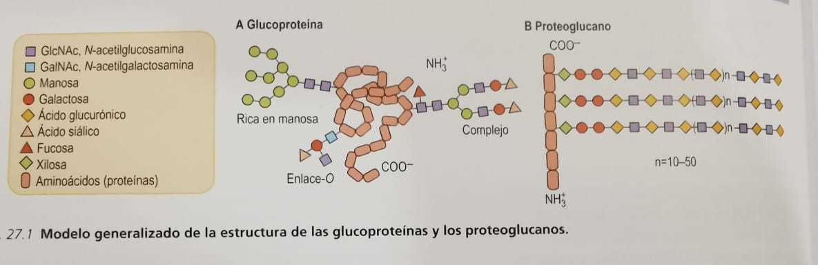
- General structures of glycoproteins
- the
- LDL receptor has two N-glucans located near the LDL-binding domain
- and
- A group of O-glucans near the membrane-spanning region
- and
- A glycoprotein can have a single N-glucan chain
- or it can have
- Several types of oligosaccharides
- or it can have
- Microheterogeneity of the oligosaccharide structures
- is due to
- Incomplete processing in some chains during their biosynthesis
- is due to
- N-glucans exposed on the surface of a folded protein are more vulnerable to processing enzymes
- while
- Those protected behind the protein structure are inaccessible to enzymes
- while
- Glycoproteins with N-glycosidic bonds also contain O-glucans
- LDL receptor has two N-glucans located near the LDL-binding domain
- the
- Structure-function relationships in mucin glycoproteins
- the
- Mucins are glycoproteins secreted by epithelial cells
- that line the
- Respiratory
- Gastrointestinal
- Genitourinary systems
- that line the
- Oligosaccharides carry a negatively charged sialic acid
- preventing
- The protein from folding, causing it to remain extended
- preventing
- Mucins are glycoproteins secreted by epithelial cells
- there
- Are many complex linear and branched oligosaccharide structures on mucins
- including
- Blood group antigens
- including
- Are many complex linear and branched oligosaccharide structures on mucins
- about
- A third of the amino acids in mucins are serines or threonines
- most of them
- Are replaced by the O-glycosidic linked oligosaccharide
- most of them
- A third of the amino acids in mucins are serines or threonines
- the
- the
- Sugars can bind to proteins
- either
- N-glycosidic bonds
- O-glycosidic bonds
- either
- Oligosaccharides with N-glycosidic bonds (N-glucans) are widely distributed in nature
- they are characteristic of
- The membrane and secretory proteins
- they are characteristic of
- Union of the oligosaccharides with the proteins involved a glucosamine union
- between
- N-acetylglucosamine residue (GlcNAc)
- The amide nitrogen of an asparagine residue
- between
- O-glycosidic linked oligosaccharides (O-glucans)
- are found
- In proteins of mucosal fluids
- are found
- O-glucans generally contain three or more sugars in straight or branched chains
- linked to proteins by
- A glycosidic bond between an N-acetylgalactosamine (GalNAc) residue
- The hydroxyl group of a serine or threonine residue in the protein
- linked to proteins by
- Glucosyl-galactose disaccharide is often attached to the hydroxyl group of hydroxylysine residues
- in
- The fibrous protein, collagen
- in
- Procollagens are usually synthesized as glycoproteins with N-glycosidic bonds
- but
- N-glucan is eliminated as part of this peptide
- but
- Only disaccharides with O-glycosidic bonds
- remain in the
- Mature collagen molecule
- remain in the
- GlcNAC modification (O-GlcNAcylation) may represent a mechanism
- that allows cells to
- Block the phosphorylation of specific serine threonine residues in specific proteins
- that allows cells to
- Donor substrate for O-GlcNAcylation
- is
- UDP-GlcNAc derived from glucose
- is
- Mannose O-glucans serve as links
- between
- The intracellular cytoskeleton
- The extracellular matrix to maintain myocyte function
- between
- Glycogenin is a self-glycosylated protein that binds a glucose
- to the
- Hydroxyl group of one of its tyrosine residues
- to the
- N-glucans have oligosaccharide chains that are branched structures
- with
- A common center of three mannose residues
- Two GlcNAC residues
- with
- Oligosaccharides give rise to a wide group of structures
- that are called
- Mannose rich chains
- Complex chains
- that are called
- N-glucans have rich structures in hands as built from a common nucleus
- O-GlcNAcylation of proteins involved in the insulin signaling pathway
- causes
- Insulin resistance
- in
- Skeletal muscle
- Adipose tissue
- Pancreatic B cells
- in
- Insulin resistance
- causes
- Sugars can bind to proteins
- General structures of glycoproteins
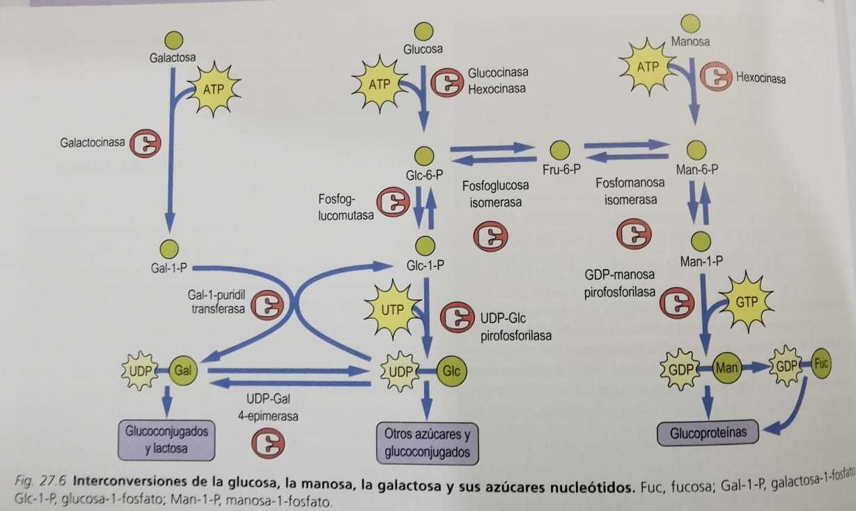
- Interconversions of sugars in the diet
- Formation of galactose, mannose and fucose from glucose
- the
- GDP-Man is the form of mannose
- that is used in the
- Formation of oligosaccharides with N-glycosidic bond
- that is used in the
- Glucose is phosphorylated
- by
- Hexokinase (or glucokinase in the liver) when it enters the cell
- by
- Glucose-6-phosphate (Glc-6-P) can then be converted by a mutase (phosphoglucose mutase)
- to form
- Glc-1-P, which reacts with uridine triphosphate (UTP) to form UDP-Glc
- catalyzed by
- UDP-Glc pyrophosphorylase enzyme
- catalyzed by
- Glc-1-P, which reacts with uridine triphosphate (UTP) to form UDP-Glc
- to form
- UDP-Glc is epimerized to UDP-galactose (UDP-Gal)
- by
- UDP-Gal 4-epimerase, providing UDP-Gal for the synthesis of glycoconjugates
- by
- Glc-6-P can also be converted to fructose-6-phosphate (Fru-6-P) by the glycolytic enzyme phosphoglucose isomerase
- and
- Fru-6-P can be isomerized to mannose-6-phosphate (Man-6 -P) by phosphomannose isomerase
- and
- Man-6-P is then converted by a mutase (phosphomannose mutase) to Man-1-P
- which
- Is condensed with GTP to form GDP-Man
- which
- GDP-Man is the form of mannose
- the
- Galactose metabolism
- the
- UDP-Gal 4-epimerase., this enzyme catalyzes the balance between UDP-Glc and UDP-Gal
- providing
- A constant source of UDP-Glc during galactose metabolism
- providing
- Galactose from the diet is transported to the liver
- where
- It is phosphorylated by a specific kinase, galactokinase
- where
- Enzyme Gal-1-P uridyltransferase catalyzes an exchange between UDP-Glc and Gal-1-P
- to form
- UDP-Gal Glc-1-P
- to form
- UDP-Gal 4-epimerase., this enzyme catalyzes the balance between UDP-Glc and UDP-Gal
- the
- Fructose metabolism
- the
- Cleavage products of fructose by aldolase B are dihydroxyacetone phosphate and glyceraldehyde
- after which
- Glyceraldehyde must be phosphorylated by triose kinase to be metabolized by glycolysis
- after which
- Fructose is a component of sucrose
- and
- Makes up about half the sugar in high-fructose corn syrup
- and
- Fructose can be phosphorylated
- by
- Hexokinase, an enzyme present in all cells
- by
- Main pathway of fructose metabolism, which is especially important in the liver after a meal involves fructokinase
- this enzyme
- Is a very specific kinase that phosphorylates fructose at carbon 1 to fructose phosphate (Fru-1-P )
- this enzyme
- Cleavage products of fructose by aldolase B are dihydroxyacetone phosphate and glyceraldehyde
- the
- the
- Cells can use glucose to form the rest of the sugars they need
- Sugars need to make glycoconjugates
- can be
- Synthesized from blood sugar
- can be
- Formation of galactose, mannose and fucose from glucose
- Other pathways of sugar nucleotide metabolism
- UDP-GIcUA
- the
- Two-stage oxidation by the enzyme UDP-Glc dehydrogenase leads to the formation of the activated
- form of
- Glucuronic acid (UDP-GlcUA), this sugar nucleotide is the glucuronic acid donor for the formation of proteoglycans
- form of
- UDP-Glc is the precursor to several other sugars, such as glucuronic acid, xylose and galactose
- which
- Are required for the synthesis of proteoglycans and / or glycoproteins
- which
- UDP-GlcUA is also the precursor to UDP-xylose. a pentose sugar nucleotide
- Two-stage oxidation by the enzyme UDP-Glc dehydrogenase leads to the formation of the activated
- the
- GDP-Man and GDP-Fuc
- the
- Resulting depletion of GDP-Fuc in the Golgi lumen blocks the biosynthesis
- of the
- Leukocyte recognition signal, the Lewis-X sialyl structure
- of the
- Guanosine diphosphate-mannose (GDP-Man) is the donor substrate for most mannosyltransferases
- Conversion of GDP-Man to GDP-fucose involves a complex series of oxidative
- and
- Reductive steps, in addition to epimerization
- and
- Resulting depletion of GDP-Fuc in the Golgi lumen blocks the biosynthesis
- the
- Amino sugars
- the
- UDP-GlcNAc can be epimerized to UDP-GalNAc
- Fru-6-P is the precursor of amino sugars
- Acetyl group is transferred from acetyl-CoA to the amino group of GlcN-6-P
- to form
- GlcNAc-6-P, which is converted to its activated form, UDP-GlcNAc
- to form
- Amino sugars in glycoconjugates are acetylated
- therefore
- They are neutral and do not contribute to the ionic charge of the glycoconjugates
- therefore
- Transfer of an amino group from the amide nitrogen of glutamine
- to
- Fru-6-P to produce glucosamine-6-P (GlcN-6-P)
- to
- the
- Sialic acid
- the
- CMP-sialic acid is the only sugar donor nucleoside monophosphate
- in the
- Metabolism of glycoconjugates
- in the
- UDP-GIcNAc is the precursor of N-acetylneuraminic acid (NeuAc)
- also called
- Sialic acid
- also called
- Sialic acid, a 9-carbon N-acetylamino-ketodeoxygluconic acid
- is produced by
- Condensation of an amino sugar with phosphoenolpyruvate
- is produced by
- CMP-sialic acid is the only sugar donor nucleoside monophosphate
- the
- UDP-GIcUA
- Oligosaccharide biosynthesis
- Oligosaccharides with O-glycosidic bonds (O-glucans)
- the
- Glycosyltransferases of the Golgi apparatus
- are
- Involved in the stepwise biosynthesis of mucin oligosaccharides
- are
- O-glycosidic linked oligosaccharides
- are
- Synthesized in the Golgi apparatus
- are
- Synthesis of the oligosaccharides with O-glycosidic bonds takes place only in the Golgi apparatus
- by
- The addition in stages of sugars from the nucleotide derivatives of sugar the protein
- by
- GalNAc is transferred from the UDP-GalNAc to serine or threonine residues in the protein
- by
- Means of a GalNAc transferase in the Golgi apparatus
- by
- Glycosyltransferases of the Golgi apparatus
- the
- the
- Final structure of the oligosaccharide chains
- depends on
- The complement of glycosyltransferases in the cell
- depends on
- Assembly of oligosaccharides with N-glycosidic bonds
- begins
- In the endoplasmic reticulum
- begins
- First sugar to be added to dolichol-P from UDP-GIcNAc is GlcNAc-1-P
- by
- A GlcNAc-1-P transferase to produce dolichol-P-P-GIcNAc
- by
- Second GlcNAc binds to the first GlcNAc
- followed by
- The addition of 4-5 mannose residues from GDP-Man
- followed by
- Presence of glucose in lipid-bound oligosaccharides
- has been shown to
- Accelerate the transfer of oligosaccharides from lipids to proteins
- has been shown to
- Glucose is important in directing protein folding in the endoplasmic reticulum
- the
- Intermediate processing continues in the endoplasmic reticulum (ER) and the Golgi apparatus
- the
- Final modifications of the N-glycosidic bond oligosaccharides
- occur
- In the Golgi apparatus
- occur
- Mannose and glucose residues are added
- to construct
- A lipid-linked oligosaccharide intermediate
- to construct
- Following ER cleavage reactions and proper folding
- the protein
- GlcNAc, is transported to the Golgi apparatus, where further modification reactions occur
- the protein
- Final structure of the oligosaccharide chains
- Oligosaccharides with O-glycosidic bonds (O-glucans)
- Functions of the oligosaccharide chains of glycoproteins
- the
- Glycoprotein ZP3, which is present in the zona pellucida of the mouse ovum
- functions as
- A receptor for sperm during fertilization
- functions as
- Proteins resident in the endoplasmic reticulum
- known as chaperones
- Help newly synthesized proteins to fold into their proper conformations
- known as chaperones
- Oligosaccharides containing Man-6-P direct lysosomal enzymes to lysosomes
- most of these lysosomal enzymes
- Are glycoproteins with N-glycosidic bonds that are synthesized and glycosylated in the endoplasmic reticulum and in the Golgi apparatus
- most of these lysosomal enzymes
- Oligosaccharide chains of glycoproteins
- frequently
- Increase the solubility and stability of proteins
- frequently
- Oligosaccharides are hydrophilic, so they increase the solubility of proteins in the aqueous medium
- Sugars are involved in chemical recognition interactions with lectins
- Glycoproteins with N-glycosidic bonds found on the cell surface of mammals
- this process is a key factor in
- Fertilization
- Inflammation
- Infection
- Development
- Differentiation
- play
- A key role in cell-cell interactions and other recognition processes
- this process is a key factor in
- Gram-negative enteric bacteria have short hair-like projections called pili on their surfaces
- these pili have
- Mannose-binding lectins at their end that can recognize and bind to mannose-rich oligosaccharides on membranes
- these pili have
- Glycoprotein ZP3, which is present in the zona pellucida of the mouse ovum
- the
- Structures and links
Proteoglycans
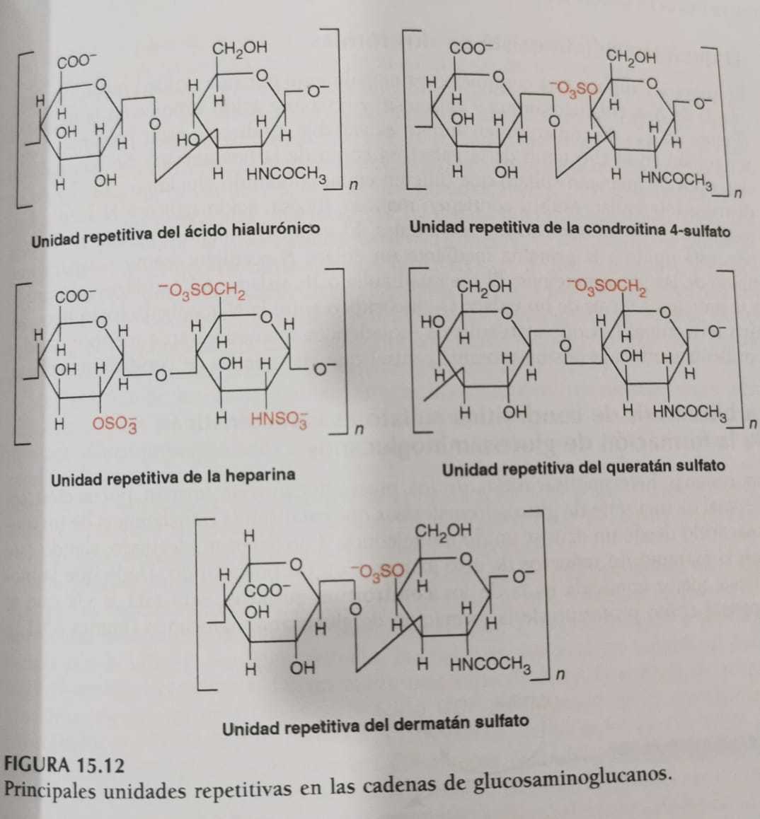
- Hyaluronate
- the
- Hyaluronate is very different from the others
- because
- It is not sulfated
- It is not covalently bound to a protein
- It is the only glycosaminoglycan that is not limited to animal tissues
- because
- Hyaluronate is very different from the others
- it
- Is found predominantly
- in the
- Synovial fluid
- Vitreous humor
- Umbilical cord
- in the
- Consists solely of repeating disaccharide units
- of
- N-acetylglucosamine
- Glucuronic acid
- of
- Is found predominantly
- the
- Chondroitin sulfates
- the
- Chondroitin sulfate proteoglycans have been shown to aggregate noncovalently
- with
- Hyaluronate, forming much larger structures
- with
- Most abundant glycosaminoglycans in the body are chondroitin sulfates
- Polysaccharide chains are linked to specific core protein serine residues of varying molecular mass
- via a
- Trisaccharide binding region
- via a
- Characteristic disaccharide repeating units formed by N-acetylgalactosamine and glucuronic acid
- are
- Covalently linked to this binding region
- are
- Disaccharides can be sulfated at the 4 or 6 position of N-acetylgalactosamine
- Chondroitin sulfate proteoglycans have been shown to aggregate noncovalently
- the
- Dermatan sulfate
- the
- Dermatan sulfate
- is found in the
- Skin
- Blood vessels
- Heart valves
- is found in the
- Dermatan sulfate differs from chondroitin 4 and 6 sulfates
- in that its
- Predominant uronic acid is L-iduronic acid
- in that its
- Glycosidic bonds are the same in position and configuration as in chondroitin sulfates
- with
- Average polysaccharide chains of molecular mass
- with
- Dermatan sulfate
- the
- Heparin and heparan sulfate
- the
- Heparan sulfate
- is found in the
- Walls of blood vessels
- Brain
- Amyloid
- is found in the
- Heparin contains α-glycosidic bonds
- can also contain
- The C-2 of uronic acid
- Sulfate on the C-3 of hexosamine
- can also contain
- Heparin is an intracellular component of mast cells
- and
- Functions predominantly as an anticoagulant and lipid clarifying agent
- and
- Heparan sulfate contains a similar disaccharide repeating unit
- but
- Has more N-acetyl groups, fewer N-sulfate groups, and a lower degree of O-sulfate groups
- but
- Heparan sulfate
- in
- Heparin, the characteristic repeating disaccharide unit is formed
- by
- Glucosamine and D-glucuronic acid
- L-iduronic acid
- by
- Heparin, the characteristic repeating disaccharide unit is formed
- the
- Keratan sulfate
- the
- Keratan sulfate is composed primarily of a repeating disaccharide unit
- of
- N-acetylglucosamine and galactose
- of
- Keratan sulfate is composed primarily of a repeating disaccharide unit
- there are
- Two types of keratin sulfate
- the
- Keratan sulfate II, isolated from cartilage, is bound to protein through an O-glycosidic bond
- between
- Serine or threonine
- N-acetylgalactosamine
- between
- Keratan sulfate I, isolated from the cornea, is bound to the protein
- by
- An N-acetylglucosamine-asparagine bond
- by
- Keratan sulfate II, isolated from cartilage, is bound to protein through an O-glycosidic bond
- the
- Two types of keratin sulfate
- the
- Chondroitin sulfate biosynthesis
- the
- Predominant extracellular species
- are
- Thrombomodulin
- Versican
- CD44
- Sirdecan
- Aggrecan
- are
- Best-known biosynthesis is that of chondroitin sulfates
- it is the
- Prototype pathway for the formation of glycosaminoglycans
- it is the
- Formation of the core proteoglycan chondroitin sulfate protein
- followed by
- The formation of polysaccharide chains catalyzed by six different glucosyltransferases
- followed by
- Polymerization then occurs
- by two glucosyltransferases
- An N-acetylgalactosaminyltransferase
- A glucuronosyltransferase
- by two glucosyltransferases
- Synthesis of the other glycosaminoglycans requires additional specific transferases
- for the
- Sugars and bonds found in these molecules
- for the
- Epimerization of D-glucuronic acid to L-iduronic acid occurs after incorporation
- into
- The polymer chain and occurs simultaneously with the sulfation process
- into
- Synthesis of both proteoglycans and glycoproteins
- is regulated by
- The same mechanism, at the level of hexosamine synthesis
- is regulated by
- Proteoglycans, like glycoproteins
- are degraded by
- The sequential action of proteases and glycosidases
- as well as
- Deacetylases and sulphatases
- as well as
- The sequential action of proteases and glycosidases
- are degraded by
- Mucopolysaccharidosis is a group of human genetic diseases
- that are characterized by
- The accumulation in the tissues and the excretion in the urine of oligosaccharide
- products derived from
- The incomplete breakdown of proteoglycans
- which is due to
- The lack of one or more lysosomal hydrolases
- which is due to
- The incomplete breakdown of proteoglycans
- products derived from
- The accumulation in the tissues and the excretion in the urine of oligosaccharide
- that are characterized by
- More proteoglycans are being defined
- based primarily on
- Their functional properties or their location
- based primarily on
- Predominant extracellular species
- the
- Introduction
- the
- Common constituents of glycosaminoglycans
- are
- Sulfate groups linked by ester bond to certain monosaccharides or by amide bond to the amino group of glucosamine
- are
- Glycosaminoglycans are the carbohydrate chains of proteoglycans
- Proteoglycans are high molecular mass polyanionic substances
- that are composed of
- Many different chains of glycosaminoglycans covalently attached to a protein core
- that are composed of
- Long linear heteropolysaccharide chains are largely made up of repeating disaccharide units
- in which
- One sugar is a hexosamine
- The other is a uronic acid
- in which
- Glycosaminoglycans are components of extracellular matrices and cell surfaces
- Common constituents of glycosaminoglycans
- the
- Hyaluronate
Glycolipids
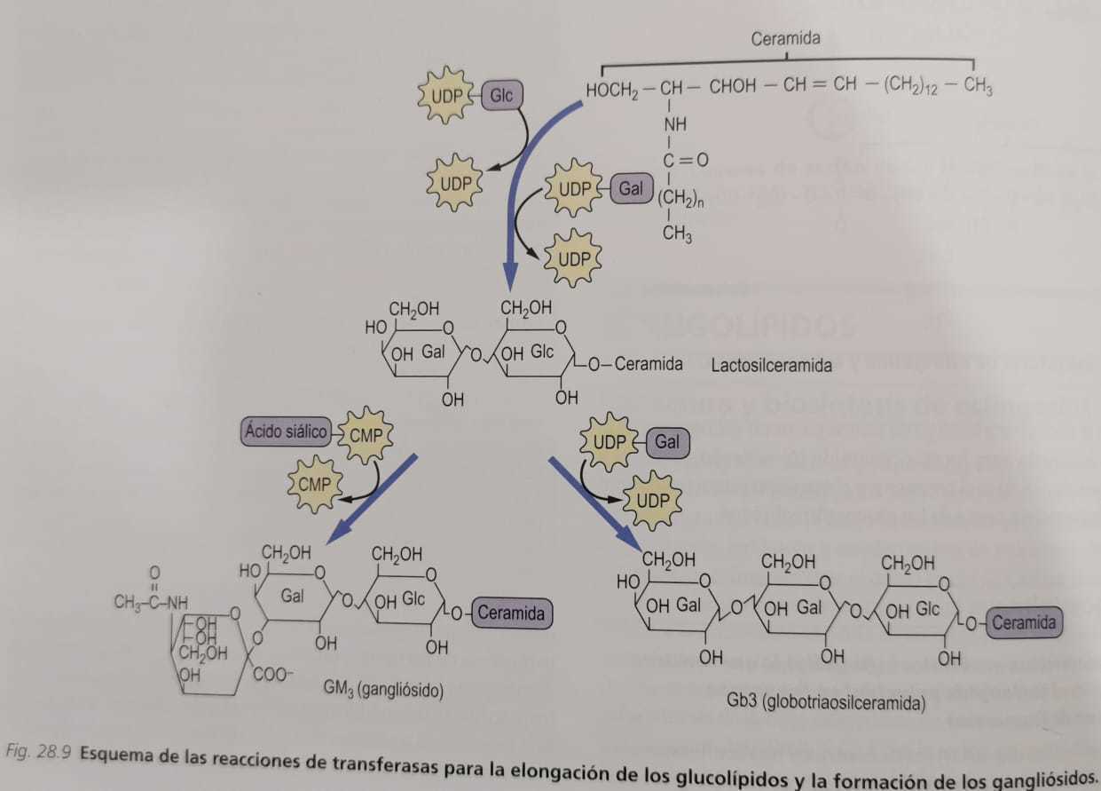
- Structure and nomenclature of gangliosides
- the
- Nomenclature used to identify the various gangliosides
- is based on the
- Number of sialic acid residues contained in the molecule and on the sequence of carbohydrates
- is based on the
- Gangliosides are glycosphingolipids that contain sialic acid (N-acetylneuraminic)
- Term ganglioside is applied to glycolipids that were originally identified
- in high concentrations in
- Ganglion cells of the central nervous system
- in high concentrations in
- Nomenclature used to identify the various gangliosides
- the
- Lysosomal storage diseases secondary to defects in glycolipid breakdown
- the
- Defects in the sequential breakdown of glycolipids
- cause
- Various lysosomal storage diseases, known as sphingolipidosis and gangliosides
- cause
- Complex oligosaccharides of glycolipids are formed
- by
- Adding a sugar residue, in the Golgi apparatus
- and
- Are degraded in the same sequential way, but in the opposite direction, by a series of deglycosylases in lysosomes
- and
- Adding a sugar residue, in the Golgi apparatus
- by
- Sphingolipidoses, like inclusion cell disease
- are characterized by
- The accumulation of undigested lipids in inclusion bodies in cells
- are characterized by
- Defects in the sequential breakdown of glycolipids
- the
- Introduction
- the
- Sulfatides are formed by adding sulfate
- from the
- Sulfate donor, 3'-phosphoadenosine-5'-phosphosulfate (PAPS), yielding, GalCer 3-sulfate
- from the
- Sphingolipids that contain sugars linked by covalent bonds
- are known as
- Glycosphingolipids or glycolipids
- are known as
- Glycolipids can be classified
- into 3 groups
- In all these compounds the polar head group
- comprising the sugars
- Is attached to the ceramide by a glycosidic bond at the terminal hydroxyl group of sphingosines
- comprising the sugars
- Neutral glycolipids
- Gangliosides
- Sulphatides
- In all these compounds the polar head group
- into 3 groups
- Neutral glycolipids contain only neutral sugars and amino sugars
- like
- Glucosylceramide (GlcCer)
- Galactosylceramide (GalCer)
- like
- Glycolipids that contain sialic acid (N-acetylneuraminic acid NeuAc) are called gangliosides
- Sulfatides are formed by adding sulfate
- the
- Structure and nomenclature of gangliosides
Made by: David Leonado López Sánchez
·
- Took from: Baynes, J., & Dominiczak, M. (2015). Bioquímica médica. En J. Baynes, & M. Dominiczak, Bioquímica médica (págs. 353-376). Barcelona: Elsevier Saunders.
·
- Took from: Schwartz, N. (2015). Metabolismo glucídico II: rutas especiales y glucoconjugados. En T. Devlin, Bioquímica libro de texto con aplicaciones clínicas (págs. 666 - 691). Reverté.
·
- Took from: Baynes, J., & Dominiczak, M. (2015). Bioquímica médica. En J. Baynes, & M. Dominiczak, Bioquímica médica (págs. 353-376). Barcelona: Elsevier Saunders.
·
- Took from: Baynes, J., & Dominiczak, M. (2015). Bioquímica médica. En J. Baynes, & M. Dominiczak, Bioquímica médica (págs. 353-376). Barcelona: Elsevier Saunders.
Imagens e arquivos do mapa
Informações
- Descrição
- Mapa Mental sobre Glycoconjugates, criado por David L Lopez Sanchez em 13-10-2020.
- Criado por
- David L Lopez Sanchez
- Visualizações
- 6
- Avaliação