
Mapa mental
Renal Pathology for Pathologists
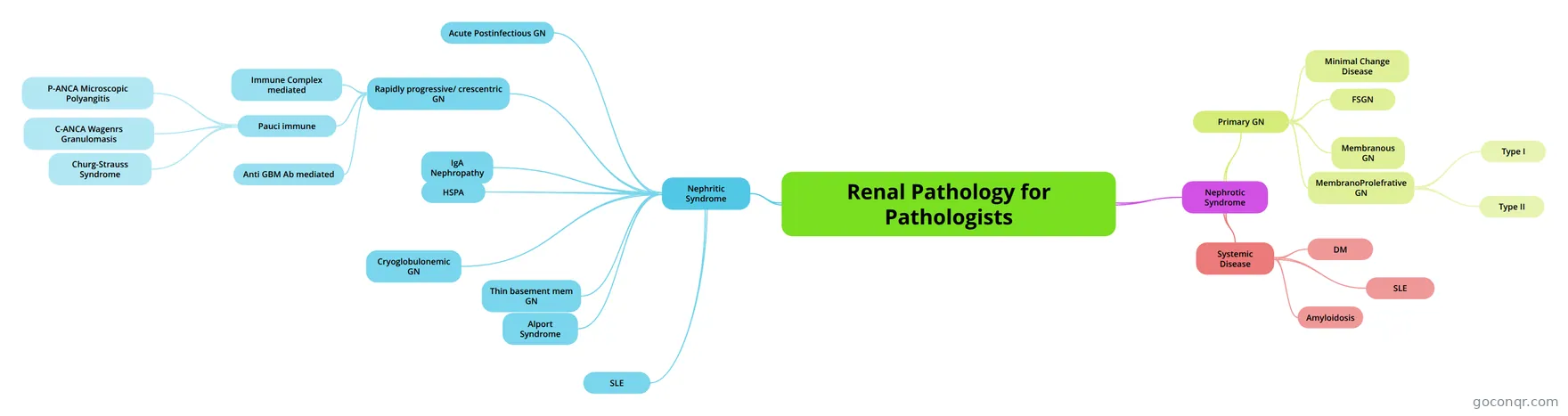
Mind map: Renal pathology for pathologists
Conteúdo do mapa
Renal Pathology for Pathologists
Nephrotic Syndrome
- PRORIEN >3.5g Hypoalbumenimia oedema
- Primary GN
- Minimal Change Disease
- Most common cause in kids LM: N IF: N EM: Diffuse effacement of the visceral epithelial cell foot processes Rx: Steroids
- FSGN
- - LM: focal and segmental sclerosis of the present glomeruli with hyaline deposition, lipid droplets may be present; eventual progression to diffuse, global sclerosis later in disease - IF: non-specific entrapment of IgM, C3, C1q in sclerotic segments; no immune complex formation - EM: diffuse effacement of foot processes and detachment from the GBM (irreversible injury) Rx: poor response to Steroids
- Membranous GN
- Most common cause in adults SUBEPITHELIAL complex deposition - LM: diffuse uniform thickening of GBM - IF: uniform GRANULAR deposits staining with IgG and C3 along the GBM, “capillary loop pattern” - EM: electron dense deposits of immune complexes in the subepithelial location with the GBM forming “spikes” between the deposits (will eventually surround the deposits), and effacement of foot processes. Rx: poor response to steroids
- MembranoProlefrative GN
- can be due to nephrotic or niphritic diseases Progressive and unremitting course Steroids are typically not effective.
- Type I
- LM- Diffuse hypercellularity with accentuation of lobular architecture, matrix is expanded, and the GBM is thickened. Silver staining of the GBM shows “tram-track” appearance. IF: granular C3 deposits, IgG, and C1q in the mesangium and along the GBM. EM: SUBENDOTHELIAL AND MESANGIAL immune complex deposition, duplication of the GBM with extension of the mesangial cells/matrix into the subendothelial space
- Type II
- LM- Diffuse hypercellularity with accentuation of lobular architecture, matrix is expanded, and the GBM is thickened. Silver staining of the GBM shows “tram-track” appearance. IF: C3 deposition as irregular granular foci along the GBM and as “rings” in the mesangium; NEGATIVE for IgG and C1q EM: dense “ribbon-like” material of unknown composition within the GBM and in the mesangium
- Minimal Change Disease
- Systemic Disease
- DM
- LM: thickening of the GBM. Diffuse mesangial expansion and sclerosis. Nodular glomerulosclerosis (Kimmelstiel-Wilson Nodules) IF: nonspecific entrapment of IgM and C3 within hyaline caps EM: thickened GBM, matrix expansion, K-W lesions, and subendothelial hyaline
- Amyloidosis
- LM: With H&E staining, these deposits resemble an amorphous, eosinophilic proteinaceous material. Stain (+) with Congo Red stain - “apple green birefringence under polarized light” LM: mesangial expansion with deposition of homogenous, eosinophilic material; with advanced cases, the GBM, interstitium, tubular basement membrane, and vessels may also be deposited with amyloid. With both PAS and silver stain, the amyloid deposits on the GBM resemble “shag carpeting.” Amyloid exhibits characteristic staining with Congo Red stain. IF: will be specific depending on type of amyloid deposited; (+)light chain staining for AL amyloidosis or (+)anti-AA antibody staining for AA amyloidosis EM: Diagnositic! Shows small randomly arranged nonbranching fibrils measuring 7.5-10 nm diameter. Fibrils are deposited in mesangium and along the GBM, again resembling “shag carpeting.”
- SLE
- DM
Nephritic Syndrome
- Acute Hematuria with RBC Casts and Dysmorphic RBCs •Oliguria •Azotemia (Increased BUN and Creatinine) •Hypertension •Mild Edema •Mild Proteinuria (<3 g/24 hrs)
- Acute Postinfectious GN
- development of nephritic syndrome 1-4 weeks after infection with group A beta-hemolytic streptococci (pharyngitis or impetigo) -Nephritogenic strains of streptococcus = Types 12, 4, and 1. •LM: global, diffuse glomerulonephritis with enlarged hypercellular glomeruli. No thickening of the GBM. •IF: “lumpy-bumpy” granular deposition of IgG and C3 along the GBM and in mesangium •EM: large subepithelial electron dense deposits, “subepithelial humps,” along the GBM and in the mesangium.
- IgA Nephropathy
- The most common type of glomerulonephritis! •Typical patient presents with recurrent self-limiting episodes of gross or microscopic hematuria, often following URI, GU, or GI infections. •Can range from mild proteinuria to nephritic syndrome. •Characterized by IgA deposition in the mesangium. - Can be Primary or Secondary Disease. •LM: variable, diagnosis is dependent on IF. • IF: strong staining of IgA and C3 granular deposits in the mesangium. •EM: electron dense deposits in the mesangium.
- HSPA
- Thin basement mem GN
- Alport Syndrome
- Rapidly progressive/ crescentric GN
- Immune Complex mediated
- Pauci immune
- P-ANCA Microscopic Polyangitis
- C-ANCA Wagenrs Granulomasis
- Churg-Strauss Syndrome
- Anti GBM Ab mediated
- SLE
- Cryoglobulonemic GN
- • Cryoglobulins = immunoglobulins that precipitate in the cold and dissolve when heated. • Associated with certain infections, especially HCV. • Circulating cryoglobulins trigger inflammation resulting in systemic small vessel vasculitis. -Renal involvement: hematuria, proteinuria, nephritic/nephrotic syndrome -Skin involvement: skin purpura -Joint involvement: arthralgias • Classification of Cryoglobulins: -Type I: single monoclonal Ig -Type II: polyclonal IgG, monoclonal IgMκ complexes -Type III: polyclonal IgG, polyclonal IgM complexes • Type II and III are termed “mixed cryoglobulinemias” and are more common, especially in pts with HCV, lupus, or RA. • LM: same as Type I MPGN due to subendothelial and mesangial cryoglobulin deposition; cryoglobulin thrombi may be visible in the capillary lumens. • IF: staining depends on type of Cryoglobulinemia -Type I: granular deposits of monoclonal Ig in mesangium, along GBM, and in areas of thrombi formation. -Type II: (+)IgG, IgM, C3, C1q, kappa, and lambda • EM: subendothelial and mesangial deposits that show microtubular structures, “thumbprint”appearance
Informações
- Descrição
- Mapa Mental sobre Renal Pathology for Pathologists, criado por Ali Al-Omari em 25-03-2014.
- Criado por
- Ali Al-Omari
- Visualizações
- 44
- Avaliação