
Mapa mental
Thigh and Knee
Conteúdo do mapa
Thigh and Knee
Anatomy
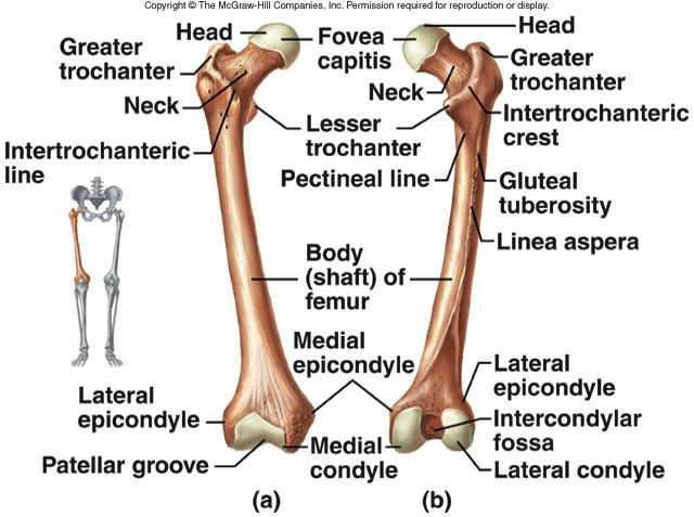
- Bones
- Femur
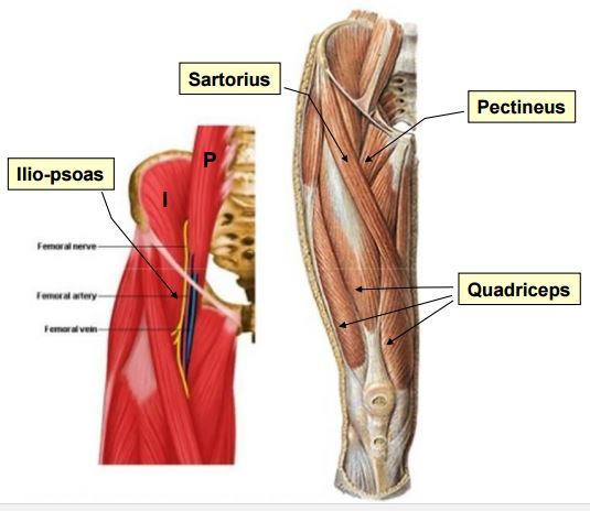
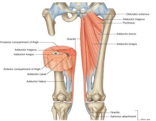
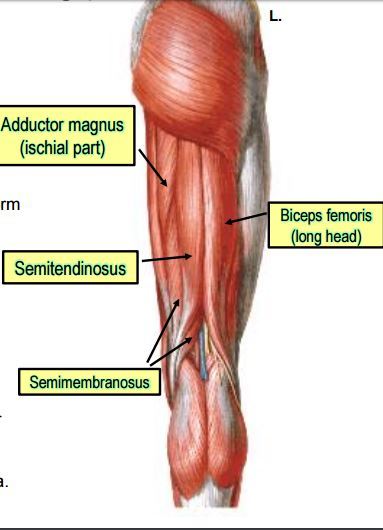
- Muscles of the thigh
- Anterior
- Medial
- Posterior
- Arteries
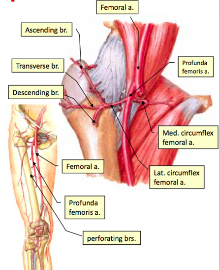
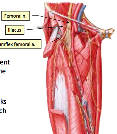
- Femoral Artery and its branches
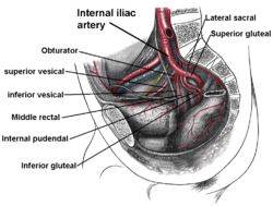
- Obturator Artery and its branches
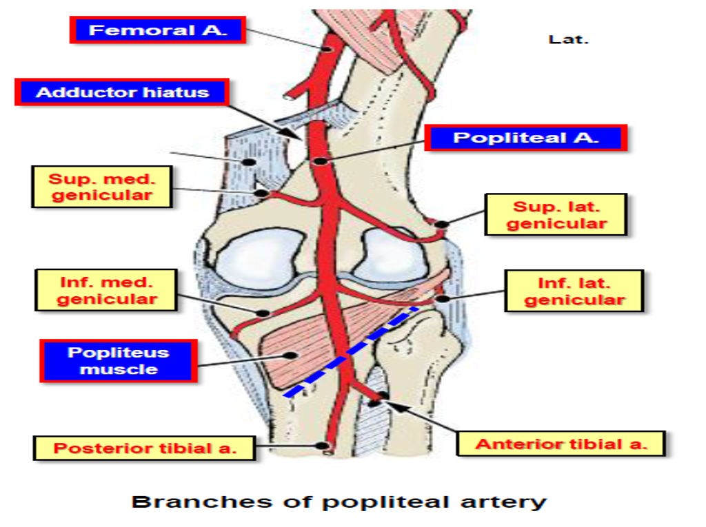
- Popliteal Artery and its branches
- Nerves
- Femoral Nerve
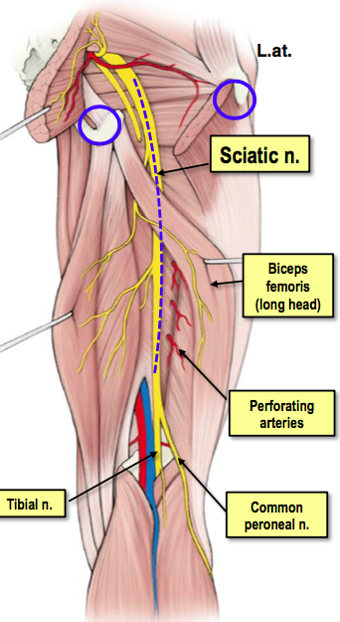
- Sciatic nerve
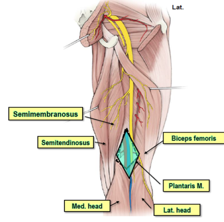
- Popliteal fossa
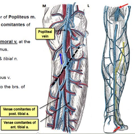
- Veins
- Popliteal vein
- Bones
Leg pain and swelling
- Boil
- deep localized infection of the hair follicle caused by an accumulation of pus and dead tissue
- Boils in the thigh will cause reddish swelling in thighs around the affected area.
- Some sufferers may experience fever, swelling around the boil, swollen lymph nodes, and fatigue.
- Ruled out because Rany has no Boils on his thigh+ his wbc count is normal
- Some sufferers may experience fever, swelling around the boil, swollen lymph nodes, and fatigue.
- Boils in the thigh will cause reddish swelling in thighs around the affected area.
- deep localized infection of the hair follicle caused by an accumulation of pus and dead tissue
- Thigh strain / injury
- a tear in the muscle that can ranges from a very mild tear to complete rupture
- Thigh strain is a common injury, especially among athletes. It usually occur when the thigh muscle is stretched excessively, tearing the muscle fibers.
- a tear in the muscle that can ranges from a very mild tear to complete rupture
- Diabetic Muscle Infarction
- longstanding diabetes mellitus.
- It does not cause any systemic signs of infection
- intense pain and localized swelling in the lower body parts,
- Ruled out because Ranny has no history triggers of diabetes
- intense pain and localized swelling in the lower body parts,
- It does not cause any systemic signs of infection
- longstanding diabetes mellitus.
- Superficial thrombophlebitis
- It is an inflammation and thrombosis (blood clot) of superficial veins
- Can be triggered by having varicose veins, intravenous injection, veins disorder, venous insufficiency.
- It is an inflammation and thrombosis (blood clot) of superficial veins
- Vasculitis
- group of uncommon disorders characterized by inflammatory destruction of blood vessels (both arteries and veins) . Depends on the type of vasculitis, leg and thigh swelling may be seen.
- immune system disorder and inflammation of blood vessels are common aspects
- Rany has no signs or symtoms of vaculitis so it is ruled out
- immune system disorder and inflammation of blood vessels are common aspects
- group of uncommon disorders characterized by inflammatory destruction of blood vessels (both arteries and veins) . Depends on the type of vasculitis, leg and thigh swelling may be seen.
- Boil
Tumors (bone tumors)
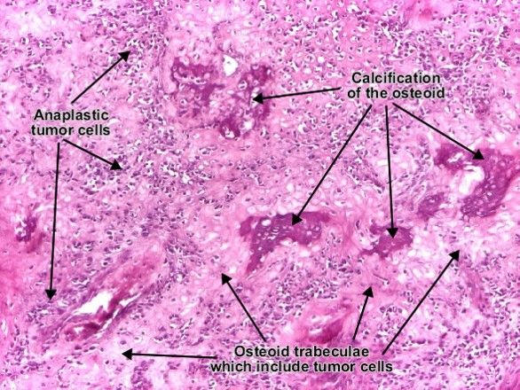
- Osteosarcoma
- Children 12-14
- Male > female
- Site: metaphyses of long bones
- Most common: Distal femur> proximal tibia > proximal humerus,
- 50% of tumours arise around the knee
- 15% osteosarcomas arise around the hip joint
- 10% arise in the shoulder
- 8% of the osteosarcomas are seen in the jaw bone
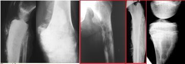
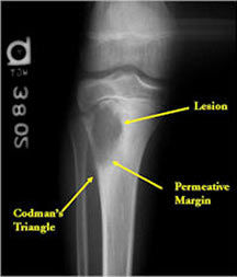
- codman’s and Sun ray
- tumour marker for osteosarcoma
- Bone-specific alkaline phosphatase in plasma
- Malignant tumor of mesenchymal cells characterized by direct formation of osteoid or bone matrix by the tumor cells
- Risk factors
- Radiation to bones
- Age
- Height
- Gender
- Race
- Genetic predisposition
- Trauma
- Complications
- Metastasis
- Movement Problems
- Chemotherapy side effects
- Loss of a Limb
- Prognosis
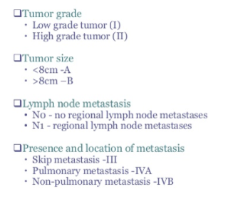
- Stage 1
- >90% with wide resection
- Stage 2
- Depends on type, site, size, and degree of necrosis because of chemotherapy.
- stage 3
- metastases depends on the resectability of the primary tumor and lung nodules, degree of necrosis of the primary tumor, and the number of metastases
- Stage 1
- Risk factors
- Clinical Picture
- pain at site of tumour; usually episodic then intense and persistent,
- swelling
- fracture of limb(rare)
- , no systemic signs usually
- bimodal age distribution
- Bimodal: 2 peaks, one at adolescence and second in older patients.
- Metastasis
- Lungs
- soft tissues
- Lymph nodes
- Types
- Primary osteosarcoma
- Most common primary malignant bone tumor in children
- Males more affected
- Metaphysis of long bones (growing area)
- Males more affected
- Affects children (12-14 yrs) and young adults
- Most common primary malignant bone tumor in children
- Secondary osteosarcoma
- Old ppl > 40 years
- Affects flat bones more than long bones
- Pre-existing bone lesions
- Affects flat bones more than long bones
- Old ppl > 40 years
- Primary osteosarcoma
- Stages
- Management
- Chemotherapy
- doxorubicin, ifosfamide, cisplatin, and high-dose methotrexate with leucovorin rescue.)
- Limb-Salvage surgery
- Autografts Allografts Rotatioplasty Prosthetic implant
- Amputation
- Chemotherapy
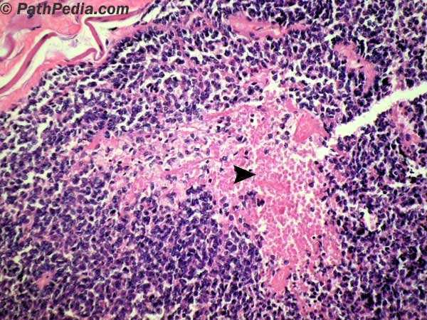
- EWING sarcoma
- children & adolescents 10-15 yrs
- Incidence: second after osteosarcoma
- Male to female 2:1
- Site: Diaphysis of long bones
- children & adolescents 10-15 yrs
- Osteosarcoma
Tumors
Physical Examination of bone tumors
- Inspect
- walking and standing as in gait
- scars, sinuses, erythema or rashes
- Posture and common deformities
- genu valgum (knock knee) or genu varum (bow legs).
- Muscle wasting
- quadriceps wasting
- Swelling
- an enlarged prepatellar bursa
- (housemaid’s knee)
- bursa enlargement in the popliteal fossa
- Baker’s cyst.
- an enlarged prepatellar bursa
- Leg length discrepancy: Apparent length is from umbilicus and true length Is from anterior superior iliac spine.
- Palpate
- Warmth: feel the skin, comparing both sides.
- Effusion
- The patellar tap
- excess fluid accumulates in or around the knee joint.
- (With the patient’s knee extended, empty the suprapatellar pouch by sliding your left hand down the thigh until you reach the upper edge of the patella. Keep your hand there and, with the fingertips of your right hand, press down briskly and firmly over the patella)
- The patellar tap
- Joint lines:
- feel the tibial and femoral joint lines. If there is tenderness.
- Synovitis:
- with the knee extended and the quadriceps relaxed, feel for sponginess on both sides of the quadriceps tendon
- The bulge or ripple test.
- (A) Empty the suprapatellar pouch, as for the patellar tap test. (B) Stroke the medial side of the joint to displace excess fluid to the lateral side of the joint. (C) Stroke the lateral side while watching the medial side closely for a bulge or ripple as fluid reaccumulates
- Move
- Active Flexion and Extension of the knee
- With the patient supine, ask him to flex his knee up to the chest and then extend the leg back down to lie on the couch (normal range 0–140‹).
- Feel for crepitus between the patella and femoral condyles
- chondromalacia patellae
- Record the range of movement:
- Ask the patient to lift the leg, keeping it straight. If the knee cannot be kept fully extended an extensor lag is present, indicating quadriceps weakness or other abnormality of the extensor apparatus.
- Passive flexion and extension of the knee
- Normally the knee can extend so that the femur and tibia are in longitudinal alignment. Record full extension as 0‹. A restriction to full extension occurs with meniscal tears, osteoarthritis and inflammatory arthritis.
- To assess hyperextension, lift both legs by the feet. Hyperextension (genu recurvatum) is present if the knee extends beyond the neutral position. Up to 10‹ is normal
- Active Flexion and Extension of the knee
- Special Tests +stability
- Collateral ligaments stress test
- Appley's distraction tests
- Anterior & posterior drawer tests (cruciate ligament)
- McMurray’s test (Menisci)
- Appley's grinding test ( Menisci)
- Inspect
Investigations
- Blood Tests
- White Blood Cell Count
- helps to count the number of white blood cells
- To rule out an infection
- helps to count the number of white blood cells
- Hemoglobin Test
- measures the amount of hemoglobin levels in blood.
- Cancer can result in anemia that can be detected through this test
- measures the amount of hemoglobin levels in blood.
- The Erythrocyte Sedimentation Rate
- help detect inflammation associated with conditions such as infections, cancers, and autoimmune diseases.
- High values means there is inflammation associated with cancer
- help detect inflammation associated with conditions such as infections, cancers, and autoimmune diseases.
- White Blood Cell Count
- Radiology
- X-rays
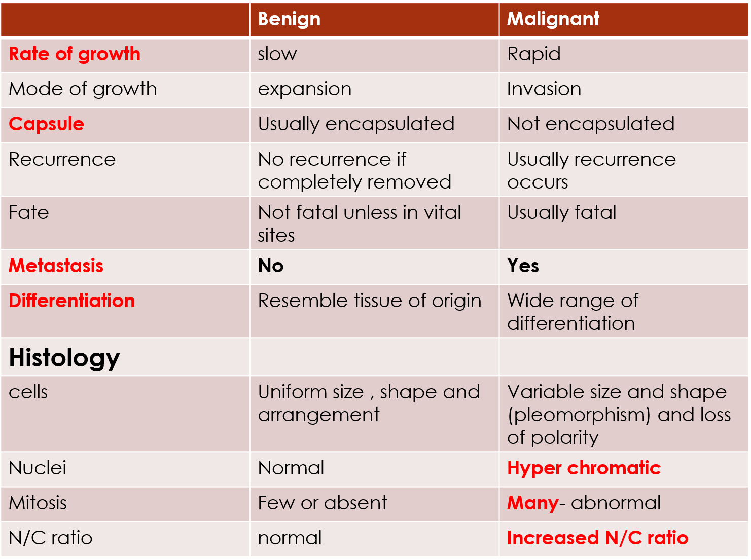
- How to differentiate between Benign and malignant tumors?
- 1.Size – The larger the lesion the more likely it is to be aggressive or malignant 2.Border Narrow zone of transition “ lesion is clear” *more likely to be benign Wide zone of transition “difficult to be certain where the lesion starts and stops” *more likely to be malignant
- Used for initial evaluation of bone lesions.
- How to differentiate between Benign and malignant tumors?
- magnetic resonance imaging
- Determine the extent of intraosseus and extraosseus spread
- computed tomography
- Mainly to detect metastatic pulmonary disease
- Positron emission tomography (PET)
- Radionuclide Bone Scan
- X-rays
- Bone Biopsy
- A bone biopsy is a procedure in which bone samples are removed (with a special biopsy needle or during surgery) to find out if cancer or other abnormal cells are present.
- Blood Tests
Imagens e arquivos do mapa
Informações
- Descrição
- Mapa Mental sobre Thigh and Knee, criado por Monspient Dono em 16-03-2020.
- Criado por
- Monspient Dono
- Visualizações
- 0
- Histórico
-
- Criado por Fatma Shwaylia · 25 mar 2017
- Copiado por Monspient Dono · 16 mar 2020
- Avaliação