
Mapa mental
CVS
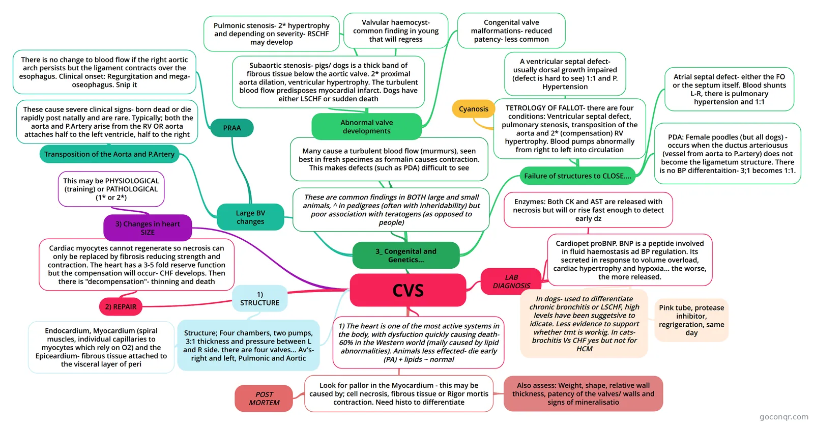
Mind map: CVS heart pathology
Conteúdo do mapa
CVS
1) The heart is one of the most active systems in the body, with dysfuction quickly causing death- 60% in the Western world (maily caused by lipid abnormalities). Animals less effected- die early (PA) + lipids ~ normal
Structure; Four chambers, two pumps, 3:1 thickness and pressure between L and R side. there are four valves... Av's- right and left, Pulmonic and Aortic
- Endocardium, Myocardium (spiral muscles, individual capillaries to myocytes which rely on O2) and the Epiceardium- fibrous tissue attached to the visceral layer of peri
- 1) STRUCTURE
Look for pallor in the Myocardium - this may be caused by; cell necrosis, fibrous tissue or Rigor mortis contraction. Need histo to differentiate
- Also assess: Weight, shape, relative wall thickness, patency of the valves/ walls and signs of mineralisatio
- POST MORTEM
2) REPAIR
- Cardiac myocytes cannot regenerate so necrosis can only be replaced by fibrosis reducing strength and contraction. The heart has a 3-5 fold reserve function but the compensation will occur- CHF develops. Then there is "decompensation"- thinning and death
3) Changes in heart SIZE
- This may be PHYSIOLOGICAL (training) or PATHOLOGICAL (1* or 2*)
LAB DIAGNOSIS
- Enzymes: Both CK and AST are released with necrosis but will or rise fast enough to detect early dz
- Cardiopet proBNP. BNP is a peptide involved in fluid haemostasis ad BP regulation. Its secreted in response to volume overload, cardiac hypertrophy and hypoxia... the worse, the more released.
- Pink tube, protease inhibitor, regrigeration, same day
- In dogs- used to differentiate chronic bronchitis or LSCHF, high levels have been suggetsive to idicate. Less evidence to support whether tmt is workig. In cats- brochitis Vs CHF yes but not for HCM
3_ Congenital and Genetics...
- These are common findings in BOTH large and small animals, ^ in pedigrees (often with inheridability) but poor association with teratogens (as opposed to people)
- Many cause a turbulent blood flow (murmurs), seen best in fresh specimes as formalin causes contraction. This makes defects (such as PDA) difficult to see
- Failure of structures to CLOSE....
- PDA: Female poodles (but all dogs) - occurs when the ductus arteriousus (vessel from aorta to P.artery) does not become the ligametum structure. There is no BP differentaition- 3;1 becomes 1:1.
- Atrial septal defect- either the FO or the septum itself. Blood shunts L-R, there is pulmonary hypertension and 1:1
- A ventricular septal defect- usually dorsal growth impaired (defect is hard to see) 1:1 and P. Hypertension
- TETROLOGY OF FALLOT- there are four conditions: Ventricular septal defect, pulmonary stenosis, transposition of the aorta and 2* (compensation) RV hypertrophy. Blood pumps abnormally from right to left into circulation
- Cyanosis
- Abnormal valve developments
- Pulmonic stenosis- 2* hypertrophy and depending on severity- RSCHF may develop
- Subaortic stenosis- pigs/ dogs is a thick band of fibrous tissue below the aortic valve. 2* proximal aorta dilation, ventricular hypertrophy. The turbulent blood flow predisposes myocardial infarct. Dogs have either LSCHF or sudden death
- Valvular haemocyst- common finding in young that will regress
- Congenital valve malformations- reduced patency- less common
- Large BV changes
- PRAA
- There is no change to blood flow if the right aortic arch persists but the ligament contracts over the esophagus. Clinical onset: Regurgitation and mega- oseophagus. Snip it
- Transposition of the Aorta and P.Artery
- These cause severe clinical signs- born dead or die rapidly post natally and are rare. Typically; both the aorta and P.Artery arise from the RV OR aorta attaches half to the left ventricle, half to the right
- PRAA
- These are common findings in BOTH large and small animals, ^ in pedigrees (often with inheridability) but poor association with teratogens (as opposed to people)