
Mapa mental
biochemistry of metastasis
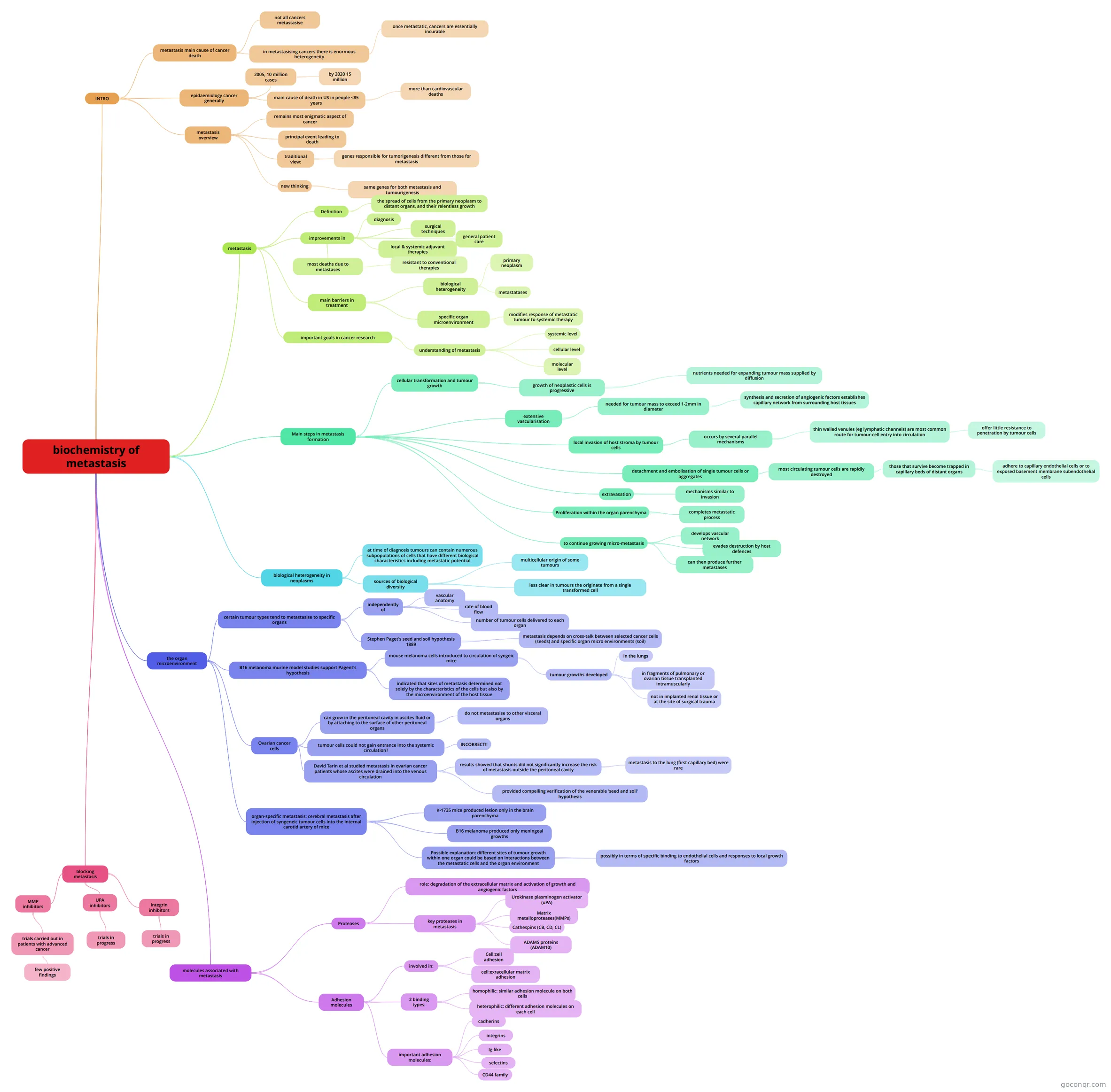
Mind map: biochemistry of metastasis
Conteúdo do mapa
biochemistry of metastasis
INTRO
- metastasis main cause of cancer death
- not all cancers metastasise
- in metastasising cancers there is enormous heterogeneity
- once metastatic, cancers are essentially incurable
- epidaemiology cancer generally
- 2005, 10 million cases
- by 2020 15 million
- main cause of death in US in people <85 years
- more than cardiovascular deaths
- 2005, 10 million cases
- metastasis overview
- remains most enigmatic aspect of cancer
- principal event leading to death
- traditional view:
- genes responsible for tumorigenesis different from those for metastasis
- new thinking
- same genes for both metastasis and tumourigenesis
- metastasis main cause of cancer death
metastasis
- Definition
- the spread of cells from the primary neoplasm to distant organs, and their relentless growth
- improvements in
- diagnosis
- surgical techniques
- general patient care
- local & systemic adjuvant therapies
- most deaths due to metastases
- resistant to conventional therapies
- main barriers in treatment
- biological heterogeneity
- primary neoplasm
- metastatases
- specific organ microenvironment
- modifies response of metastatic tumour to systemic therapy
- biological heterogeneity
- important goals in cancer research
- understanding of metastasis
- systemic level
- cellular level
- molecular level
- understanding of metastasis
- Definition
Main steps in metastasis formation
- cellular transformation and tumour growth
- growth of neoplastic cells is progressive
- nutrients needed for expanding tumour mass supplied by diffusion
- growth of neoplastic cells is progressive
- extensive vascularisation
- needed for tumour mass to exceed 1-2mm in diameter
- synthesis and secretion of angiogenic factors establishes capillary network from surrounding host tissues
- needed for tumour mass to exceed 1-2mm in diameter
- local invasion of host stroma by tumour cells
- occurs by several parallel mechanisms
- thin walled venules (eg lymphatic channels) are most common route for tumour-cell entry into circulation
- offer little resistance to penetration by tumour cells
- thin walled venules (eg lymphatic channels) are most common route for tumour-cell entry into circulation
- occurs by several parallel mechanisms
- detachment and embolisation of single tumour cells or aggregates
- most circulating tumour cells are rapidly destroyed
- those that survive become trapped in capillary beds of distant organs
- adhere to capillary endothelial cells or to exposed basement membrane subendothelial cells
- those that survive become trapped in capillary beds of distant organs
- most circulating tumour cells are rapidly destroyed
- extravasation
- mechanisms similar to invasion
- Proliferation within the organ parenchyma
- completes metastatic process
- to continue growing micro-metastasis
- develops vascular network
- evades destruction by host defences
- can then produce further metastases
- cellular transformation and tumour growth
biological heterogeneity in neoplasms
- at time of diagnosis tumours can contain numerous subpopulations of cells that have different biological characteristics including metastatic potential
- sources of biological diversity
- multicellular origin of some tumours
- less clear in tumours the originate from a single transformed cell
the organ microenvironment
- certain tumour types tend to metastasise to specific organs
- independently of
- vascular anatomy
- rate of blood flow
- number of tumour cells delivered to each organ
- Stephen Paget's seed and soil hypothesis 1889
- metastasis depends on cross-talk between selected cancer cells (seeds) and specific organ micro environments (soil)
- independently of
- B16 melanoma murine model studies support Pagent's hypothesis
- mouse melanoma cells introduced to circulation of syngeic mice
- tumour growths developed
- in the lungs
- in fragments of pulmonary or ovarian tissue transplanted intramuscularly
- not in implanted renal tissue or at the site of surgical trauma
- tumour growths developed
- indicated that sites of metastasis determined not solely by the characteristics of the cells but also by the microenvironment of the host tissue
- mouse melanoma cells introduced to circulation of syngeic mice
- Ovarian cancer cells
- can grow in the peritoneal cavity in ascites fluid or by attaching to the surface of other peritoneal organs
- do not metastasise to other visceral organs
- tumour cells could not gain entrance into the systemic circulation?
- INCORRECT!!
- David Tarin et al studied metastasis in ovarian cancer patients whose ascites were drained into the venous circulation
- results showed that shunts did not significantly increase the risk of metastasis outside the peritoneal cavity
- metastasis to the lung (first capillary bed) were rare
- provided compelling verification of the venerable 'seed and soil' hypothesis
- results showed that shunts did not significantly increase the risk of metastasis outside the peritoneal cavity
- can grow in the peritoneal cavity in ascites fluid or by attaching to the surface of other peritoneal organs
- organ-specific metastasis: cerebral metastasis after injection of syngeneic tumour cells into the internal carotid artery of mice
- K-1735 mice produced lesion only in the brain parenchyma
- B16 melanoma produced only meningeal growths
- Possible explanation: different sites of tumour growth within one organ could be based on interactions between the metastatic cells and the organ environment
- possibly in terms of specific binding to endothelial cells and responses to local growth factors
- certain tumour types tend to metastasise to specific organs
molecules associated with metastasis
- Proteases
- role: degradation of the extracellular matrix and activation of growth and angiogenic factors
- key proteases in metastasis
- Urokinase plasminogen activator (uPA)
- Matrix metalloproteases(MMPs)
- Cathespins (CB, CD, CL)
- ADAMS proteins (ADAM10)
- Adhesion molecules
- involved in:
- Cell:cell adhesion
- cell:exracellular matrix adhesion
- 2 binding types:
- homophilic: similar adhesion molecule on both cells
- heterophilic: different adhesion molecules on each cell
- important adhesion molecules:
- cadherins
- integrins
- Ig-like
- selectins
- CD44 family
- involved in:
- Proteases
blocking metastasis
- MMP inhibitors
- trials carried out in patients with advanced cancer
- few positive findings
- trials carried out in patients with advanced cancer
- UPA inhibitors
- trials in progress
- Integrin inhibitors
- trials in progress
- MMP inhibitors
Informações
- Descrição
- 4th year Adv Pharmacology of Cancer Mapa Mental sobre biochemistry of metastasis, criado por aoife.lacey.1 em 04-05-2013.
- Criado por
- aoife.lacey.1
- Visualizações
- 101
- Tags
- Avaliação