
Notas
Digestive System LAB
Conteúdo das notas (3)
Página 1
Whats in it?
Alimentary Canal/Gastrointestinal tract
hollow tube from mouth to anus
various accessory organs or glands empty secretions here.
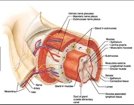
Has 4 layers/tunics: (inner to outer)
mucosa: epithelium, lamina propia, muscularis mucosae. Absorbs and secrete
submucosa: vascular supply for mucosa, protection
muscularis externa: longitudinal muscle, circular muscle. Churning, mixing, propulsion of food
serosa/adventitia: epithelium. Protection
Accessory organs (secrete products to alimentary canal)
teeth
salivary glands
parotid gland: secrete serous
submandibular: mucin and serous components
sublingual : mixed gland makes most mucin
Saliva: has mucin (glycoprotein) creates bolus
gallbladder
when there is no digestive activity, the gallbladder sucks up the bile through cystic duct. When bile is needed its released.
liver
largest gland in body
four lobes, suspended by falciform ligament
produces bile (fat emulsifier), leaves through common hepatic duct and enters duodenum through bile duct
important in inital processing of nutrient rich blood draining digestive organs.
lobules: structural and functional units exagonal consisting of hepatocytes (liver cells). At each 6 corner of lobule is a protal triad which contains portal arteriole, portal venule, and bile duct. Between each cell there is a sinusoid
stellate/hepatic macrophage: phagocytic cells that line sinusoid and remove debris such as bacteria from blood as it goes past while hepatocytes pick up oxygen and nutrients.
Sinusoid empty to central vein and blood drains from liver via hepatic veins. blood filled space that drain into central vein.
acinar cell
pancreas
horizonal across posterior ab wall from spleen to duodenum.
retroperitoneal organ
Both endocrine (insulin/glucagon) and exocrine function of hydrolytic enzymes made by acinar cells which secrete to duodenum through pancreatic duct.
Pancreatic juice: very alkaline, concentration of bicarbonate ion (HCO3-). It neutralizes chyme when entering the duodenum from stomach.
Oral Cavity
Palate: roof of mouth. Hard palate (palatine process of maxillae/palatine bones. Soft palate (fibromuscular no bone)
Uvula: fingerlike projection of soft palate
Tongue: has mylohoid muscle. Attaches to hyoid bone, mandible, styloid process, pharynx
lingual frenulum: secure inferior midline of tongue to floor of mouth
oral vestibule: area between teeth and cheek
oral cavity proper: space within teeth and gums
Palatine tonsil: mass of lymphoid tissue
Palatoglossal arch:
lingual tonsil: lymphoid tissue, part of defense system
tonsilitis: inflammed tonsil blocking entrance to pharnyx making swallowing hard.
Salivary amylase: enzyme that breaks down starch
Pharynx
nasopharynx
oropharynx
largyngopharnx
walls have two layers of skeletal muscle:
inner layer of longitudinal muscle
outer layer of circular constrictor muscles.
Reason is to initiate wavelike contractions that move food into esophagus
mucosa of oro and laryngo have protected squamous epithelium
Esophagus (gullet)
pharynx to diaphagm to gastroesophageal sphincter in superior stomach.
25 cm
food passage by way of peristalsis (wavelike movement)
gastroesophageal sphincter: slight thickening of smooth muscle layer between esophagus and stomach. Controls food passage to stomach
Stomach
mesentary: double layer of peritoneum from digestive organs to body wall
greater omentum: extends from greater curvature of stomach to most of abdominal organs
lesser omentum: liver to lesser curvature of stomach
contains third layer of smooth muscle in muscularis externa allowing it to churn, mix, and pummel food.
Gastric glands: secrete hydrochloric acid and hydrolytic enzymes
Mucosal glands: secrete viscous mucus to prevent stomach from being digested from proteolytic enzymes.
MOST DIGESTION HAPPENS IN PYLORIC PART OF STOMACH
Chyme: creamy mass of food after being in stomach, goes through pyloric sphincter to small intestine
Parts of stomach: Cardial part, fundus, body, pyloric part
Small intestine
20 feet, 6 to 7 meters
pyloric sphincter to ileocecal valve
suspended by mesentary
3 divisions:
duodenum: pyloric sphincter and curves around head of pancreas retroperitoneal position.
intestinal crypts: invaginated area that produces intestinal juice
ileum: terminal portion, joins large intestine at ileocecal valve. Inferior and right in ab cavity, mostly in hypogastric region
jejunum: continues with duodenum, occupies umbilical region of ab cavity
surface area of mucosa:
microvilli: microscopic projections of surface plasma membrane of columnar epithelial
villi: fingerlike projection of mucosa tunic giving velvety appearance
circular folds: deep folds of mucosa and submucosal layers that force chyme to spiral though intestine. Mixing it and slowing progress. Decreases in size and frequency
Peyer's patches: lymphoid nodules
Large Intestine
ileocecal valve to anus
Subdivisions:
cecum
appendix: hangs from cecum. Generally twisted, ideal location for bacteria to multiply
colon: ascending colon, right colic (hepatic) flexure, transverse colon, left colic (splenic) flexure, decending colon, sigmoid colon. CONSIDERED RETROPERITONIAL (except sigmoid and transverse)
rectum,
anal canal: internal anal sphincter (smooth muscle) external anal (skeletal)
longitudinal muscle layer of muscularis externa is reduced to three longitudinal muscle bands called teniae coli, cause wall to pucker into small pocketlike sacs called haustra
Goblet cells: mucus is produced and secreted by them.
Página 2
Role/Processes
Role of Digestive System
provide body w/nutrients, water, electrolytes.
Organs ingest/digest food,absorb, eliminate undigested as feces
Digestion
where ingested food is broken to smaller molecules to be available for cells. Then goes to absorption
In small intestine
2 enzymes:
brush border enzymes: hydrolytic enzyme in microvilli of columnar epithelium cells
enzymes made by pancreas and ducted in duodenum via main pancreatic duct.
Bile from liver enters duodenum through bile duct .
At Duodenum ducts join to form hepatopancreatic ampulla and empty product to duodenal lumen through major duodenal papilla, an orifice controlled by muscular valve called hepatopancreatic sphincter.
Absorption
Digested end products pass through epithelial cells lining the tract into the blood for distribution of body cells.
Large intestine
consolidates and propels unusable fecal matter toward anus and eliminate it from body
provides a site where intestinal bacteria manufacture vitamin b and k
reclaim most of water from ndigested food, conserving body water
Movement of bile
made by hepatocytes in liver.
flows through tiny canals called bile canaliculi which run retween adjacent cells toward bile duct branches in triad regions where it leaves liver.
Página 3
Diseases
Cirrosis: when liver is severely damaged and becomes hard/fibrous
hepatitis: inflammation of liver
A blocked common hepatic or bile duct:
Jaundice: bile cant enter small intestine, accumulates, and backs up in liver. Causes pressure on liver cells and bile enters bloodstream. As it circulates tissues become yellow or jaundiced.
Informações
- Descrição
- Notas sobre Digestive System LAB, criado por Stacylynn Conceicao em 01-05-2018.
- Criado por
- Stacylynn Conceicao
- Visualizações
- 0
- Avaliação