
Topic 1.3 Skill 1: Drawing and labeling a Fluid Mosaic Model
Slides do conjunto (11)
-
- The Fluid Mosaic Model was created by Singer and Nicholson in 1966.
- This model includes a phospholipids bilayer, cholesterol, and peripheral and integral proteins within the cell membrane.
- This model also shows the differences in proteins and their positioning within the cell membrane.
- This model is considered "fluid" because it allows the phospholipids and proteins within the cell bilayer to freely move around.
- There are several different proteins that go into a cell membrane. Peripheral and integral proteins are the two main types of proteins included in the cell membrane.
- All of the images shown in this slide show are ways in which each parts of the fluid mosaic model may be drawn. It is recommended to stick to the simplistic drawing and examples of each protein, and the cholesterol and phospholipids.
-
- Peripheral proteins are attached to the inner or outer surface of the phospholipids bilayer of a cell membrane.
- These proteins do not go all the way through the cell membrane, but instead are on the edges of the membrane.
- Think of these proteins as your Peripheral Vision, they are only located on the outside ring of your vision.
- The image to the left shows a peripheral protein (pink). As you can see, it is on the outside edge of the phospholipids (orange and yellow).
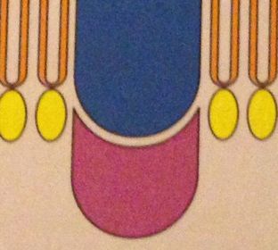
- Legenda: This is an example of what a peripheral protein would look like in a model drawing. If you see any structures like this label them as a peripheral protein.
-
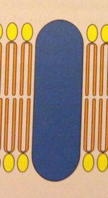
- Integral proteins are within the phospholipids bilayer. They go all the way through the bilayer.
- Sometimes integral proteins, such as Oligosaccharides, protrude from the cell membrane.
- Integral Proteins can also be "transport channels" that allow specific molecule to go through the cell membrane.
- An example of an integral protein (blue) is to the left. As you can see the protein goes right through the phospholipids bilayer (yellow and orange).
-
- Cholesterol is NOT a fat despite common belief, but is instead a Steroid.
- Cholesterol keeps the phospholipids bilayer of cells fluid and running correctly making it necessary for all animal cells to have.
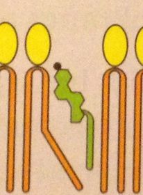
- Cholesterol is always located in the phospholipids bilayer and when drawn looks like one as well with a different "head" attached.
- An example of what cholesterol looks like in a drawn representation is shown at the left. The cholesterol is green and as you can see also has a tail similar to the phospholipid's tails (orange). The top of the cholesterol is different, however, because it is made up of a different chemical composition.
-
Labeling the Fluid Mosaic Model
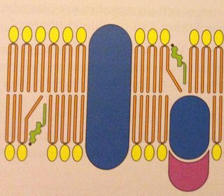
- Take time now and label what you think is the:
- Integral Protein
- Peripheral Protein
- Cholesterol
- Phospholipids (Heads and Tails along with their attraction to water)
- Legenda: This is a model drawing of a phospholipids bilayer. This picture includes integral and peripheral proteins, cholesterol, and phospholipids.
-
Labeling the Fluid Mosaic Model
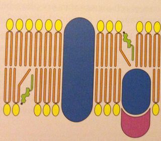
- By color:
- Green= Cholesterol
- Blue=Integral Protein
- Pink= Peripheral Protein
- Yellow and Orange= Phospholipid
- Yellow= Phospholipids Head; Hydrophilic
- Orange= Phospholipids Tail; Hydrophobic
- Legenda: Note: This is also an example of how you might draw a fluid mosaic model.
-
Labeling the Fluid Mosaic Model
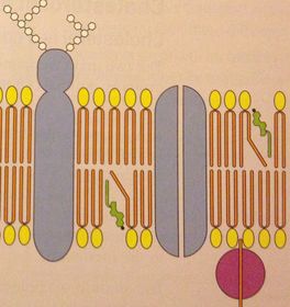
- This one is a bit harder, but try to label:
- Integral Proteins
- Peripheral Proteins
- Cholesterol
- Phospholipids- parts and water attraction of each part
-
Labeling the Fluid Mosaic Model
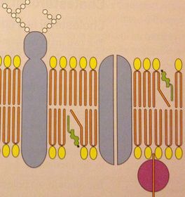
- By color:
- Blue: Integral Proteins
- Pink: Peripheral Proteins
- Green: Cholesterol
- Yellow and Orange: Phospholipids
- Yellow: Phospholipid Heads; Hydrophilic
- Orange: Phospholipid Tails; Hydrophobic
- Important:
- The integral proteins here are shown different because these are specific proteins within the cell membrane. For instance, the integral protein on the left is an oligosaccharide and the one to the right is a transport channel.
- Note that the peripheral is also barely hanging on to the cell membrane, sometimes peripheral proteins barely hang on to the cell membrane. The attachment that this protein has does not make it a different protein. It is still a peripheral.
-
- You just learned how to label a fluid mosaic model!
-
Drawing the Fluid Mosaic Model
- Create a phospholipids bilayer leaving room for labeling and at least one of the following: integral and peripheral protein and cholesterol.
- Draw each of these parts in the gaps you provided when you drew the phospholipids bilayer.
- Label each part.
- Check that there are no gaps within your bilayer (i.e. no gaps in between phospholipids and proteins or other phospholipids.
- Done!
Informações
- Descrição
- This is a slide show about how to label and draw a Fluid Mosaic Model.
- Criado por
- book.consumer.19 · out 2015
- Atualizado
- 19 fev 2016
- Visualizações
- 108
- Avaliação