
Quiz
Lower Limb
This quiz has question types that open in the usual player.
Questions in this quiz (167)
1The gluteal muscles have the following actions:
- AAll make external rotation of the inferior limb
- BThe gluteus maximus muscle makes abduction, respectively the medius and minimus muscles make adduction
- CThe gluteus maximus muscle makes external rotation, respectively the medius and minimus muscles make internal rotation
- DHaving the same innervation, all gluteal muscles make similar actions
- EThe gluteus maximus muscle is mainly extensor, respectively the medius and minimus are flexors
2The following statements about gluteal muscles insertion and topography are truth:
- ABetween gluteal muscles, the maximus is the deepest
- BBetween gluteal muscles, the minimus is the deepest
- CAll have the distal insertion on the greater trochanter and the iliotibial tract
- DAll have the origin on the sacroiliac ligaments and the posterior iliac surface
- ECovering the other muscles, the gluteus maximus area corresponds for intramuscular injections
3Vascular and nerve supply of gluteal muscles is made by:
- AInferior gluteal bundle for gluteus maximus muscle
- BSuperior gluteal bundle for gluteus medius and minimus muscles
- CInferior gluteal bundle for gluteus medius and minimus muscles
- DSuperior gluteal bundle for gluteus maximus muscle
- EThe medius and minimus muscles have different blood supply
4The following statements about piriform muscle are truth:
- AHas the origin on the anterior surface of the sacrum
- BHas the origin on the posterior surface of the sacrum
- CHas a triangular shape with the apex on the lesser trochanter
- DHas a triangular shape with the apex on the greater trochanter
- EHas inferior relations with the sacrospinous ligament and anterior relationships with the greater ischiatic notch, from the posterior border of the hip bone
5Obturator internus muscle:
- AHas insertions on the medial surface of the obturator membrane and the lesser trochanter
- BThe ischiatic bursae is between this muscle and inferior border of the hip bone
- CBy insertion of levator ani muscle, the obturator internus is a muscle of both pelvis and perineum regions
- DIts main action is external rotation of the lower limb together with both gemelli muscles
- EIts innervation is made by obturator nerve, branch of the lumbar plexus
6Quadratus femoris muscle:
- AHas the distal insertion on the intertrochanteric crest
- BAt the level of the thigh is located deeper to the obturator externus muscle
- CHas posterior relations with sciatic nerve and gluteus maximus muscle
- DIt’s an internal rotator of the inferior limb
- EIt’s innervated by a branch from the sacral plexus
7Obturator externus muscle:
- AThrough the direction of its muscular fibers, it intersects inferior the neck of the femur
- BHas anterior relations with the hip joint
- CAt the thigh is more superficial than the obturator internus muscle
- Dt’s innervated by the obturator nerve, branch of the sacral plexus
- EIts action is external rotator and maintaining of the femoral head into the hip joint
8About superior and inferior gemelli muscles, the following statement is false:
- ATheir origin is on the spine and respectively on the ischiatic tuberosity
- BHave a common distal insertion with the obturator externus muscle
- CTheir action is external rotation of the inferior limb
- DBoth are innervated by branches from the sacral plexus
- EHave relations with inferior gluteal bundle, respectively with the sciatic and the posterior cutaneous nerve of the thigh
9Gluteus maximus muscle has the following attributes
- AIs a deep muscle
- BLies most superficially
- CIs a powerful abductor of the lower limb
- DExtend the trunk on the femur
- EIs an important adductor muscle
10Gluteus medius and minimus muscles have the following attributes
- AAttaches proximally to the outer surface of the ilium
- BAttaches proximally to the inner surface of the ilium
- CAttaches distally to the greater trochanter of the femur
- DAttaches distally to the lesser trochanter of the femur
- EAttaches distally to the intertrochanteric line of the femur
11Gluteus medius and minimus muscles have the following attributes
- AAre abductors of the hip
- BAre adductors of the hip
- CThe most important action of this muscles acts during locomotion
- DTheir most important action is to stabilize the pelvis on the femur
- EThey are helped in this function by tensor fasciae latae
12The following muscles arise from within the pelvis:
- AGemelli muscles
- BObturator externus muscle
- CQuadratus femoris muscle
- DPiriformis muscle
- EObturator internus muscle
13The following muscles arise externally of the pelvis:
- AGemelli muscles
- BObturator externus muscle
- CQuadratus femoris muscle
- DPiriformis muscle
- EObturator internus muscle
14Extension of the lower limb is made by:
- AGluteus minimus muscle
- BPiriformis muscle
- CGluteus maximus muscle
- DObturator internus muscle
- EGemelli muscles
15The following muscle make lateral rotation of the lower limb:
- AObturator internus muscle
- BPiriformis muscle
- CGemelli muscles
- DGluteus medius muscle
- EQuadratus femoris muscle
16The following muscles are medial rotators of the lower limb:
- AGluteus minimus muscle
- BGemelli muscles
- CGluteus maximus muscle
- DObturator internus muscle
- EGluteus medius muscle
17Abduction of the lower limb is made by:
- AObturator internus muscle
- BGluteus minimus muscle
- CGemelli muscles
- DGluteus medius muscle
- EPiriformis muscle
18The following muscles are innervated from L5 segment:
- AGluteus maximus muscle
- BObturator internus muscle
- CPiriformis muscle
- DGluteus medius muscle
- EGluteus minimus muscle
19The following muscles are innervated from S1 segment:
- AGluteus maximus muscle
- BObturator internus muscle
- CPiriformis muscle
- DGluteus medius muscle
- EGluteus minimus muscle
20The cutaneous nerve supply of the gluteal region is ensured by
- AIliohypogastric nerve
- BIlioinguinal nerve
- CObturator nerve
- DDorsal rami of the 1-3 spinal lumbar nerves
- EDorsal rami of the 1-3 spinal sacral nerves
21Most of the skin of the buttock is blood supplied by:
- ASuperior gluteal artery
- BExternal pudendal artery
- CSubcostal artery
- DInferior gluteal artery
- EInferior epigastric artery
22Gluteus maximus muscle has the following attributes:
- AIs thicker in humans than in any non-human primate
- BIs less extensive in humans than in any non-human primate
- CIs involved in transition to bipedality and a permanently upright posture
- DIs the largest muscle in the gluteal region
- EIs the deepest muscle in the gluteal region
23Lymphatic drainage from the gluteal region is made in:
- AMedial group of the proximal superficial inguinal nodes
- BLateral group of the proximal superficial inguinal nodes
- CThe distal group of the superficial inguinal nodes
- DExternal iliac nodes
- EUsually drain in the deep inguinal nodes
24Identify which of the following muscles are located in the anterior compartment of the thigh.
- ASartorius muscle
- BRectus femori muscle
- CGracilis muscle
- DVastus intermedius muscle
- ETensor fasciae latae muscle
25Which of the following muscles present insertions at the level of the greater trochanter?
- AGluteus maximus muscle
- BPiriformis muscle
- CObturator externus muscle
- DGluteus medius muscle
- EQuadratus femori muscle
26Which of the muscles from the medial compartment of the thigh are adductors of the thigh?
- AAdductor brevis muscle
- BObturator externus muscle
- CAdductor magnus muscle
- DGracilis muscle
- ESartorius muscle
27Which of the following statements regarding the origins and insertions of extensor muscles of the posterior compartment of the thigh are false?
- AIschial tuberosity - origin: semitendinosus, semimembranosus, biceps femoris (long head) muscles
- BLinea aspera + supracondylar line – origin: biceps femoris (long head) muscle
- CMedial tibial condyle - insertion: semitendinosus, semimembranosus muscles
- DHead of fibula - insertion: biceps femoris (short head) muscle
- EFibula and lateral surface of the proximal epiphysis of tibia - insertion: biceps femoris (long head) muscle
28Identify the correct territory of distribution for the obturator nerve (L2- L4):
- ASensory: inferomedial thigh
- BMotor: Gracilis muscle
- CMotor: Sartorius muscle
- DMotor: Vastus medialis muscle
- EMotor: Adductor magnus muscle
29Which of the following nerves are not branches of the lumbar plexus?
- AIlioinguinal
- BGenitofemoral
- CObturator
- DPosterior cutaneous femoral
- EPerforant cutaneous
30Which of the following vessels are branches of the femoral artery?
- ASuperficial circumflex iliac artery
- BDeep inferior epigastric artery
- CDeep circumflex iliac artery
- DSuperficial inferior epigastric artery
- EDescending genicular artery
31Which of the following arteries supply the proximal epiphysis of the femur:
- AMedial circumflex femoral artery: ascending, descending, and transverse branches
- BLateral circumflex femoral artery
- C1st perforator – descending branch
- DObturator artery
- ESuperior and inferior gluteal arteries
32Which of the following statements regarding the origin, path and divisions of the sciatic nerve are correct?
- AOrigin: L5-S3
- BReaches the gluteal region via the grater sciatic foramen superior to the piriform muscle
- CIn the gluteal region is located deep to the gluteus maximus muscle
- DIn the thigh is located deep in between the muscles of the posterior compartment and adductor longus muscle
- EIt has the following divisions: posterior tibial nerve + common peroneal nerve (superficial and deep)
33Which of the following statements regarding the borders of the femoral triangle are false?
- AThe base is located superior and is formed by the inguinal ligament
- BMedially is bordered by the adductor magnus muscle
- CLaterally is bordered by the medial border of the sartorius muscle
- DThe base is formed by the following structures: laterally the pectineus and adductor longus muscles, medially by the iliopsoas muscle
- EThe apex is located superior and continues with the Hunter canal
34Which muscles have their origin on the anterior surface of the body of pubic bone?
- AAdductor brevis muscle
- BAdductor magnus muscle
- CAdductor longus muscle
- DSartorius muscle
- ERectus femoris muscle
35Which of the following muscles present origins/insertions at the level of the medial femoral epicondyle?
- AAdductor longus muscle
- BAdductor magnus muscle
- CGastrocnemius muscle
- DPopliteus muscle
- ESartorius muscle
36Which of the following muscles have their origin at the level of the ischial tuberosity?
- AAdductor magnus muscle
- BRectus femoris muscle
- CVastus lateralis muscle
- DBiceps femoris (short head) muscle
- ESemitendinosus muscle
- FSemimembranosus muscle
37Which nerve located in the anterior compartment of the thigh is responsible for the innervation of the quadriceps muscle?
- AObturator nerve
- BSaphenous nerve
- CFemoral nerve
- DCommon fibular nerve
- ESciatic nerve
38Which arteries provide blood supply for the posterior compartment of the thigh?
- APerforator vessels from the posterior tibial artery
- BPerforator vessels from profunda femoris artery
- CDirect perforator branches from the femoral artery
- DPerforators from the medial circumflex femoral artery
- EPerforators from the obturator artery
39Which blood vessels are responsible for the cutaneous vascularization of the anterolateral region of the thigh?
- APerforators from the descending branch of the lateral circumflex femoral artery
- BPerforators from the transverse branch of the lateral circumflex femoral artery
- CDirect perforators from the femoral artery
- DPerforators from the descending genicular artery
- EPerforators from the profunda femoris artery
40Which muscles are responsible for the flexion of the thigh at the level of the hip?
- AIliopsoas muscle
- BBiceps femoris muscle
- CSartorius muscle
- DRectus femoris muscle
- EGracilis muscle
41Which muscles are responsible for the external rotation of the thigh at the level of the hip?
- AGluteus maximus muscle
- BGluteus medius muscle
- CPiriformis muscle
- DTensor fasciae latae muscle
- EInternal obturator muscle
42Which muscles are responsible for the extension of the knee?
- ARectus femoris muscle
- BVastus lateralis muscle
- CAdductor magnus muscle
- DSartorius muscle
- EBiceps femoris muscle
43The small saphenous vein drains into the:
- AThe femoral vein
- BThe deep inferior epigastric veins
- CThe basilic vein
- DThe posterior tibial vein
- EThe popliteal vein
44Which muscles are responsible for the extension of the thigh at the level of the hip?
- AVastus lateralis muscle
- BGluteus maximus muscle
- CIliopsoas muscle
- DGracilis muscle
- EBiceps femoris muscle
45The greater saphenous vein drains into the:
- AInferior vena cava
- BRight atrium
- CPopliteal vein
- DFemoral vein
- ETibial veins
46The femoral artery has the following origin and collaterals:
- AIs the terminal branch of the internal iliac artery
- BIs the terminal branch of the external iliac artery
- CIts largest collateral branch is the profunda femoris artery
- DDivides into the anterior and posterior tibial arteries
- EThe fibular artery is a direct branch of the femoral artery
47The obturator nerve provides motor innervation for the following muscles:
- ASartorius muscle
- BGracilis muscle
- CRectus femoris muscle
- DAdductor magnus muscle
- EBiceps femoris muscle
48The Sciatic nerve provides motor innervation for the following muscles:
- AGluteus maximus muscle
- BPiriformis muscle
- CQuadratus femoris muscle
- DSemitendinosus muscle
- ESemimembranosus muscle
49What are the actions of the soleus muscle?
- APlantar flexion
- BPlantar inversion
- CPlantar eversion
- DKnee flexion
- EKnee extension
50Pes anserinus refers to the following conjoined tendons of muscles:
- ASemimembranosus muscle
- BSemitendinosus muscle
- CSartorius muscle
- DGracilis muscle
- EBiceps femoral muscle
51Which of the following muscles are part of the posterior compartment of the calf?
- ATriceps surae muscle
- BSoleus muscle
- CPosterior tibial muscle
- DBiceps femoral muscle
- EFlexor digitorum longus muscle
52Which of the following statements about the great saphenous vein are true?
- AIt has its origin at the medial end of the dorsal venous plexus on the dorsum of the foot
- BIt has its origin at the level of the plantar part of the foot
- CAscends of the anterior side of the tibia
- DLocated underneath the subcutaneous layer of the medial surface of the calf
- ELocated in the deep layer of the calf
53Which of the following statements about the posterior tibial artery are true?
- AIt has its origin at the level tendinous arch of the soleus muscle
- BIs a branch of the femoral artery
- CIs continued with anterior tibial artery
- DIt has close relationship with the flexor digitorum longus muscle
- EIs located between the two layers of the posterior compartment of the calf
54Which of the following statements about the posterior tibial artery are true?
- AIt is the terminal branch of the posterior tibial artery
- BIt is a collateral branch of the popliteal artery
- CIt has close relationship with the anterior tibial muscle
- DIt descends on the anterior side of the interosseous membrane
- EIt has a common course with the small saphenous vein
55Which of the following statements are true?
- AMalleolar arteries are branches of the anterior tibial artery, with the origin at the level superior side of the ankle
- BDorsal artery of the foot is a branch of the saphenous artery
- CAnterior and posterior tibial arteries are branches of the popliteal artery
- DFibular artery is the terminal branch of the popliteal artery
- EPosterior tibial artery is giving off the medial and lateral plantar arteries
56Which of the following statements about the common fibular nerve are true?
- AIt has its origin at the level of the lumbar plexus
- BAscends through the lateral compartment of the calf, going around the neck of the fibula
- CIt is a branch of the ischiatic nerve
- DAt the level of the popliteal fossa is giving off the medial cutaneous sural nerve
- EIts terminal branches are deep fibular nerve and tibial nerve
57Tibial nerve has the following features:
- AIt is a branch of the femoral nerve
- BGives off the medial cutaneous sural nerve
- CInnervates all the muscles from the posterior compartment of the calf
- DIt is part of the popliteal neuro-vascular bundle, being the deepest structure
- EGiving off 4 terminal branches
58The calf has the following features:
- AIts proximal border is represented by a transverse plane through tibial tuberosity
- BIts distal border is represented by a transverse plane through the medial and lateral malleolus
- CTibial nerve innervates all the muscles from the posterior compartment of the calf
- DComposed by 4 topographic regions
- EPosterior tibial artery is supplying the posterior compartment of the calf and foot
59The small saphenous vein:
- AForms on the lateral side of the foot
- BAscends laterally to the medial malleolus
- CDrains in the popliteal vein at the level of the popliteal fossa
- DAscends superficially on the posterior face of the calf
- EAscends deep in the posterior compartment of the calf
60Which of the following statements in regard with the knee articulation are true:
- AIs formed by: distal femoral diaphysis, proximal tibial diaphysis and the patella
- BIs formed by: distal femoral diaphysis, proximal tibial diaphysis and the fibula
- CThe fibula is not part of the knee articulation
- DThe articular meniscus has a role in enhancing the articular surface
- EThe articular capsule unites 3 bones: the femur, the tibia and the patella
61Which of the following statements in regard with the knee articulation are true:
- AThere are 3 cruciate ligaments: anterior, posterior and intermediary
- BThe anterior cruciate ligament is attached on the anterior intercondylar area of the tibia
- CThe posterior cruciate ligament is attached on the anterior intercondylar area of the tibia
- DThe medial collateral ligament is also called the tibial collateral ligament and it attaches superiorly above the medial femoral condyle
- EThe intermediary cruciate ligament is inserted proximally on the medial condyle
62Which of the following statements about the saphenous nerve are true:
- AIs a branch of the sciatic nerve
- BInnervates the lateral muscular compartment of the leg
- CArises from the sacral nervous plexus
- DAccompanies the great saphenous vein
- EIs a branch of the femoral nerve
63Which of the following statements about the popliteal fossa are true:
- AIt represents the anterior region of the knee
- BIt is triangular shaped
- CThe inferior border is represented by the 2 gastrocnemius heads
- DThe supero-lateral border is represented by the biceps femoris head
- EThe semimembranosus and semitendinosus muscles are not part of the popliteal fossa borders
64Which of the following statements about the popliteal fossa are true:
- AIt consists of the femoral nerve, artery and vein
- BThe femoral nerve divides in the tibial and peroneal nerve
- CThe small saphenous vein perforates the popliteal fascia in order to drain in the popliteal vein
- DThe tibial nerve is located superficially to the popliteal artery
- EAt this level, the femoral artery changes its name in the popliteal artery
65Which of the following statements about the popliteal fossa are true:
- AThe common peroneal nerve is a branch of the sciatic nerve
- BThe sciatic nerve divides in 2 terminal branches at the superior angle of the popliteal fossa
- CThe tibial nerve gives of the sural nerve which is a superficial branch
- DThe common peroneal nerve passes under the tendinous arch of the soleus muscle
- EThe sciatic nerve descends through the fossa
66Which of the following characteristics are common for both gastrocnemius and soleus muscles:
- AThey are innervated by the tibial nerve
- BThey are part of the posterior compartment of the calf
- CThey are responsible with the flexion of the knee joint
- DThey are innervated by the femoral nerve
- EThey represent the base of the Scarpa triangle
67Which of the following statements are true?
- AThe muscles that represent the superior border of the popliteal fossa, are responsible with the flexion of the knee
- BThe quadriceps muscle is responsible with the extension of the knee
- CThe biceps femoris muscle is innervated by the tibial and peroneal nerves
- DThe superior insertions of the gastrocnemius muscle are the tibia and fibula
- EThe sciatic nerve passes medial to the patella
68The structures that pass through the popliteal fossa have the following characteristics:
- AThe patella does not articulate with the tibia
- BThe popliteal vein is located superficial and passes medially to the knee
- CThe popliteal artery is the terminal branch of the femoral artery
- DThe common peroneal nerve is not part of the popliteal fossa
- EThe sural nerve is a collateral branch of the sciatic nerve
69The following statements about the vessels that are located at the level of the popliteal fossa are true:
- AThe femoral vein represents the continuation of the popliteal vein
- BThe femoral and popliteal vein have nothing in common
- CThe femoral vein is part of the popliteal fossa
- DThe popliteal vein is part of the superficial drainage system of the inferior limb
- EThe femoral artery and vein are not the vascular structures of the popliteal fossa
70Which of the following statements are true?
- AThe popliteal fossa has no lymph nodes
- BThe popliteal vein passes through the opening of the adductor magnus muscle to become the femoral vein
- CThe popliteal lymph nodes receive superficial lymph vessels that accompany small saphenous vein
- DDamage of the popliteal artery has no implications on the posterior tibial artery
- EThe popliteal artery divides in two tibial arteries
71Biggest cringelord
- ACF
- BJJ
72Which of the following statements are true?
- AThe tarsal artery is a direct branch of the popliteal artery
- BThe popliteal artery gives articular branches
- CThe gluteal arteries are not related the popliteal artery
- DThe sciatic nerve is located anterior to the popliteal artery
- EThe sciatic nerve is located lateral to the popliteal artery
73Which of the following statements are false?
- AThe popliteal fossa is diamond shaped
- BThe popliteal nerve is part of the popliteal fossa
- CThe popliteal vein is located superficially to the popliteal fascia
- DThe popliteal vein drains in the small saphenous vein
- EThe small saphenous vein drains in the great saphenous vein
74The following statements regarding the posterior compartment of the leg are false:
- AThe great saphenous vein and the small saphenous vein are situated in the subcutaneous plane
- BThe crural fascia splits in the inferior part of the leg and includes the calcaneal tendon
- CThe plantar muscle is situated in the superficial muscular layer
- DThe deep muscular layer contains the anterior tibial artery and the tibial nerve
- EIt contains the extensor muscles of the foot
75The following statements regarding the anterior tibial muscle are false:
- AIt inserts distally on the first cuneiform and the base of the first metatarsal bone
- BIt is the satellite muscle of the posterior tibial artery
- CIt covers the medial surface of the tibia
- DIt is the strongest extensor of the leg
- EIt is innervated by the superficial peroneal nerve
76The following statements about the muscles of the lateral compartment of the leg are true:
- AAre represented by the two peroneal muscles
- BThe short peroneal muscle covers the long peroneal muscle
- CThe long peroneal muscle is perforated by the common peroneal nerve
- DThey have as action the plantar flexion and pronation of the foot
- EThey are innervated by the deep peroneal nerve
77The triceps sural muscle:
- AIs formed by the gastrocnemius and popliteal muscles
- BIt inserts on the calcaneal tuberosity through the Achilles tendon
- CThe soleus muscle is situated superficial to the gastrocnemius muscle
- DIt is the strongest plantar extensor
- EIt is innervated by the tibial nerve
78The great saphenous vein:
- AIs a deep vein
- BStarts from the medial extremity of the dorsal venous arch of the foot and ascends along the medial side of the leg
- CDoes not have tricuspid venous valves
- DIt has anastomoses with the small saphenous vein
- EEmpties itself into the popliteal vein
79The following statements regarding tibial arteries are true:
- AIts proximal segment is situated at the level of the anterior compartment of the leg
- BThe anterior tibial has relationships with the anterior tibial nerve
- CFlexor digitorum longus muscle is the satellite muscle of the posterior tibial artery
- DThey are accompanied throughout their trajectory by two satellite veins
- EThe peroneal artery is a collateral branch of the anterior tibial artery
80The following statements are false:
- AThe superficial and deep peroneal nerves are the bifurcation branches of the common peroneal nerve
- BThe common peroneal nerve has relationships with the neck of the fibula
- CThe skin of the lateral compartment of the leg is innervated by the saphenous nerve
- DThe pedis artery is the continuation of the posterior tibial artery
- EThe anterior tibial artery bifurcates into two plantar arteries – medial and lateral
81The following statements are true:
- APeroneal veins drain into the anterior tibial veins
- BAnterior tibial veins accompany the anterior tibial artery, then they join the posterior tibial veins to form the popliteal vein
- CThe small saphenous vein crosses the posterior compartment of the leg and empties into the femoral vein
- DThe flexor hallucis longus muscle is the satellite muscle of the posterior tibial artery
- EThe lateral sural cutaneous nerve innervates the skin of the lateral leg compartment
82The following statements regarding the popliteal artery are true:
- AIt continues the femoral artery from the tendinous hiatus of the adductor brevis muscle
- BIt is situated in the popliteal fossa, bordered superomedial by the femoral biceps muscle
- CIt runs between the medial and lateral heads of the gastrocnemius muscle
- DAlong with the popliteal vein and the tibial nerve, it forms the femoral vasculo nervous bundle
- EThrough its collateral branches, it participates in the formation of the vascular network of the knee
83The following statements regarding the collaterals of the anterior tibial artery are true:
- AThe recurrent posterior tibial artery participates in the formation of the arterial network of the knee
- BThe muscular branches supply the muscles of the anterior calf region
- CThe lateral malleolar artery anastomoses with the malleolar branch of the fibular artery
- DIt sends nutrient arteries for the calf bones
- EThe dorsal pedis artery reaches the dorsal region of the foot, passing superficial to the flexor retinaculum
84The posterior tibial artery:
- AIt supplies the anterior calf region
- BIt extends from the tendinous arch of the soleus muscle to the carpal canal
- CIt becomes superficial in the medial retro malleolar region
- DRetro malleolar, it is in an anterior relationship with the flexor hallucis longus muscle
- EAlong with two comitans veins and the tibial nerve it forms the anterior tibial vasculo-nervous bundle
85The following statements regarding the collaterals of the posterior tibial artery are false:
- AIt sends muscle branches for the gastrocnemius muscle
- BIt does not supply the tibia
- CThe medial malleolar artery is a branch of the fibular artery
- DThe medial malleolar artery anastomoses to the malleolar branch of the anterior tibial artery
- EThe posterior tibial artery continues in the plantar region with the plantar arteries - medial and lateral
86The following statements regarding the small saphenous vein are false:
- AIt originates in the medial segment of the dorsal venous arch of the foot
- BIt is situated posteriorly to the lateral malleolus
- CIn the popliteal space, it pierces the calf fascia
- DThe superficial venous network of the calf establishes many anastomoses with the deep venous network
- EIt drains into the popliteal vein
87The following statements regarding the tibial veins are false:
- AAnterior, they result from the confluence of plantar veins
- BPosterior, they run anteriorly to the medial malleolus
- CPosterior, they are situated in the space between the superficial and deep muscles plane of the anterior calf region
- DAnterior, they cross the interosseous membrane between the tibia and the fibula
- EAnterior, they join the posterior tibial veins and form the portal vein
88The calf lymphatics:
- AAre organized into two networks: superficial and deep
- BAccompany the arterial system
- CDrain into the lymph nodes of the popliteal space
- DDrain directly into the inguinal lymph nodes
- EWill get larger in size in infections of the toes
89The common peroneal nerve:
- AIt originates from the neuromeres L5-S3
- BIt is the medial bifurcation branch of the ischiatic nerve
- CIt is situated on the medial side of the brachial biceps muscle tendon
- DIt runs around the lateral femoral condyle and the fibular head (lateral
- EAfter penetrating the short peroneal muscle, it bifurcates into a deep and a superficial branch
90The following statements regarding the sensitive innervation of the calf and foot are true:
- AThe sural nerve supplies the skin of the posterior calf region
- BThe skin of the lateral side of the hallucis is supplied by a branch of the superficial peroneal nerve
- CThe anterior branch of the lateral sural cutaneous nerve supplies the lateral region of the knee and calf
- DThe third and fourth interdigital spaces are supplied by branches of the deep peroneal nerve
- EInjury to sensitive branches may cause hypoesthesia, anesthesia, or hyperesthesia
91The following statements regarding the calf muscles are true:
- AThe extensor digitorum longus is not supplied by the deep peroneal nerve
- BThe flexor digitorum longus is supplied by the tibial nerve
- CThe soleus arch extends between the fibular head and the soleus line on the medial tibial side
- DThrough the contraction of the gastrocnemius muscle, the calf can be rotated medially, and laterally respectively
- EThe anterior tibial muscle forms with the long peroneal muscle a sling that supports the plantar vault
92The following statements are false:
- AThe popliteal muscle has a triangular shape and is covered by the gastrocnemius muscle
- BThe anterior tibial muscle is the strongest extensor muscle of the foot
- CThe extensor hallucis longus muscle inserts on the second phalanx of the hallucis
- DAchilles’ tendon is the most voluminous tendon in the human body
- EThe long peroneal muscle lifts the lateral margin of the foot
93Calcaneus (tarsal) canal:
- ABelongs only to the hindfoot
- BBelongs only to the plantar part of the foot
- CBelongs also to the hind foot and plantar part of the foot
- DRepresent a elongation of the posterior compartment of the leg
- EContain the tendons of the deep flexor muscles cover by a synovial fascia together with the posterior tibial artery and vein
94 The tibial nerve:
- AIs the only terminal branch of the ischiatic nerve
- BInnervates all the muscles of the posterior compartment of the leg
- CIt divides into medial and lateral plantar nerves posterior to the lateral malleolus
- DAt the level at the ankle, it passes between the tendons of flexor digitorum longus and flexor hallux longus muscles
- EIt innervates all the muscle from the posterior compartment of the leg and the tibialis anterior muscle
95Regarding the peroneal retinaculum the following statements are true:
- AThe role of this retinaculum is to maintain the tendons of the fibular muscles all together, after which they change their direction
- BIs located on the lateral side of the ankle
- CIs located on the medial side of the ankle
- DIs formed by two bands: superior and inferior peroneal retinaculum
- EIt also covers the tibial nerve and the posterior tibial artery and vein
96The flexor retinaculum has the following characteristics:
- AIs located on the medial part of the ankle
- BFrom the deep part it gives up three bands that limit different spaces for the muscles and the neurovascular bundle
- CCovers the tendon of tibialis posterior muscle, flexor hallucis and flexor digitorum longus muscles
- DSends a septum that covers the peroneal muscles tendons
- EIts origin is on the medial side of the lateral malleolus, and it attaches to the lateral surface of the calcaneus
97The following anatomical elements are located at the level of the hindfoot:
- ASubtalar joint
- BAchilles’ tendon
- CAbductor digiti minimi muscle
- DPlantar fascia
- EAbductor hallux muscle
98Select the muscles with the main action the dorsiflexion of the foot:
- AGastrocnemius muscle
- BSoleus muscles
- CExtensor digitorum longus muscle
- DFlexor digitorum longus muscle
- ETibialis anterior muscle
99Select the muscles with the main action the plantarflexion of the foot:
- AExtensor digitorum longus muscle
- BFlexor digitorum longus muscles
- CFibularis longus muscles
- DGastrocnemius muscles
- ESoleus muscle
100When a person is not able to make plantar flexion, the following structures can be damaged:
- ATibial nerve
- BSpinal level damaged L1-L2
- CDeep fibular nerve
- DSciatic nerve
- ESpinal level damaged S1-S2
101Select the structures located on the medial side of the ankle:
- ASaphenous nerve
- BSmall saphenous vein
- CTibial nerve
- DPosterior tibial artery
102The proper plantar digital arteries:
- AThere are two for each finger
- BThe first three originate from the medial plantar artery
- CThe first two originate from the medial plantar artery
- DThey are direct branches of the dorsalis pedis artery
- EThey are located in the subcutaneous tissues of the plantar region of the fingers
103The following statements about the innervation of the dorsal region of the foot are true:
- AThe saphenous nerve innervates the skin of the medial surface of the foot
- BThe saphenous nerve innervates the lateral edge of the foot
- CThe sural nerve innervates the skin of the lateral edge of the foot
- DThe medial dorsal cutaneous nerve is the terminal branch of the superficial peroneal nerve
- EThe sural nerve innervates the medial edge of the foot
104The muscular layer of the dorsal region of the foot is formed by the:
- AExtensor digitorum brevis muscle
- BExtensor hallucis brevis muscle
- CAdductor digiti minimi muscle
- DFlexor digitorum longus muscle
- EFlexor digitorum brevis muscle
105The posterior tibial vascular-nervous bundle:
- AIn the plantar area is located deep in confront with the tendinous plane of this area
- BHas relationships with the lateral malleolus
- CHas relationships with the medial malleolus
- DIs located between the tendon of flexor digitorum longus muscle and the tendon of flexor hallucis longus muscle
- EAlso contains the posterior tibial artery on which the pulse can be palpated
106The following statements are true about the dorsalis pedis artery:
- AIt is the terminal branch of the anterior tibial artery
- BIt is the terminal branch of the posterior tibial artery
- CIt is located between the tendon of the flexor digitorum longus muscle and the tendon of the flexor hallucis longus muscle
- DIt gives up the medial tarsal arteries
- EIt gives up the arcuate artery which runs under the extensor digitorum brevis muscle
107The lateral plantar artery:
- AAnastomosis with the deep branch of the dorsalis pedis artery and forms the plantar arch
- BIs the terminal branch of the anterior tibial artery
- CHas relationships with the lateral malleolus
- DIs accompanied by the lateral plantar nerve
- EThe peripheral pulse can be measured by palpation of this artery
108The interosseous muscles:
- AThere are 10 of them
- BThere are 7 of them
- CThey are grouped in two categories –plantar and dorsal interosseous muscles
- DThey have insertions at the level of the metatarsal bones and at the level of the cuneiform bones
- ETheir tendons are inserted at the level of the proximal phalange
109The dorsal digital nerves:
- AThere are two for each finger
- BThey originate from the superficial peroneal nerve
- CThey originate from the superficial peroneal nerve and from the saphenous nerve
- DThey have relationships with the dorsal digital arteries
- EThey come from the deep branch of the lateral plantar nerve
110Plantar aponeurosis is:
- AMade of longitudinally oriented collagen fibers
- BMade of collagen fibers oriented longitudinally and transversely
- CDivided into 2 parts
- DDivided into 3 parts
- EThe lateral part is inserted on the lateral process of the calcaneal tubercle
111 The deep plantar arch:
- AIs extending from the fifth metatarsal base to the proximal end of the first intermetatarsal space
- BIs extending from the fifth metatarsal base to the proximal end of the second intermetatarsal space
- CGives rise to three perforating and four plantar metatarsal branches
- DThe plantar metatarsal arteries are four in number
- EThe plantar metatarsal arteries are five in number and divides into two plantar digital arteries
112The following statements are true about the posterior tibial artery:
- AIt is divided into 3 main branches
- BIt is divided into 2 main branches
- CThe lateral plantar artery passes distally and laterally to the fifth metatarsal base
- DThe medial plantar artery is thicker than the lateral plantar artery
- EMedial plantar artery it arises midway between the medial malleolus and the medial calcaneal tubercle
113Which of the following statements is true about the calcaneus tendon (Achilles tendon)?
- AIs the common tendon of gastrocnemius muscle
- BIs the common tendon of gastrocnemius and soleus muscles
- CIt becomes attached to the midpoint of the posterior surface of the calcaneus bone
- DIt becomes attached to the midpoint of the anterior surface of the calcaneus bone
- EIt is the thickest and strongest tendon in the human body
114The following statements are true about the medial plantar nerve:
- AIs the larger terminal division of the tibial nerve
- BIs located medial to the lateral plantar artery
- CIts origin is under the flexor retinaculum
- DAbductor hallucis, flexor hallucis brevis and the first lumbrical muscles are all supplied by the medial plantar nerve
- EIt supplies flexor accessorius and abductor digiti minimi muscles
115The following statements are true about the lateral plantar nerve:
- AIt supplies the skin of the fifth toe
- BIt supplies the lateral half of the fourth toe, and most of the deep muscles of the foot
- CIt ends between flexor digiti minimi brevis and abductor digiti minimi muscles
- DPerforates the flexor retinaculum to supply the skin
- EThe superficial branch splits into two common plantar digital nerves
116Regarding the abductor hallucis muscle, the following can be stated:
- AArises principally from the flexor retinaculum
- BArises principally from the extensor retinaculum
- CLies immediately deep to the central part of the plantar aponeurosis
- DHas a bifurcate tendon of origin
- EArises also from the medial process of the calcaneal tuberosity, the plantar aponeurosis, and the intermuscular septum, between this and flexor digitorum brevis muscle
117Name the structures pointed in
the picture at the level of the
ankle: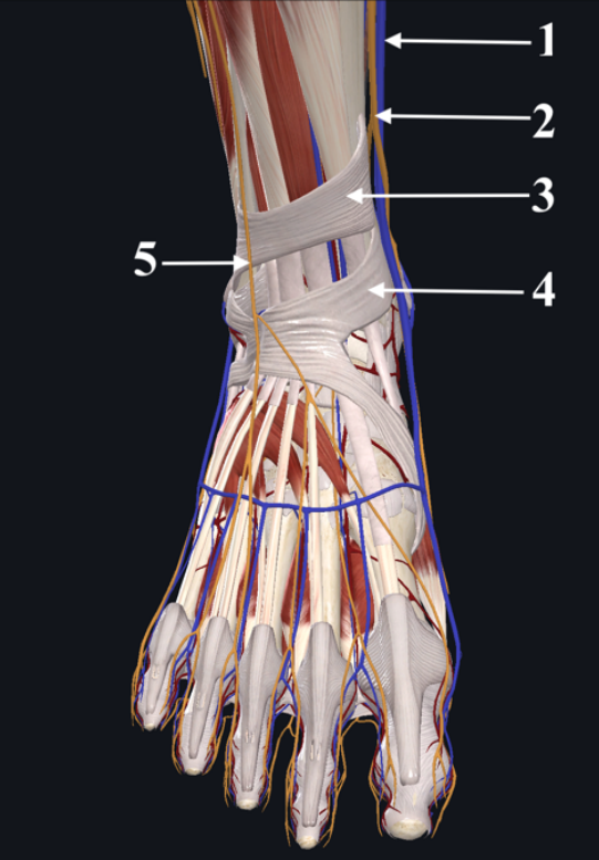
- AGreat saphenous vein
- BSaphenous nerve
- CSuperior extensor retinaculum
- DInferior extensor retinaculum
- ESuperficial fibular nerve
118name
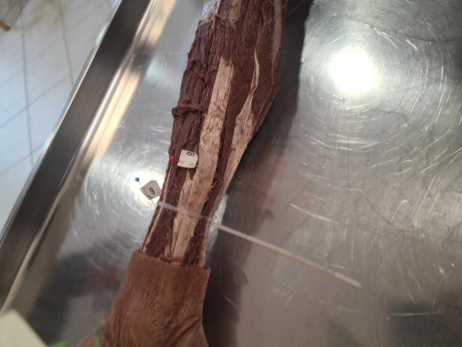
- Atibialis anterior muscle
- Btendon of fibularis longus
119name
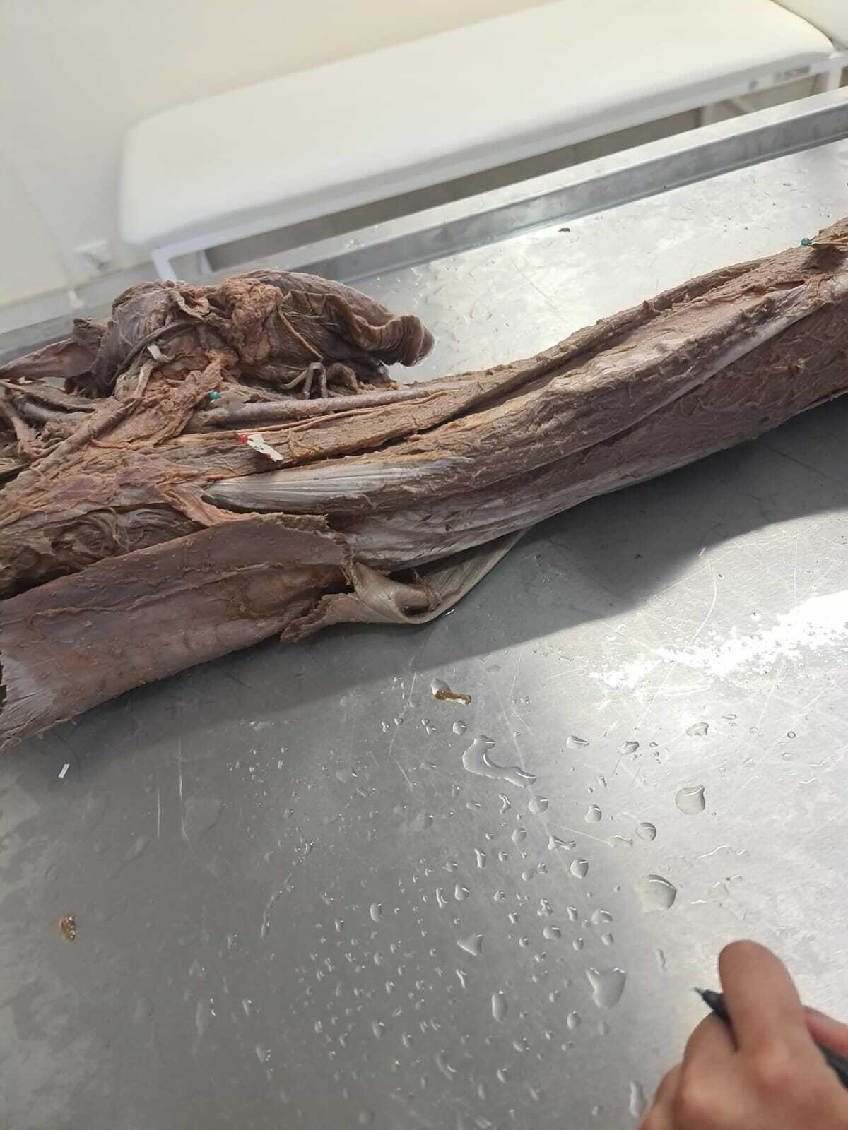
- Asartorius muscle
- Bfemoral artery
120name
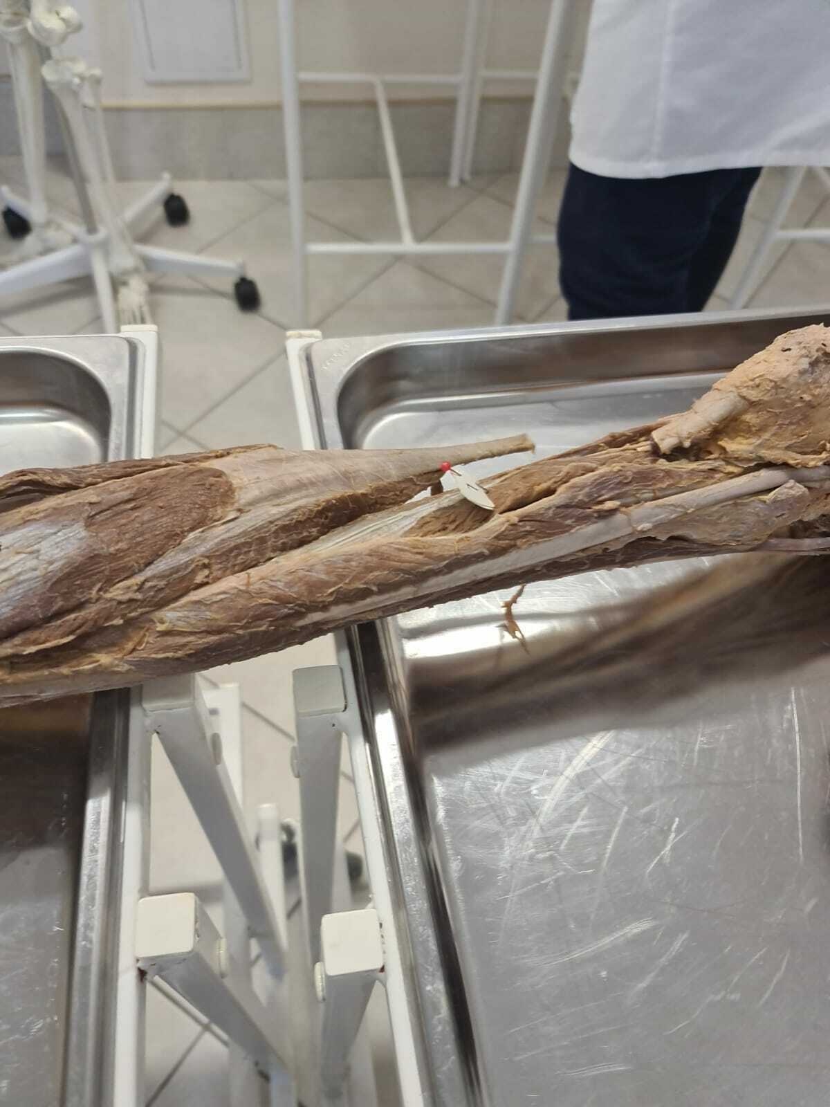
- Aflexor hallucis longus
121name
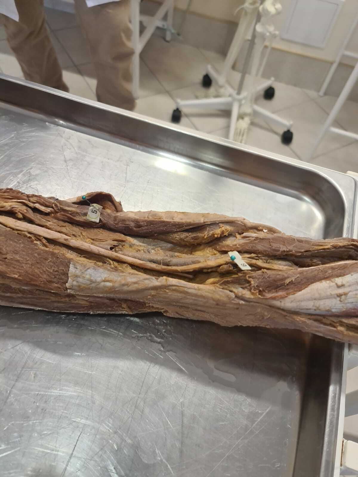
- Atibial nerve
- Bsemitendinous muscle
122name
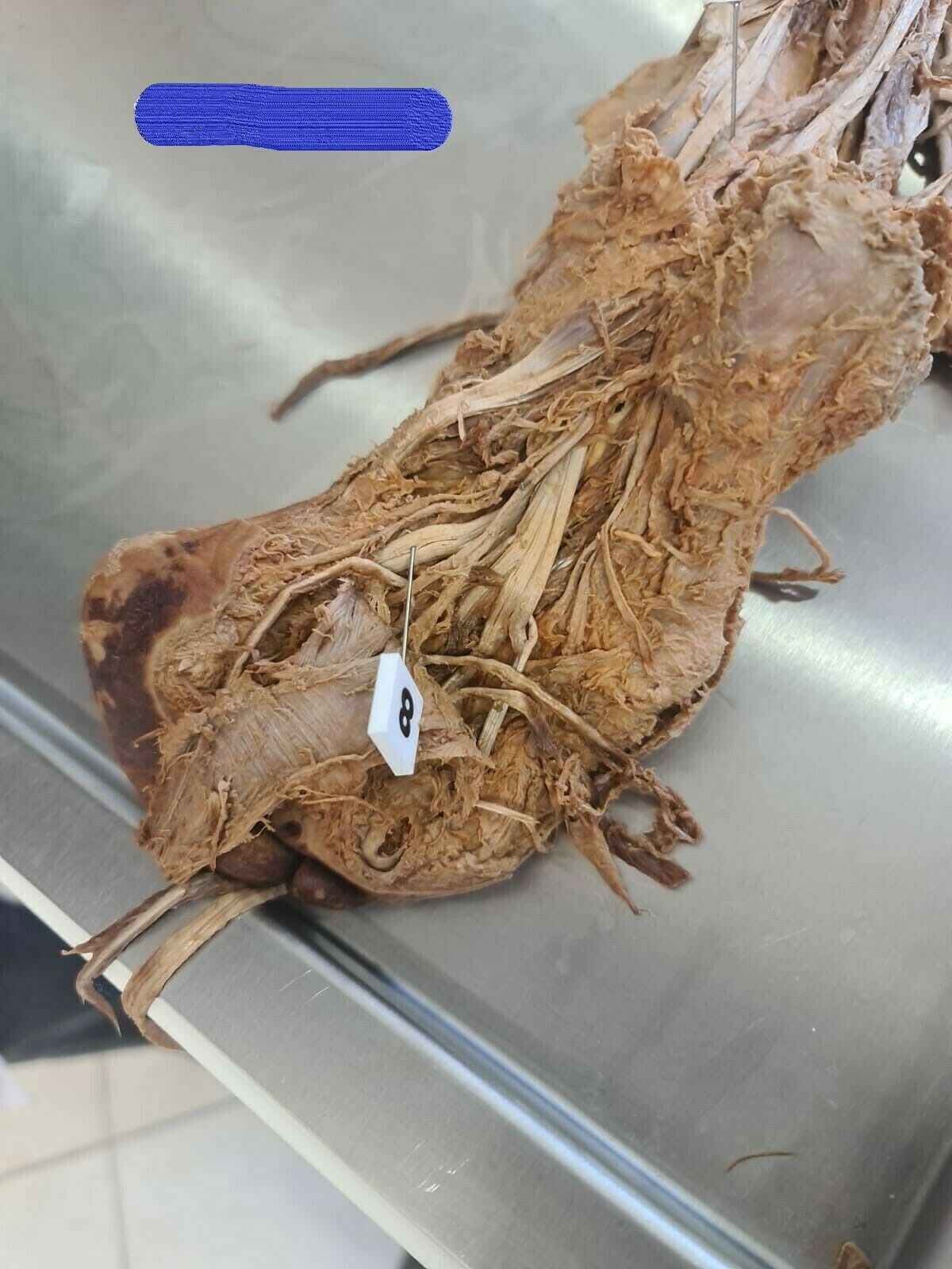
- Aflexor hallucis longus
123name
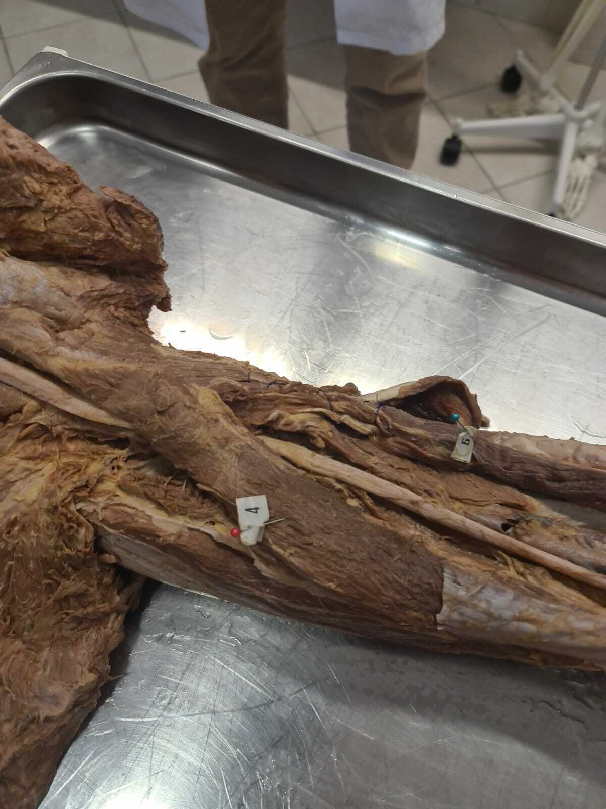
- Asemitendinous muscle
- Blong head of biceps femoris
124name
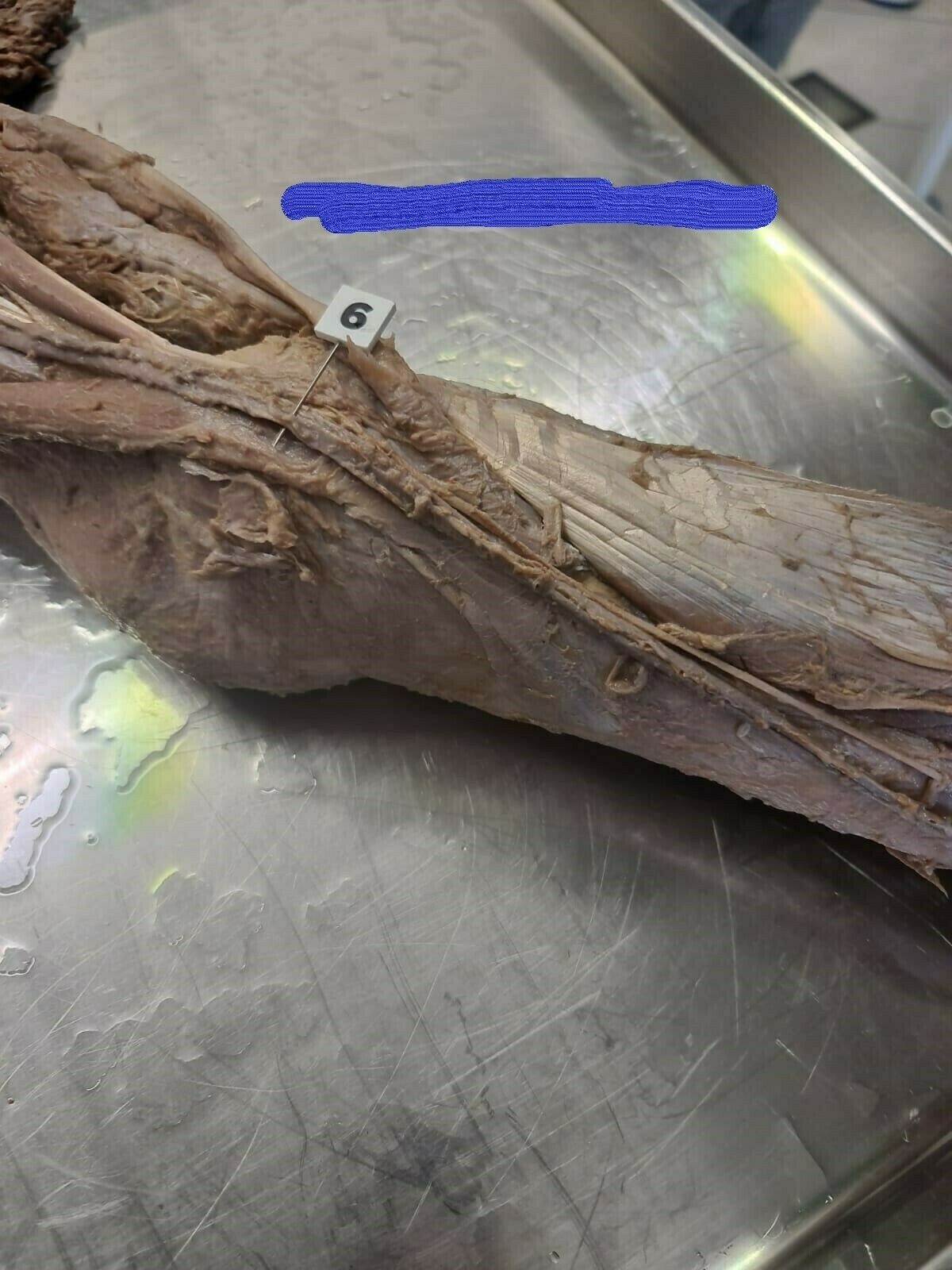
- Agreater saphenous vein
125name
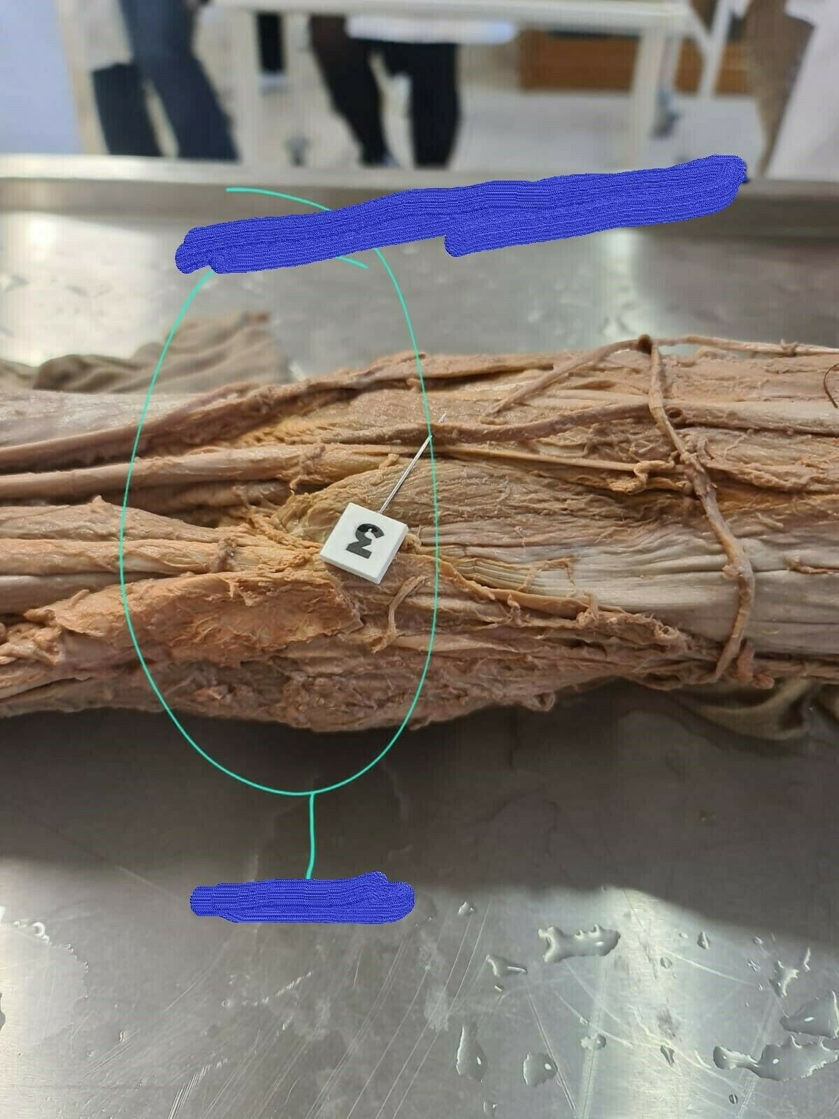
- Apopliteal fossa
- Bsmall saphenous vein
126name
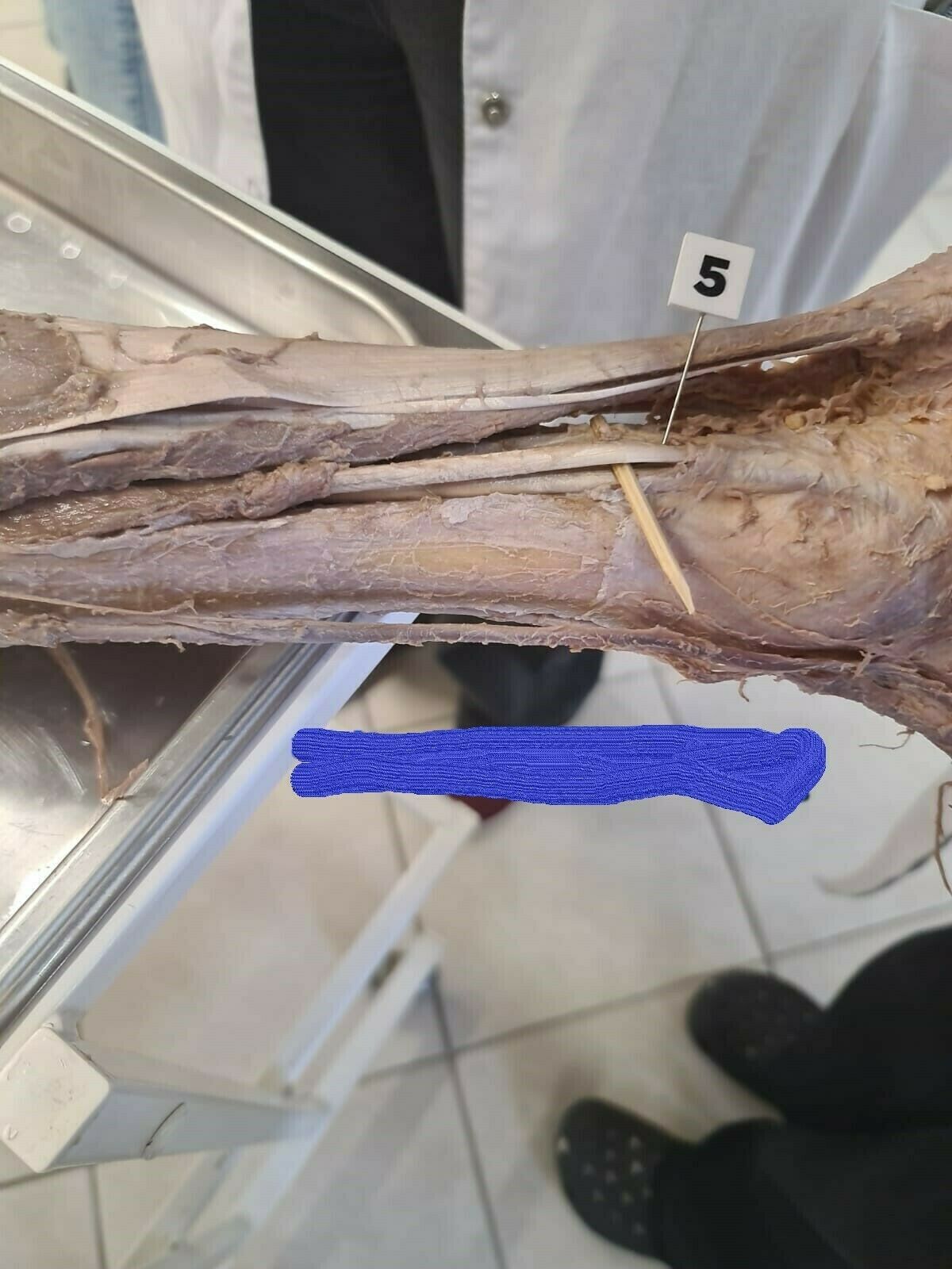
- Atendon of flexor digitorum longus
127name
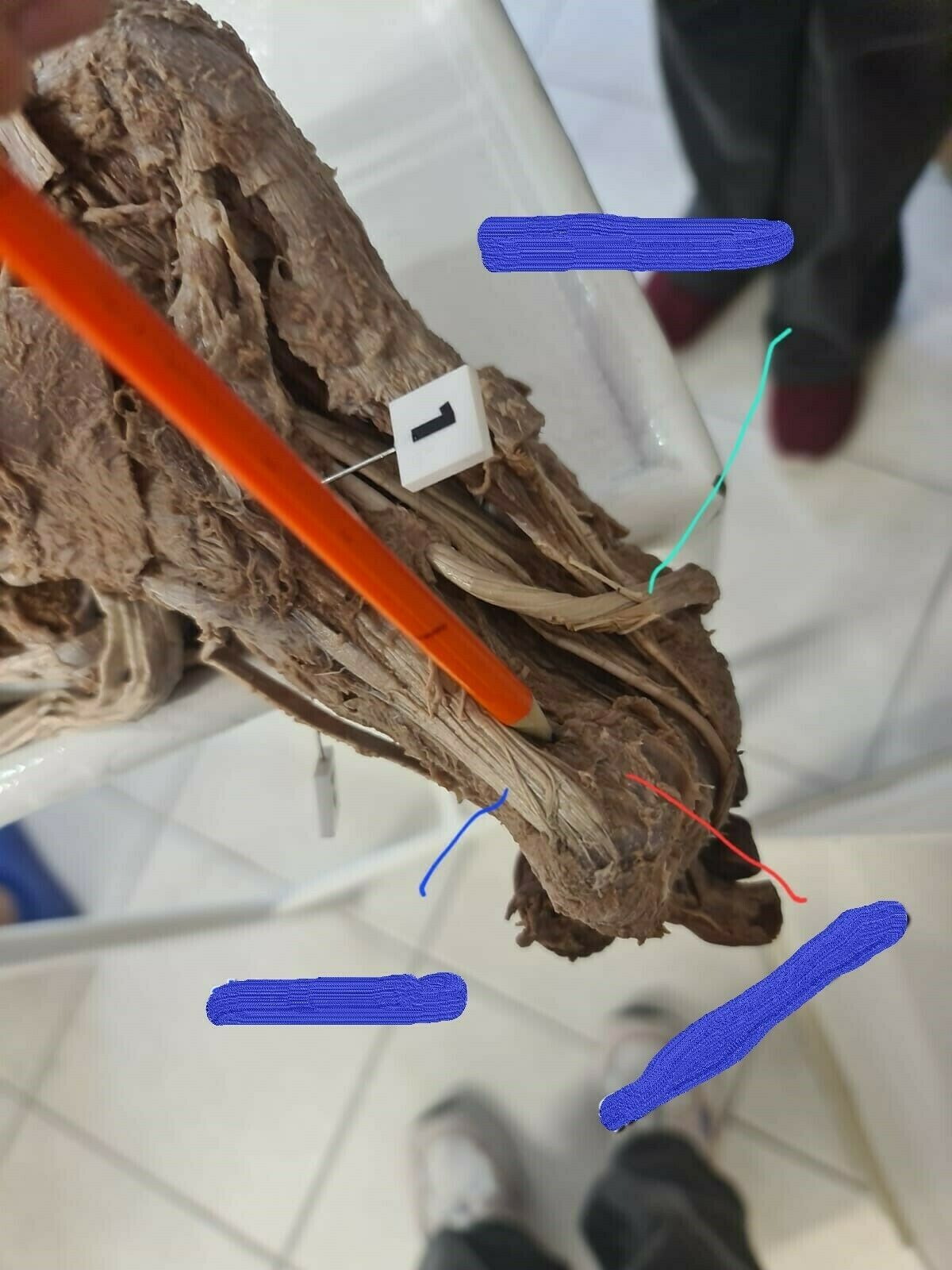
- Aflexor hallucis longus
- Bflexor hallucis brevis
- Cabductor hallucis
128name
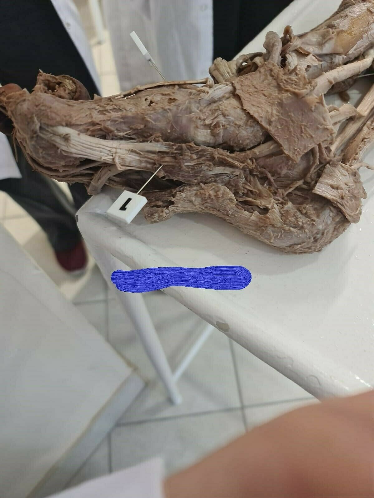
- Aabductor hallucis
129name
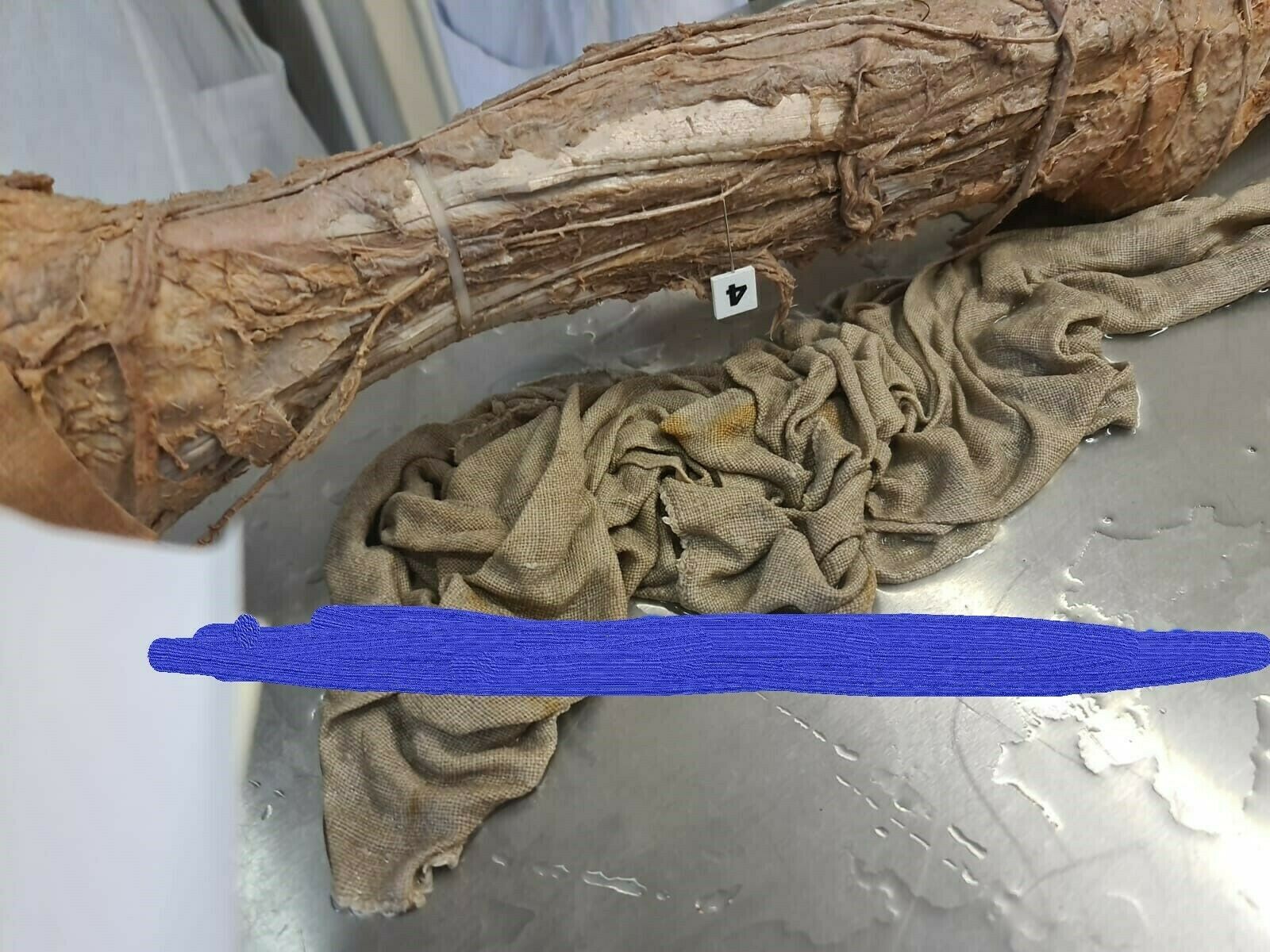
- Asuperficial fibular nerve
130name
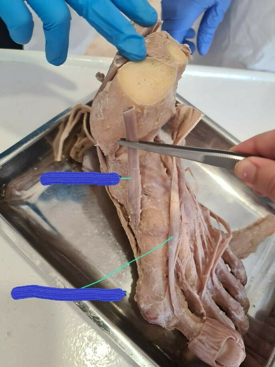
- Aextensor hallucis longus
- Btendon of tibialis anterior
131name
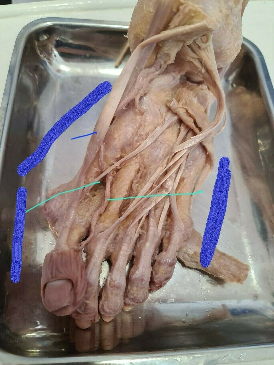
- Atibialis anterior
- Bextensor hallucis brevis
- Cdorsalis pedis artery
132name
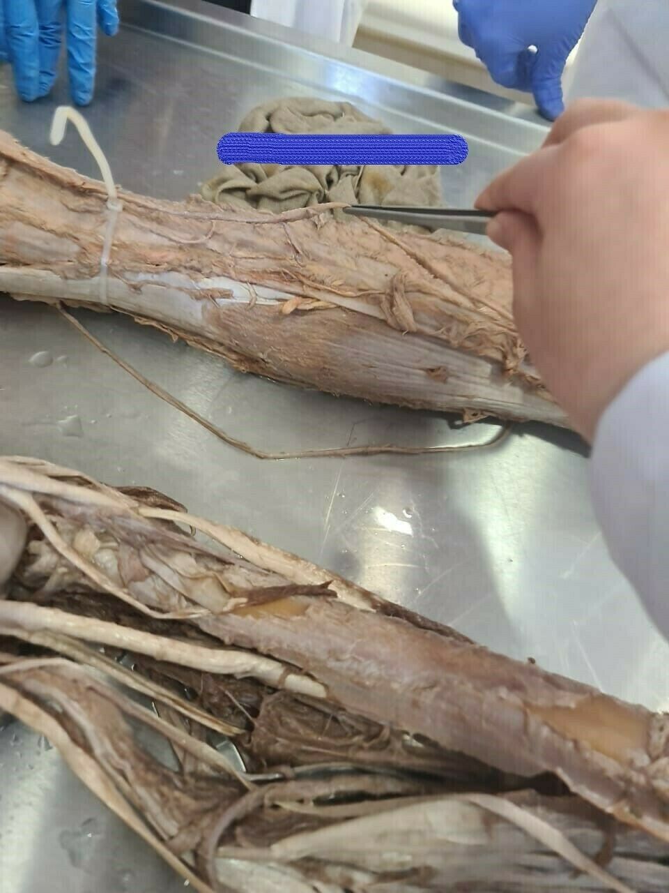
- Agreat saphenous vein
133name
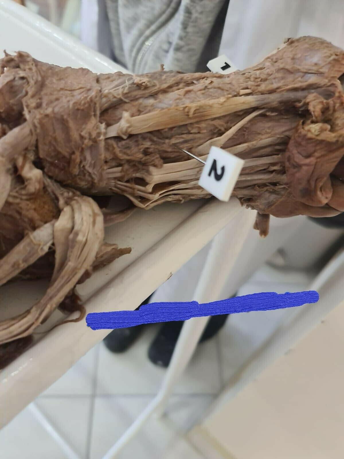
- Adorsal artery of foot
134name
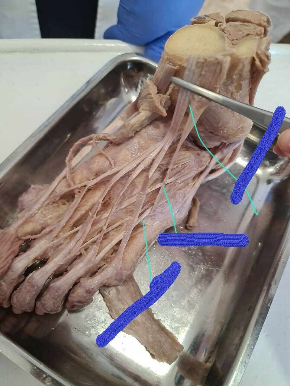
- Aextensor digitorum brevis
- Bfibularis tertius
- Cextensor digitorum longus
135n
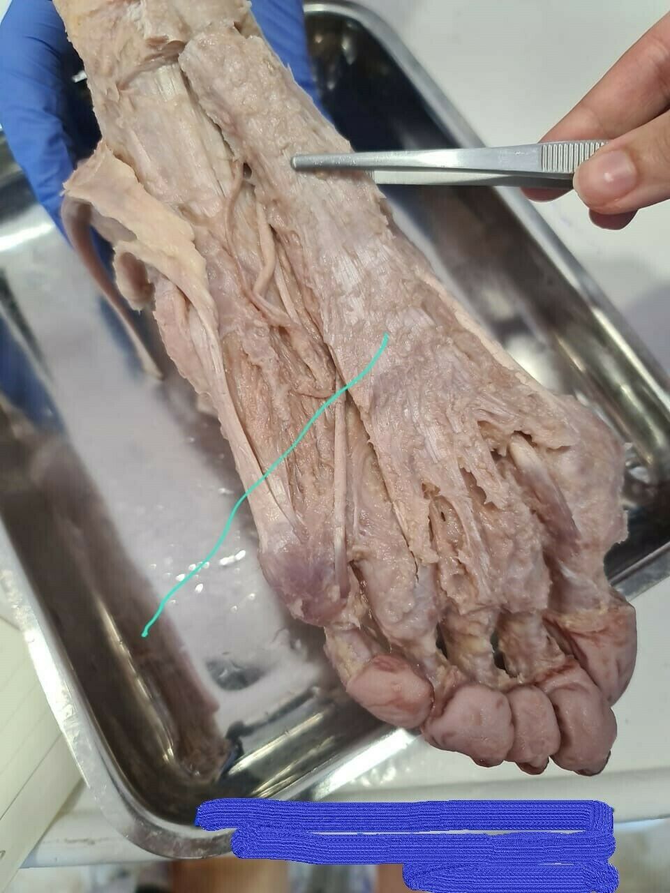
- Aplantar aponeurososis
136n
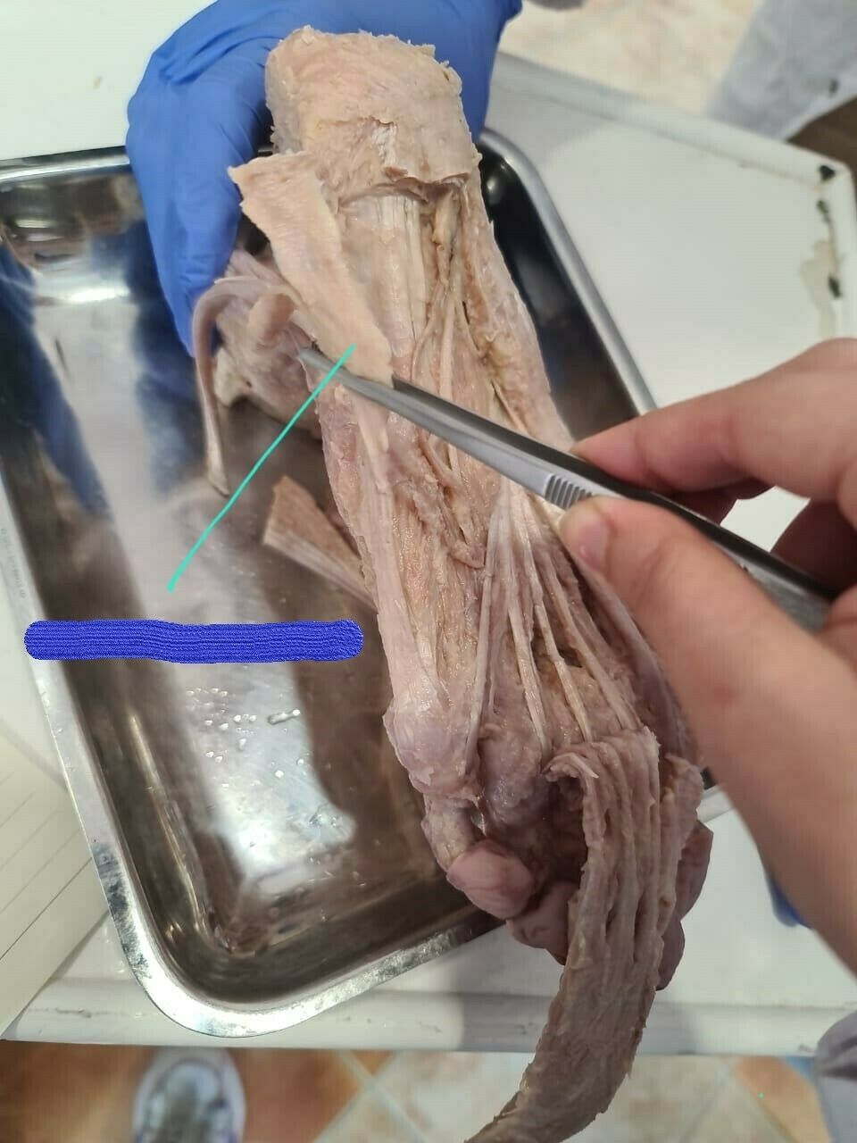
- Aabductor digiti minimi
137name
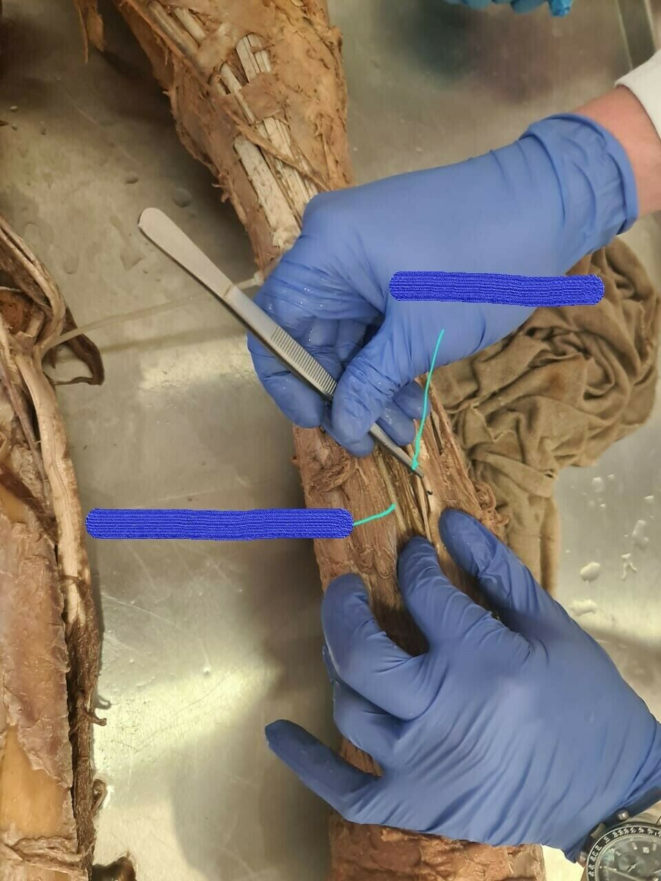
- Adeep fibular nerve
- Banterior tibial artery
138N
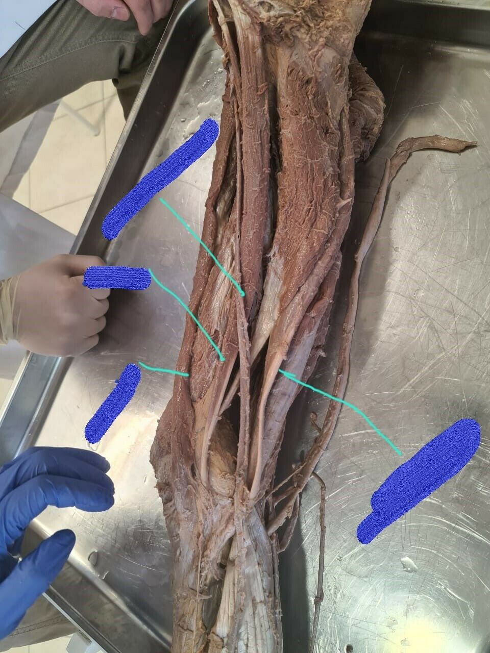
- Asartorius muscle
- Bsemitendinous muscle
- Cgracilis muscle
- Dgreat saphenous vein
139N
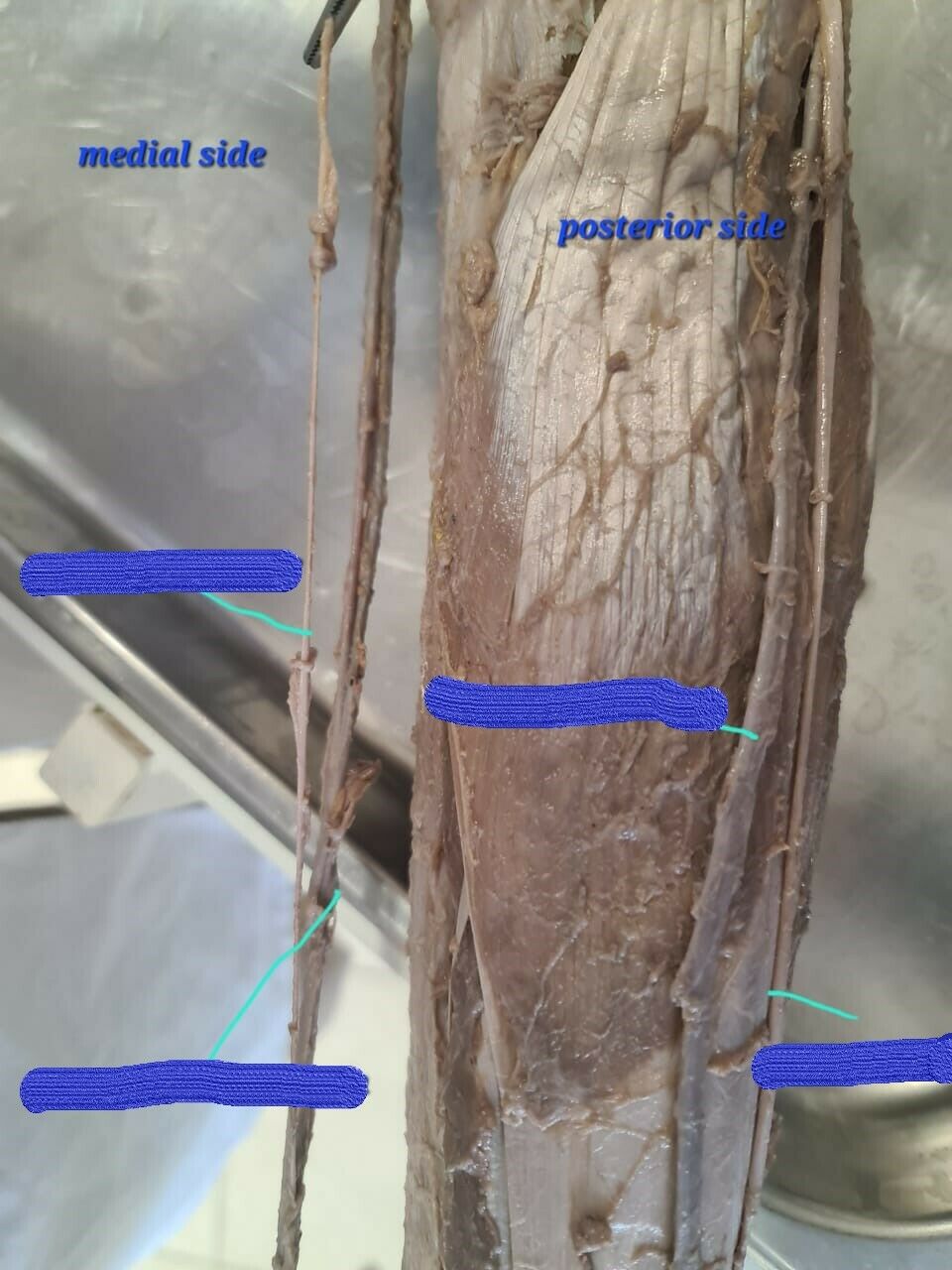
- Asaphenous nerve
- Bgreat saphenous vein
- Csmall saphenous vein
- Dsural nerve
140N
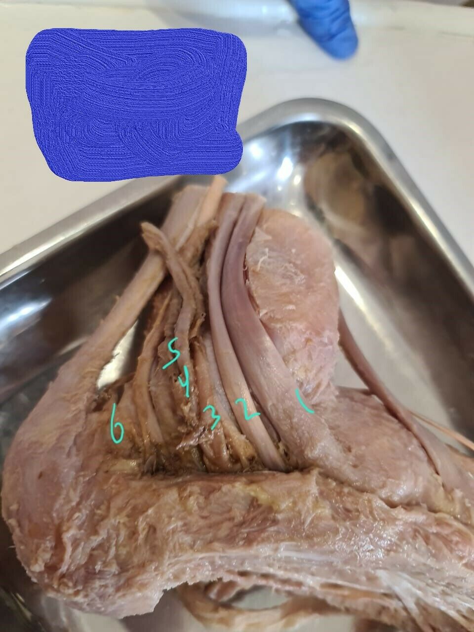
- Atibialis posterior
- Bflexor digitorum longus
- Cposterior tibial artery
- Dposterior tibial vein
- Etibial nerve
- Fflexor hallucis longus
141N
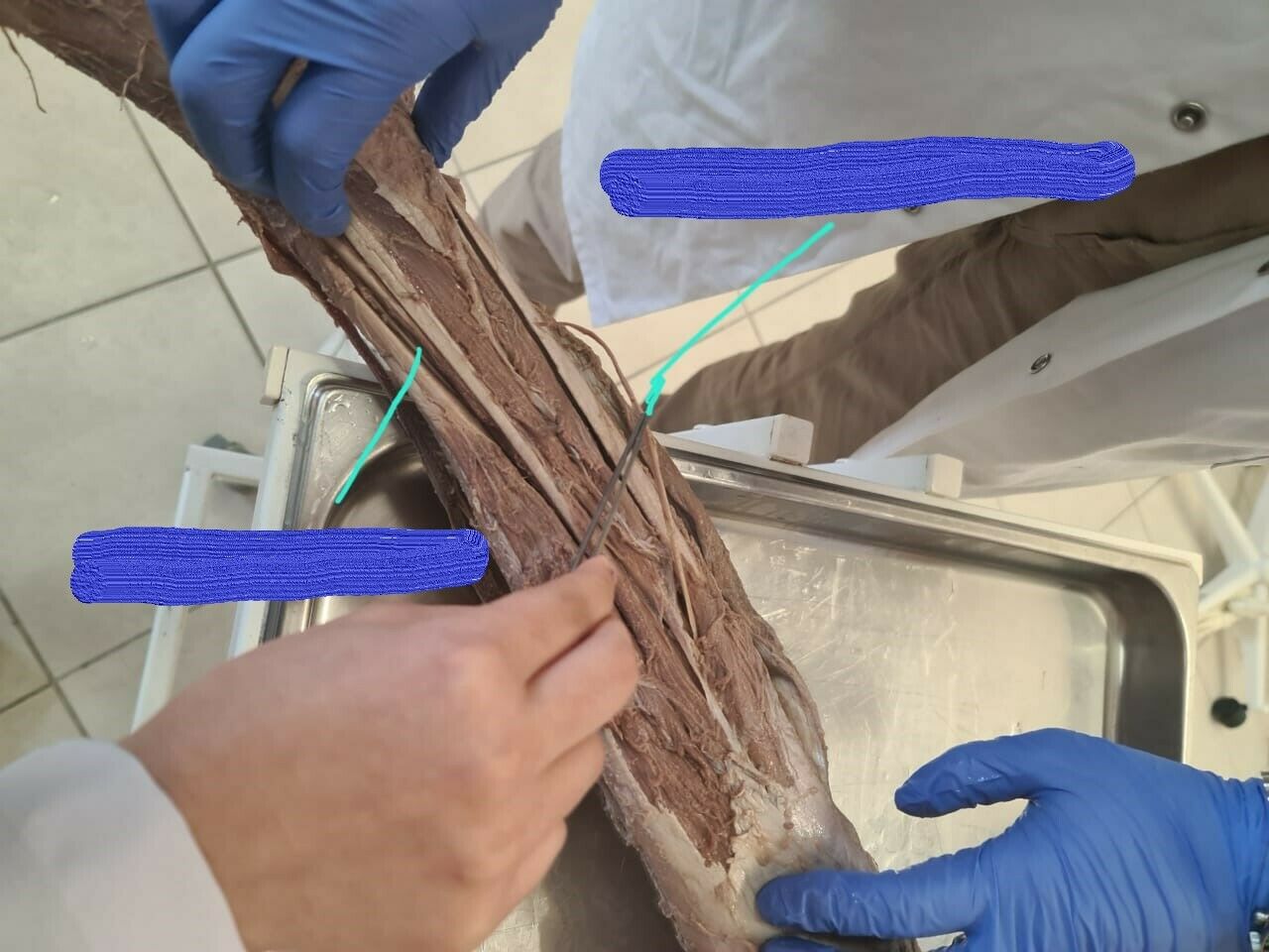
- Asuperficial fibular nerve
- Bdeep fibular nerve
142N
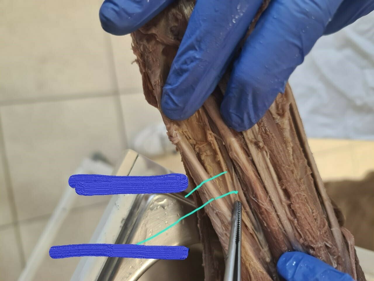
- Adeep fibular nerve
- Banterior tibial artery
143N
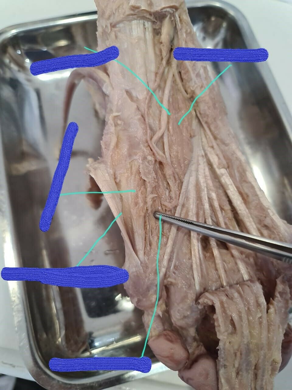
- Aabductor digiti minimi
- Bflexor digiti minimi
- Copponens digiti minimi
- Dlateral plantar nerve
- Equadratus plantae
144N
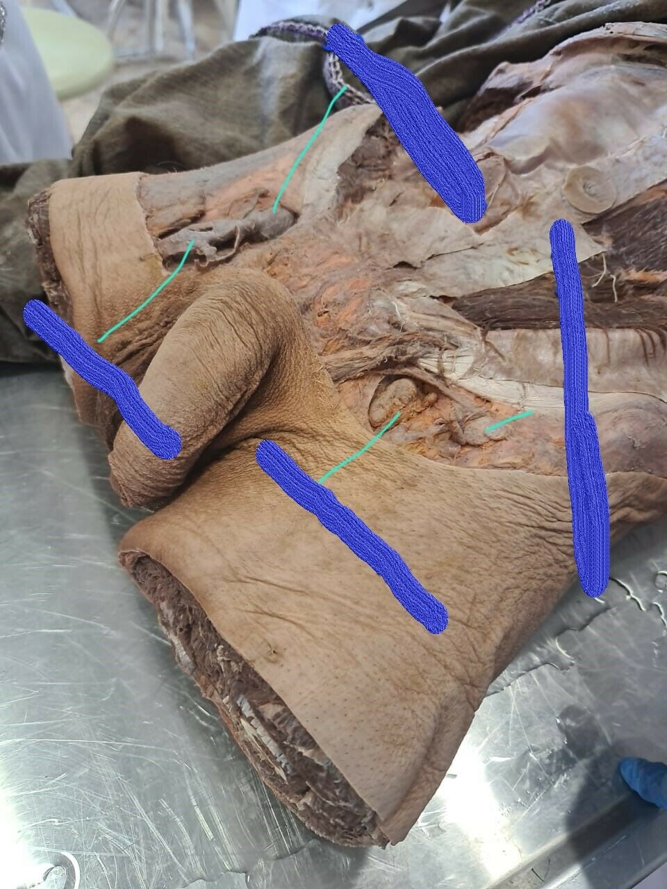
- Adeep inguinal lymph node
- Bsaphenous vein
- Cfermoral vein
- Dsurperficial inguinal lymph node
145N
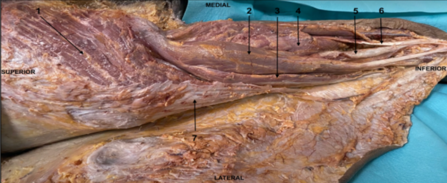
- Agöluteus maximus muscle
- Bsemitendinosous
- Cbiceps femoris
- Dadductor magnus
- Esemimembranosus
- Fgracilis
- Gtensor fascia latae
146N
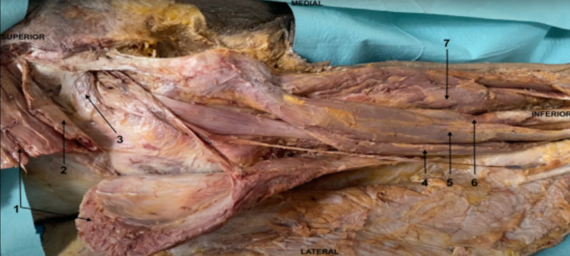
- Agluteus maximus
- Bgluteus medius
- Cpiriformis
- Dbiceps femoris
- Esemitendinosus
- Fsemimembranosus
- Gadductor magnus
147N
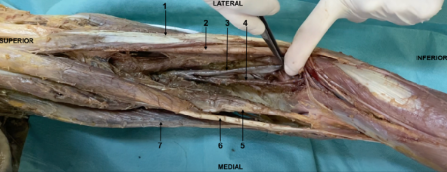
- Abiceps femoris
- Bsciatic nerve
- Cpopliteal vein
- Dpopliteal artery
- Esemimembranosus
- Fsemitendinosus
- Ggracilis
148N
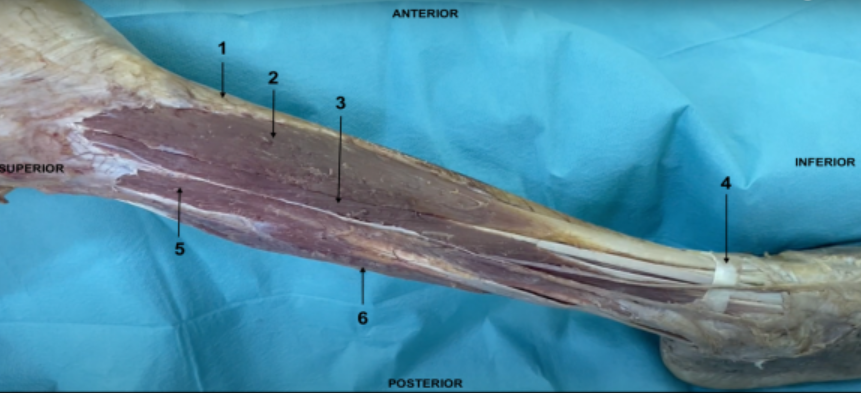
- Atibia
- Btibialis anterior
- Cextensor digitorum longus
- Dextensor retinaculum
- Efibularis longus
- Fgastrocnemius
149N
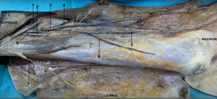
- Afemoral vein
- Bgreat saphenous vein
- Cfemoral artery
150N
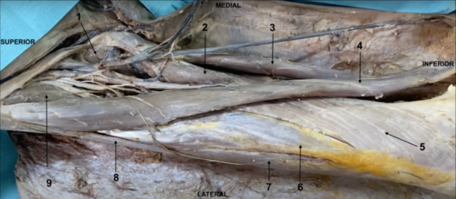
- Apectineus
- Badductor longus
- Cgracilis
- Dsartorius
- Evastus medialis
- Frectus femoris
- Gvastus lateralis
- Htensor fascia latae
- Iiliopsoas
151N
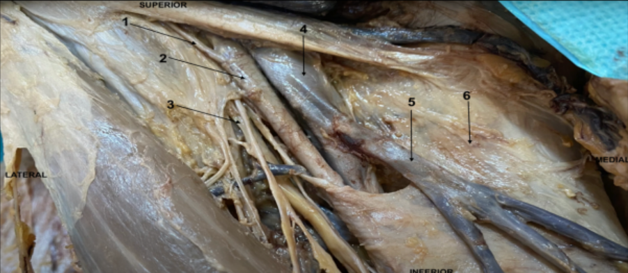
- Asuperficial circumflex artery
- Bfemoral artery
- Cfemoral nerve
- Dfemoral vein
- Egreat saphenous vein
- Ffascia latae
152N
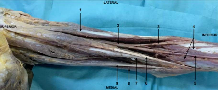
- Abiceps femoris
- Bsciatic nerve
- Ccommon fibular
- Dgastrocnemius
- Etibial nerve
- Fsemitendinosus
- Gsemimembranosus
- Hsartorius
- Igracilis
153Lumbar plexus
- Aformed by ant rami L1-L5
- Bgives off the genitofemoral nerve that emorges from the lat border of the psoas major
- Cgives off the lat cutaneous nerve of thigh which contains only sensory branches
- Dgives off the obturator nerve which emerges from the medial border of psoas major
- Efemoral nerve is the largest branch of this plexus and is formed by ant rami L2-L4
154Great saphenous vein
- Acould be used for coronary arterial bypass
- Bpasses aöllong the lateral border of the calcaneal tendon
- Cis accompanied by the vertial group of the deep inguinal lymph nodes
- Dempties into the femoral vein
- Eascends as a continuation of the lateral marginal vein
155adductor canal does NOT contain
- Afenoral artery
- Bfemoral vein
- Cdescending genicular artery
- Ddeep artery of thigh
- Esaphenous nerve
156ONE FALSE statement.
To the ischial tuberosity attaches- Asemimembranosus
- Badductor magnus
- Csuperior gemellus
- Dlong head of biceps femoris
- Equadratus femoris
157Medial boundery of femoral triangle is formed by
- Asartorus
- Badductor longus
- Cvastus lateralis
- Dsemitendinosus
- Eiliopsoas
158Deep muscle of posterior compartment of leg does NOT contain
- Atibialis post
- Bplantaris
- Cpopliteus
- Dflexor digitorum longus
- Eflexor hallucis longus
159Dermatome of the 5th digit ( digiti minimi) is supplied by root of the spinal nerve
- AL4
- BL5
- CS1
- DS2
- ES3
160ONE FLASE STATEMENT. Pes anserinus
- Ais a common tendinus insertion at the superior part of the medial surface of the tibia
- Bis formed by the tendon of the gracilis
- Cis formed by the muscles that flex a leg at the knee joint and rotate it medially
- Dis formed by the thendon of the biceps femoris
- Eis formed by the tendon of the sartorius
161In the case of damage to the obturator nerve one of the following muscles is completely paralyzed
- Asartorius
- Bgracilis
- Cobturator internus
- Dbuceps femoris
- Esemimembranosus
162to the calcaneal tuberosity DOES NOT ATTACH
- Aplantar aponeurosis
- Bflexpr digitorum brevis
- Cgrastrocnemius
- Dflexor retinaculum
- Efibularis longus
163Sciatic nerve
- Aforms the greatest part of the sacral plexus and is formed by anterior rami L4-S3
- Bits dibial division innervates all of the post. thigh muscles
- Cit innervates the gluteus maximus
- Dat the thigh rests on the adductor magnus and is covered by long head of biceps femoris
- Epasses through teh greater sciatic foramen above the piriformis
164Choose TRUE ANSWERS
- Athe deep artery of thigh arises from the femoral artery in the femoral triangle
- Bpost tibial pulse can usually be palpated btw the post surface of the medial malleolus and the nedial border of the calcaneal tendon
- Cdeep veins located internally to the deep fascia accompany all arteries and their branches of the lower limb
- Dpopliteal artery gives off the fibular artery which supplies the muscles of the lat and post compartement of the leg
- Ethe dorsalis pedis pulse can be palpated medially to then tendon of extensor hallucis longus
165Dermatome of the great toe (hallux) is supplied by the root of spinal nerve
- AL4
- BL5
- CS1
- DS2
- ES3
166One false answer regarding femoral sheath
- Alateral compartment contains the femoral artery
- Bis subdivided into three smaller subcompartments
- Cpasses deep to the inguinal ligament
- Dencloses femoral nerve
- Efemoral canal is located at the medial side of the femoral vein
167which of the following muscles could be paralyzed in the case of fracture of the necvk of the fibula
- Agastrocnemius
- Bpopliteus
- Cextensor digitorum longus
- Dflexor digitorum longus
- Eplantaris
Information
- Description
- Quiz on Lower Limb, created by Nils Bgee on 21/05/2023.
- Author
- Nils Bgee
- Language
- Deutsch
- Views
- 20
- Rating
- No ratings yet Rate this quiz