
Quiz
Protein section 4
This quiz has question types that open in the usual player.
Questions in this quiz (56)
1The binding of other molecules or [blank_start]LIGANDS[blank_end] is key to the action of most proteins. This involves molecular recognition of [blank_start]the ligand[blank_end] by the protein and this is usually very specific using the same type of [blank_start]non-covalent[blank_end] bonds involved in stabilising protein structure. The following points are generally true of most binding interactions/sites in proteins/ligands.
- ALIGANDS
- BENZYMES
- CATOMS
- DINHIBITORS
- Ethe ligand
- Fthe enzyme
- Gthe atom
- Hthe inhibitor
- Inon-covalent
- Jcovalent
2On what does molecular recognition depend?
- Aon complementarity of shape and charge distribution between the ligand and the protein binding site.
- Bon the formation of specialised microenvironments formed in the tertiary structure of the proteins called ligand binding sites or in enzymes active sites.
- Con the molecular weight of the ligand and the amino acids in the protein binding site
- Don the presence of the non-covalent bonds in both the ligand and the protein binding site
3Protein flexibility inherant in many proteins structures enables proteins to adapt to ligand on binding. And this is often essential for protein function. This mechanism is called.
- AInduced fit
- BLock and key mechanism
- CRecognition transformation
- DLIgand adaptation
4The binding of a ligand to a protein can be expressed mathematically and a constant can be derived which describes the tightness of binding of a ligand to a protein. What constant is it?
- AKd, the equilibrium constant for the release of ligand.
- BKcat, the turnover number -- the number of substrate molecule each enzyme site converts to product per unit time
- CKm, the substrate concentration at which the reaction rate is at half-maximum
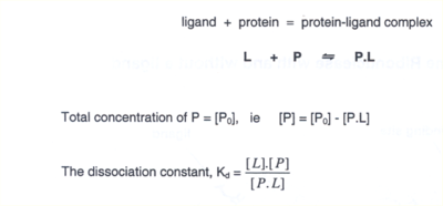
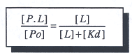
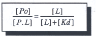
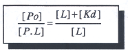
5FInish the equation transformation
- A
- B
- C
- D
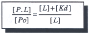
- A
6Binding sites will be lined with appropriate amino acids or binding elements which interact specifically with the [blank_start]ligand[blank_end].
Binding si Binding sites can be different shapes, for [blank_start]macromolecules[blank_end] they can be concave, convex or flat and may be grooves, [blank_start]for small molecules[blank_end] they can be clefts, pockets or cavities.
tes and more specifically catalytic [blank_start]active[blank_end] sites often occur at the interface between subunits or domains in proteins.
Binding sites generally have a higher than average amount of exposed [blank_start]hydrophobic[blank_end] groups.
[blank_start]Weak[blank_end] interactions between ligand and protein can favour easy exchange of molecules.
The energy for driving binding events is often provided by the displacement of [blank_start]water molecules[blank_end] from the ligand binding site.- Aligand
- BInhibitor
- Ccoenzyme
- Dcofactor
- Emacromolecules
- Fsmall molecules
- Gfor small molecules
- Hfor macromolecules
- Iactive
- Jbinding
- KInhibition
- Lenzyme
- Mhydrophobic
- Nhydrophilic
- OWeak
- PStrong
- Qwater molecules
- Rhydrophobic amino acids
- Shydrophilic amino acids
- Tcoordination atoms
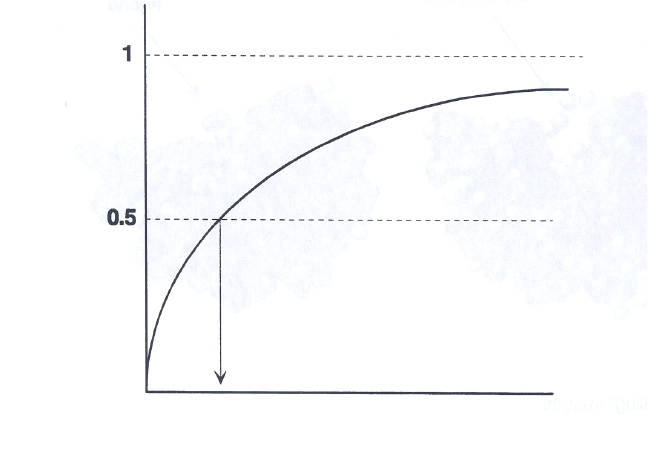
7Label the graph
- AKd
- B[L]
- C[P.L] / [Po]
8The dissociation constant [blank_start]Kd[blank_end] represent the equilibrium constant for the release of ligand and that when the concentration of [blank_start]L[blank_end] is equal to Kd [blank_start]half[blank_end] of the ligand binding sites are occupied.
- AKd
- BKm
- CKcat
- DKa
- EL
- FPo
- G1/2
- H3/4
9 In practice Kd is used to express the [blank_start]affinity[blank_end] of a protein for a ligand.
The units of Kd are molar concentration ie. [blank_start]Moles/litre[blank_end]
When [blank_start]concentration[blank_end] of L is [blank_start]lower[blank_end] than Kd then very little ligand is bound.
In order for 90% of ligand binding sites to be occupied [L] must be [blank_start]nine times[blank_end] Kd.
The lower the value of Kd the [blank_start]higher[blank_end] the affinity of the ligand for the protein.- Aaffinity
- BMoles/litre
- Cconcentration
- Dlower
- Ehigher
- Fnine times
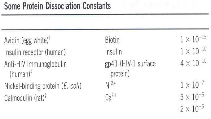
10Identify each column
- AProtein
- BLigand
- CKd (M)*
11the most studied and best understood proteins with a well defined structure/function relationship established. These are excellent proteins to use as a model to show the complexities and intricacies of protein ligand binding and regulation. What are these proteins ?
- AInsulin and glycogen
- Bhaemoglobin and myoglobin
- Ccollagen and keratin
12 Oxygen is very soluble in the bloodstream and to allow it to be transported and carried into tissues it is bound to proteins. These are Haemoglobin and Myoglobin.
- ATrue
- BFalse
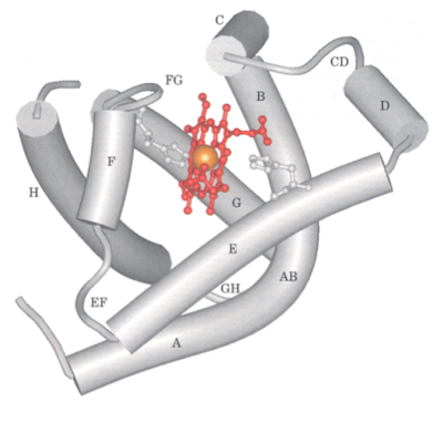
13Myoglobin is predominantly beta-sheet (80%) compact single polypeptide of 153 aminoacids
- ATrue
- BFalse
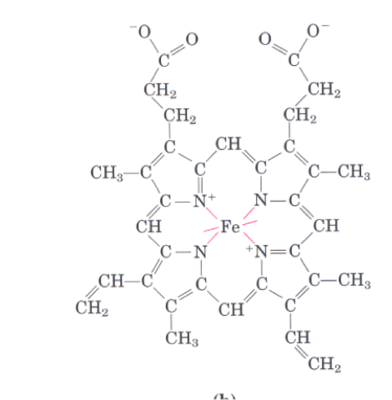
14Myoglobin contains a haem group comprising [blank_start]protoporphyrin[blank_end] IX with a bound iron in the Ferrous state. The iron has [blank_start]six[blank_end] coordination bonds, [blank_start]four[blank_end] to the flat porphyrin ring system and [blank_start]two[blank_end] perpendicular to it. One is associated with a [blank_start]histidine[blank_end] (proximal) on the F- helix, the other is free to bind [blank_start]oxygen[blank_end].
- Aprotoporphyrin
- Bphophtalein
- Cphotosystem
- Dsix
- Eeight
- Fseven
- Gfive
- Hten
- Ifour
- Jthree
- Ktwo
- Lone
- Mtwo
- None
- Othree
- Pfour
- Qhistidine
- Rglycine
- Slysine
- Tadenine
- Uoxygen
- Vcarbon
- Wiron
- Xnitrogen
- Ycalcium
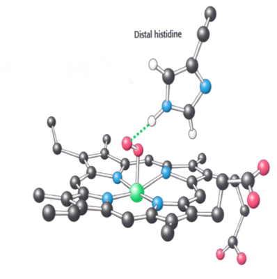
15[blank_start]Oxygen[blank_end] binds to the [blank_start]iron[blank_end] in haem. This pulls the iron into the plane of the porphyrin ring and moves the [blank_start]F-helix[blank_end]. [blank_start]The oxygen[blank_end] is stabilised in the binding site by a hydrogen bond donated by the histidine (distal) on the [blank_start]E-helix[blank_end].
- AOxygen
- BCarbon
- CNitrogen
- DHydrogen
- Eiron
- Fcalcium
- Gmercury
- Hsodium
- Ipotassium
- JF-helix
- KAlpha-helix
- LC-helix
- MB-helix
- NX-helix
- OThe oxygen
- PThe nitrogen
- QThe carbon
- RThe hydrogen
- SE-helix
- TL-helix
- UM-helix
- VZ-helix
- WH-helix
16THE BINDING OF OXYGEN TO MYOGLOBIN 1
The protein contains a haem group with a bound ferrous [blank_start]iron[blank_end].
The iron had [blank_start]six[blank_end] coordinating bonds, four are satisfied by binding to [blank_start]the plane[blank_end] of the porphyrin ring.
One other is bound to [blank_start]a histidine[blank_end] on the F-helix of [blank_start]myoglobin[blank_end]. This is the [blank_start]proximal[blank_end] histidine.
In [blank_start]deoxymyoglobin[blank_end] the sixth is free.
In deoxymyoglobin the interaction of the iron with the proximalal histidine induces a puckering of [blank_start]the porphyrin ring system.[blank_end]- Airon
- Bsix
- Cthe plane
- Da histidine
- Emyoglobin
- Fproximal
- Gdeoxymyoglobin
- Hthe porphyrin ring system.
17The binding site for oxygen is actually deeply buried and local breathing of the protein, allowed because of the [blank_start]flexibility[blank_end] of the molecule allows oxygen to enter the binding site by moving through local cavities in the protein
When oxygen binds to form [blank_start]oxymyoglobin[blank_end] it does so by binding to the [blank_start]sixth[blank_end] coodinating bond of the haem bound iron.
This binding results in the partial transfer of an electron from [blank_start]thje ferrous ion to oxygen[blank_end] creating a superoxide ion. This is not stable.
However the location of the [blank_start]distal[blank_end] histidine on [blank_start]helix-E[blank_end] creates an H bond to the bound oxygen and stabilises it thus preventing release of unreactive species.
This h bonding pulls the oxygen into the plane of the porphyrin ring and the whole structure changes [blank_start]shape[blank_end] slightly via movement of [blank_start]the H-helix[blank_end] to tighten up round the bound oxygen molecule.- Aflexibility
- Bcharge
- Celectronegativity
- Dmass
- Ereactivity
- Foxymyoglobin
- Ghaemoglobin
- Hmyoglobin
- Ideoxymyoglobin
- Jsixth
- Kfifth
- Lfourth
- Mfirst
- Nthird
- Othje ferrous ion to oxygen
- Pthje oxygen to ferrous ion
- Qthje myoglobin to haemoglobin
- Rthe haemoglobin to myoglobin
- Sdistal
- Tproximal
- Uhelix-E
- Vhelix-F
- Wthe H-helix
- Xthe F-helix
- Ythe E-helix
- Zshape
- AApolarity
- ABdirectionality
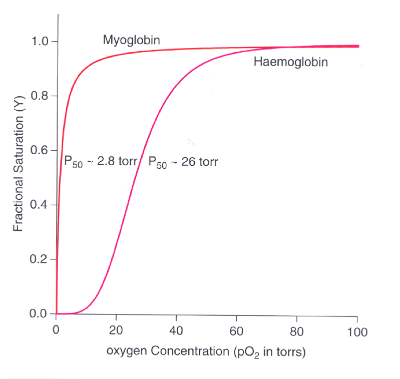
18[blank_start]Myoglobin[blank_end] has a very high affinity for oxygen and its binding is described by the classical hyperbolic binding curve, the high affinity reflected in the fact that the molecule is half saturated at very [blank_start]low[blank_end] oxygen concentrations. [blank_start]Haemoglobin on the other hand[blank_end] exhibits very different and anomalous binding characteristics. The curve for haemoglobin is [blank_start]sigmoidal[blank_end] indicating a different affinity profile [blank_start]to myoglobin[blank_end].
- AMyoglobin
- BHaemoglobin
- Clow
- Dhigh
- EHaemoglobin on the other hand
- FMyoglobin on the other hand
- Gto myoglobin
- Hto haemoglobin
- Isigmoidal
- Jepispiral
- Kparabolic
19Label the diagram
- AHyperbolic binding curve
- BSigmoidal binding curve
20THE STRUCTURE OF HAEMOGLOBIN
Haemoglobin has [blank_start]quarternary[blank_end] structure
It has [blank_start]four[blank_end] subunits [blank_start]2α[blank_end] and [blank_start]2β[blank_end]
Each subunit has a bound haem containing an [blank_start]iron[blank_end] in the ferrous form
Each subunit is capable of binding [blank_start]a molecule[blank_end] of oxygen
Each subunit whilst sequentially [blank_start]disimilar[blank_end] to myoglobin is structurally [blank_start]very similar[blank_end] to myoglobin
Key residues in each subunit are highly [blank_start]conserved[blank_end] and identical to myoglobin.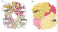
- Aquarternary
- Bprimary
- Ctertiary
- Dsecondary
- Efour
- Fsix
- G2α
- H3α
- I2β
- J3β
- Kiron
- Lcalcium
- Mlithium
- Nmercury
- Omagnesium
- Pa molecule
- Qtwo molecules
- Rdisimilar
- Ssimilar
- Tvery similar
- Uvery dissimilar
- Vconserved
- Wreactive
- Xhydrophobic
- Ypolar
21THE MYOGLOBIN MOLECULE AND THE HAEMOGLOBIN SUBUNITS ARE STRUCTURALLY VERY SIMILAR
The polypeptide chains of Myoglobin (pink), the α-subunit (purple) and the β-subunit (yellow) are almost superimposable showing the similarity of tertiary structure. The proximal and distal histidines are conserved as are the haem binding pocket interactions.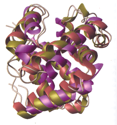
- ATrue
- BFalse
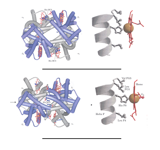
22The studies of Max Perutz in the 60’s and 70’s show3ed that there are two major conformations of haemoglobin. These he called the T state and the R state originally denoted ‘tense’ and ‘relaxed’
- ATrue
- BFalse
23 Oxygen can bind to haemoglobin in either the T or the R state.
Oxygen has a much [blank_start]higher[blank_end] affinity for haemoglobin in the R state and actually [blank_start]stabilises[blank_end] the R state.
The predominant conformation of deoxyhaemoglobin in the [blank_start]T state[blank_end].
The binding of oxygen to a haemoglobin subunit in the [blank_start]T[blank_end] state triggers a change in conformation of the subunit to that of [blank_start]the R state.[blank_end]
This causes the subunits to change position [blank_start]relative to one another[blank_end].- Ahigher
- Blower
- Cstabilises
- Ddisrupts
- ET state
- FR state
- GT
- HR
- Ithe R state.
- Jthe T state.
- Krelative to one another
- Lof each other
24The [blank_start]T to R[blank_end] transition is caused by oxygen binding.
As in myoglobin oxygen binds to the [blank_start]sixth uncoordinated position[blank_end] of the haem bound ferrous iron.
This pulls the iron into the plane of the [blank_start]porphyrin ring[blank_end] and because this iron is also coordinated to the proximal histidine attached to [blank_start]helix-F[blank_end] it slightly changes the position of helix F in the subunit.
In [blank_start]myoglobin[blank_end] this has no discernable effect other than to stabilise oxygen binding, but in [blank_start]haemoglobin[blank_end] this and other small changes cause the subunit to rotate relative to the other subunits in the structure and the [blank_start]overall shape[blank_end] of the molecule changes from T to R conformation.- AT to R
- Bsixth uncoordinated position
- Cporphyrin ring
- Dhelix-F
- Emyoglobin
- Fhaemoglobin
- Goverall shape
25Find the T state and the R state
- AT state
- BR state
26HAEMOGLOBIN BINDS OXYGEN COOPERATIVELY
- ATrue
- BFalse
27Haemoglobin must bind oxygen efficiently in the lungs at [blank_start]high partial pressures[blank_end] of oxygen and release it at the tissues in [blank_start]low partial pressures of oxygen[blank_end].
A protein like myoglobin that exhibits a [blank_start]hyperbolic[blank_end] binding curve would not release oxygen very effectively at the tissues and a protein that has sufficiently [blank_start]low[blank_end] affinity to allow relaese at the tissues would not pick up much oxygen in the lungs. HAEMOGLOBIN SOLVES THIS PROBLEM BY UNDERGOING THE TRANSITION FROM A [blank_start]LOW AFFINITY[blank_end] T STATE TO A [blank_start]HIGH AFFINITY R STATE.[blank_end]- Ahigh partial pressures
- Blow partial pressures
- Clow partial pressures of oxygen
- Dhigh partial pressures of oxygen
- Ehyperbolic
- Fsigmoidal
- Glow
- Hhigh
- ILOW AFFINITY
- JHIGH AFFINITY
- KHIGH AFFINITY R STATE.
- LLOW AFFINITY R STATE.
- MHIGH AFFINITY T STATE.
- NLOW AFFINITY T STATE.
28COOPERATIVE BINDING SHOWS A...
- ASIGMOID BINDING CURVE
- BHYPERBOLIC BINDING CURVE
29Oxygen binding to individual subunits in haemoglobin can alter the affinity for oxygen in [blank_start]adjacent subunits[blank_end]
The first molecule of oxygen that interacts with [blank_start]deoxyhaemoglobin[blank_end] binds weakly since it binds to a subunit in [blank_start]the T state[blank_end].
This leads to a conformational change (outlined previously) which is communicated to adjacent subunits of the [blank_start]quarternary structure[blank_end] making it easier for additional oxygen molecules to bind.
The T to R transition occurs more readily in [blank_start]the second subunit[blank_end] once oxygen is bound to the first.
Binding of oxygen to [blank_start]the haem[blank_end] in the final subunit is high affinity because it is already in [blank_start]the R state[blank_end]- Aadjacent subunits
- Bdeoxyhaemoglobin
- Cthe T state
- Dquarternary structure
- Ethe second subunit
- Fthe R state
- Gthe haem
30Label the diagram
The classical sigmoid or cooperative binding curve shown for haemoglobin is actually a composite of the binding curves shown by the T state and the R state.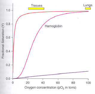
- AR form
- BT form
31Haemoglobin is an ALLOSTERIC protein
- ATrue
- BFalse
32Allosteric proteins are those which have other conformations induced by the binding of ligands referred to as modulators. The conformational changed induced by the modulator interconvert more or less inactive forms of the protein.
- ATrue
- BFalse
33When the normal ligand and modulator are the same molecule the interaction is termed [blank_start]HOMOTROPIC[blank_end] but when the modulator is a different molecule to the normal ligand the interaction [blank_start]is termed HETEROTROPIC.[blank_end]
In [blank_start]haemoglobin[blank_end] the binding of one ligand affects the affinities of any remaining unfilled binding sites and oxygen is both a normal ligand and an activating homotropic modulator. It activates by causing conformational changes in the [blank_start]first[blank_end] subunit transmitted to the rest of the molecule by subunit to subunit interactions.- AHOMOTROPIC
- BHETEROTROPIC
- Cis termed HETEROTROPIC.
- Dis termed HOMOTROPIC.
- Ehaemoglobin
- Fmyoglobin
- Gfirst
- Hsecond
34For most proteins some sort of binding event is necessary for their action and that the binding is specific and relies of complementarity, flexibility and the dynamic nature of the proteins structure.
- ATrue
- BFalse
35What is a prerequisite for the action of most proteins.?
- ALigand binding
- BHydrophobic core
- CPolar amino outside on the surface of the protein
- DCatalytic activity
36[blank_start]Carrier proteins[blank_end] must bind a ligand eg. Myoglobin and Haemoglobin
[blank_start]Receptors[blank_end] must bind a ligand agonist or antagonist to elicit their effect.
[blank_start]Proteins involved in switching[blank_end] must bind a trigger molecule which may cause a conformational change leading to activation/deactivation of another component of a signalling system.
[blank_start]Structural proteins[blank_end] often bind other monomeric proteins of the same type to form polymeric structures such as F-actin and tubulin.
[blank_start]DNA and RNA binding protein[blank_end]s and ribosomal proteins must effectively bind macromolecular structures or associate with other proteins in large assemblies.
Enzymes responsible for dramatic rate enhancements in biological systems must effectively bind [blank_start]substrates[blank_end] to elicit their effect.- ACarrier proteins
- BReceptors
- CProteins involved in switching
- DStructural proteins
- EDNA and RNA binding protein
- Fsubstrates
37THE LARGEST CLASSES OF PROTEINS THAT BIND LIGANDS AND CATALYSE THEIR CHEMICAL REACTION, WHERE LIGAND IS A SUBSTRATE. THESE PROTEINS ARE CALLED....
- Aenzymes
- Bcarrier proteins
- Creceptors
- Dinhibitors
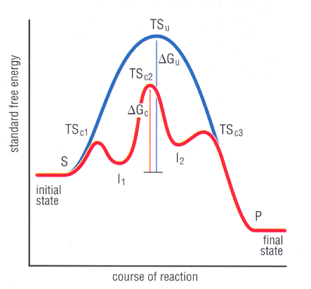
38Label the image
- AEnergy barrier
- BTurnover point
- CT-R transition state
- DThe state of highest entropy
39For an enzyme reaction to occur the [blank_start]substrate(ligand)[blank_end] must undergo chemical transformations perhaps in stereochemistry, charge configuration or [blank_start]covalent bonding[blank_end].
There will be a free energy barrier to overcome to achieve this. The higher the barrier the [blank_start]slower[blank_end] the reaction and the more [blank_start]difficult[blank_end] the chemical step to be catalysed.- Asubstrate(ligand)
- Badjacent subunits
- Ccovalent bonding
- Dnon-covalent
- Eslower
- Ffaster
- Gdifficult
- Heasy
40The energy required to overcome this barrier is the [blank_start]ACTIVATION energy.[blank_end]
The [blank_start]transition state[blank_end] is the highest point in the free energy of reaction pathway from [blank_start]substrate[blank_end] to product.
Chemically the transition state is where the bonds are being made or broken and represents a reaction that has not reached completion. It only exist for about the time it takes for [blank_start]a bond vibration[blank_end] (10-15sec.)
Enzymes work by lowering this [blank_start]activation barrier[blank_end] and thereby speeding up the rate of reaction- AACTIVATION energy.
- Btransition state
- Csubstrate
- Da bond vibration
- Eactivation barrier
41HOW DO ENZYMES SPEED UP REACTION RATES?
- Aby raising the free energy of the ground state
- Bby lowering the free energy of the transition state (stabilising it)
- Cby providing an alternative lower energy path for the reaction
- Dby increasing the entropy of the reaction
- Eby increasing the concentration of the product
42ENZYMES BINDING SITES FOR LIGANDS ARE CALLED ACTIVATION SITES
- ATrue
- BFalse
43All [blank_start]enzymes[blank_end] bind their substrate in an active site and the residues in the active site are [blank_start]complementary[blank_end] to the structure of the substrate and thus promote binding. The [blank_start]catalytic residues[blank_end], the ones actually involved in the chemical transformations that the enzyme will carry out are also in the [blank_start]active[blank_end] site and will be in the correct [blank_start]ionisation[blank_end] state or chemical [blank_start]configuration[blank_end] to carry out the reaction.
- Aenzymes
- Bcatalytic residues
- Ccomplementary
- Dactive
- Eionisation
- Fconfiguration
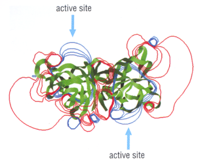
44Here we see [blank_start]superoxide[blank_end] dismutase the active sites ([blank_start]blue[blank_end]) present [blank_start]positive[blank_end] charged fields that attract the [blank_start]negatively[blank_end] charged substrate. The substrate is directed to the [blank_start]active[blank_end] site by repulsive forces generated by the [blank_start]large[blank_end] negatively charged fields presented by the rest of the protein.
- Asuperoxide
- Bperoxide
- Cmonoxide
- Dblue
- Ered
- Fgreen
- Gpositive
- Hnegative
- Inegatively
- Jpositively
- Kactive
- Lionisation
- Mlarge
- Nsmall
45Enzymes exhibit pH optima, at this pH
- Aenzymes work best
- Bthe binding and catalytic groups are correctly ionised for binding and carrying out the chemistry of catalysis.
- Call of the side chains are fully ionised
- Dthe protein becomes inert
46 Reactive groups in the active site are optimally positioned to interact with the [blank_start]substrate[blank_end]. This effectively [blank_start]increases[blank_end] the concentration of the reacting species in the confines of the active site.
Substrate is bound in the correct orientation this can add a massive factor to the [blank_start]catalytic[blank_end] efficiency. Eg. Holding two very [blank_start]reactive[blank_end] molecules next to each other in the active site in the perfect orientation for reaction may be all they need to react without any special chemistry.
Some enzymes bind substrates in a way that [blank_start]destabilises[blank_end] the ground state for the substrate, in other words a substrate may be bound in a form less stable than when in solution and this makes the passage to product [blank_start]less[blank_end] energetic.
Some enzymes stabilise the [blank_start]transition[blank_end] state or preferentially bind the transition state structure. This means that the passage through the [blank_start]highest[blank_end] energy barrier on the pathway is made easier and the reaction goes much faster.- Asubstrate
- Benzyme
- Cincreases
- Ddecreases
- Ecatalytic
- Finhibiting
- Gligand binding
- Hreactive
- Iinert
- Jdestabilises
- Kstabilised
- Lless
- Mmore
- Ntransition
- Oactivation
- Phighest
- Qlowest
47Binding substrate often displaces water from the active site and this leads to local movement of amino acid side chains. This often results in the correct alignment and orientation of the substrate relative to the catalytic residues thereby promoting catalytic events.
- ATrue
- BFalse
48Most biological transformations catalysed by enzymes can be accounted for by a relatively small number of chemical reactions. 75% of metabolic reaction driven by enzymes can be described by just six general types of chemical transformations.
- ATrue
- BFalse
49four general types of chemical transformations
[blank_start]Oxidation/reduction (redox) reactions[blank_end] involving the transfer of electrons.
[blank_start]Addition reactions[blank_end]: The addition of atoms or chemical groups to double bonds and [blank_start]Elimination reactions[blank_end] removing atoms or chemical groups to form double bonds.
The [blank_start]cleavage of esters, amides or acetals[blank_end] by reaction with water. The formation of esters, amides and acetals requires the removal of water.
[blank_start]Removal of a single carbon atom[blank_end] from a molecule by loss of carbon dioxide.- AAddition reactions
- BOxidation/reduction (redox) reactions
- CElimination reactions
- Dcleavage of esters, amides or acetals
- ERemoval of a single carbon atom
50Most active sites promote acid base chemistry using the conjugate base of acidic amino acids such as [blank_start]aspartic acid[blank_end] or [blank_start]glutamic acid[blank_end] or the conjugate acid of more basic amino acids. [blank_start]Histidine[blank_end] as mentioned previously can act as both an acid and a base and is found in the active sites of many enzymes.
- Aaspartic acid
- Baspargine
- Csulphuric acid
- Dthreonine
- Eglutamic acid
- Ftryptophan
- Gproline
- Hhydrochloric acid
- IHistidine
- JMethionine
- KArginine
- LCysteine
- MIsoleucine
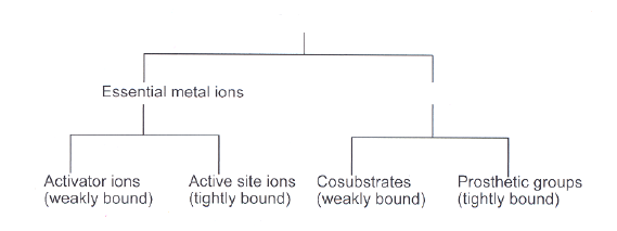
51THERE IS LIMITED CHEMICAL REACTIVITY IN THE ACTIVE SITES OF ENZYMES and many enzymes have to recruit other molecules to assist in catalysis. THESE MOLECULES ARE CALLED
- Acofactors
- Bcatalytic moleculed
- Cvitamins
- Dassistance molecules
- Ecoligands
52Cofactors can be as small as a metal ion or as a large as large a heam. Cofactors that are inorganic compounds and assist in catalytic chemistry are termed COENZYMES.
- ATrue
- BFalse
53Label the image
- ACofactors
- BCoenzymes
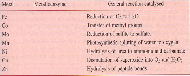
54Identify the metalloenzyme
- ACytochrome oxidase
- BVitamin B12
- CSulfite oxidase
- DWater splitting enzyme
- EUrease
- FSuperoxide dismutase
- GCarboxypeptidase A
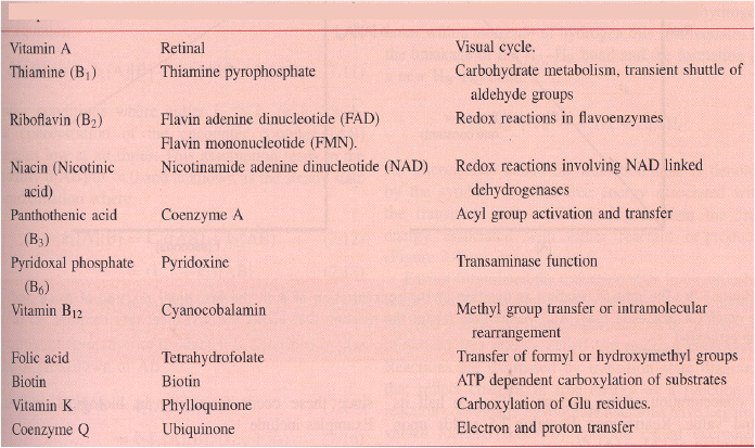
55Identify the columns
- AComponent
- BCo-factor
- CMetabolic role
56Classify the reaction
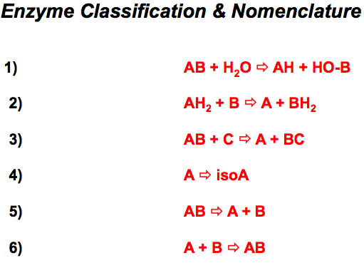
- AHydrolases
- BOxidoreductases
- CTransferases
- DIsomerases
- ELyases
- FLigases
Information
- Description
- 1st year Biochemistry and molecular biology Quiz on Protein section 4, created by MrSujg on 29/11/2015.
- Author
- MrSujg
- Language
- English
- Views
- 13
- Tags
- Rating
- No ratings yet Rate this quiz