6671312
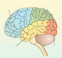
Question 1
Answer
-
frontal lobe
-
temporal lobe
-
occipital lobe
-
parietal lobe
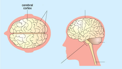
Question 2
Answer
-
cerebral cortex
-
cerebral hemispheres
-
brainstem
-
cerebellum
-
spinal cord
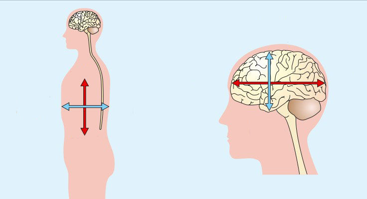
Question 3
Answer
-
rostral
-
caudal
-
ventral
-
dorsal
-
rostral
-
caudral
-
dorsal
-
ventral
-
rostral
-
caudral
-
dorsal
-
ventral
-
rostral
-
caudral
-
dorsal
-
ventral
-
rostral
-
caudral
-
dorsal (posterior)
-
ventral (anterior)
-
rostral
-
caudal
-
dorsal (posterior)
-
ventral (anterior)
-
rostral
-
caudal
-
dorsal (superior)
-
ventral (inferior)
-
rostral
-
caudal
-
dorsal (superior)
-
ventral (inferior)
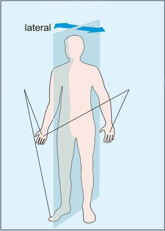
Question 4
Answer
-
medial plane
-
lateral
-
ipsilateral
-
contralateral
Question 5
Answer
-
horizontal (or axial/transverse)
-
coronal
-
sagittal
Question 6
Question
The nervous system can be divided into the [blank_start]central[blank_end] and peripheral nervous systems (CNS and PNS, respectively). The CNS consists of the [blank_start]brain and the spinal cord[blank_end] while the PNS comprises all the [blank_start]peripheral nerves[blank_end].
The brain is housed within the [blank_start]skull[blank_end] and protected by the [blank_start]meninges[blank_end] and bone. It is divided into two [blank_start]hemispheres[blank_end] and its outer structure is dominated by the [blank_start]cerebral cortices[blank_end], which can be divided into a number of [blank_start]functionally distinct[blank_end] areas.
The spinal cord consists of a [blank_start]tubular bundle[blank_end] of nerve tissue within the vertebrae which has a series of [blank_start]repeating segments[blank_end], each with associated [blank_start]spinal nerves[blank_end] and dorsal [blank_start]root ganglia[blank_end].
Answer
-
central
-
brain and the spinal cord
-
peripheral nerves
-
skull
-
meninges
-
hemispheres
-
cerebral cortices
-
functionally distinct
-
tubular bundle
-
repeating segments
-
spinal nerves
-
root ganglia
Question 7
Question
Which of these are specific to neurons?
Answer
-
plasma membrane
-
cell body
-
axon
-
dendrites
-
mitochodria
-
lysosome
Question 8
Question
Ogliodendrocyte functions:
Answer
-
Make myelin in the brain and spinal cord
-
Make myelin in the PNS
-
Structural support
-
Provide nutrients to neurons
Question 9
Question
The cells within the nervous system can be broadly classed as either n[blank_start]eurons[blank_end] or g[blank_start]lia[blank_end].
[blank_start]Neurons[blank_end] are the main signalling cells and have specialised processes, called a[blank_start]xons[blank_end] and d[blank_start]endrites[blank_end] for this purpose. They are functionally connected to one another by [blank_start]synapses[blank_end]. Signalling across synapses may be e[blank_start]lectrical[blank_end] or c[blank_start]hemical[blank_end]. The [blank_start]postsynaptic[blank_end] neuron will ultimately respond with a changed membrane potential called a [blank_start]synaptic potential[blank_end].
If the combined input of synaptic potentials is sufficient then the neuron will produce an [blank_start]action potential[blank_end] which differs in a number of ways from the synaptic potential, not least because it is [blank_start]‘all-or-nothing’[blank_end] .
Although the [blank_start]frequency[blank_end] of action potentials may vary, the speed of [blank_start]conduction[blank_end] of action potentials is constant along the length of an axon and depends on a number of factors including whether or not it is [blank_start]myelinated[blank_end]: myelinated axons conduct action potentials [blank_start]faster[blank_end] than unmyelinated axons.
[blank_start]Glia[blank_end] provide support and [blank_start]structure[blank_end] to the nervous system. They have [blank_start]‘housekeeping’[blank_end] duties, nourishing nervous tissue by regulating the supply of [blank_start]nutrients[blank_end], including oxygen, and also a [blank_start]protective[blank_end] role by mopping up debris and buffering ions in the extracellular fluid. They also provide [blank_start]myelin[blank_end]: [blank_start]oligodendrocytes[blank_end] in the CNS; [blank_start]Schwann cells[blank_end] in the PNS.
Answer
-
eurons
-
lia
-
Neurons
-
xons
-
endrites
-
synapses
-
lectrical
-
hemical
-
postsynaptic
-
synaptic potential
-
action potential
-
‘all-or-nothing’
-
frequency
-
conduction
-
myelinated
-
faster
-
Glia
-
structure
-
‘housekeeping’
-
nutrients
-
protective
-
myelin
-
oligodendrocytes
-
Schwann cells
Question 10
Answer
-
cell body
-
dendritic spines
-
dendrite
-
axon hillock
-
axon
-
axon branches
-
axon terminal
Question 11
Question
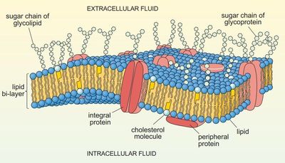
The [blank_start]neuronal membrane[blank_end], similar to the membrane surrounding other cells, is a highly dynamic structure composed of a [blank_start]phospholipid bi-layer[blank_end] in which are immersed glycolipids, [blank_start]glycoproteins[blank_end] and other protein molecules.
The neuronal membrane acts as a [blank_start]selective barrier[blank_end] separating the internal chemical environment from that outside the neuron.
Some of the [blank_start]glycolipid[blank_end], glycoprotein and protein molecules embedded in the cell membrane act as [blank_start]chemical receptors[blank_end] for [blank_start]signalling molecules[blank_end].
Answer
-
neuronal membrane
-
phospholipid bi-layer
-
glycoproteins
-
selective barrier
-
glycolipid
-
chemical receptors
-
signalling molecules
Question 12
Question
There are three main transport mechanisms across the neuronal membrane:
◦ passive [blank_start]diffusion[blank_end]
◦ transport by [blank_start]membrane proteins[blank_end]
◦ in membrane-bound [blank_start]vesicles[blank_end].
The unequal distributions of K+ and Na+ on the inside and outside of the neuronal membrane produce [blank_start]concentration[blank_end] and potential [blank_start]gradients[blank_end].
When these concentration and potential gradients are at an [blank_start]electrochemical equilibrium[blank_end], there is a potential difference (a voltage) across the neuronal membrane of about [blank_start]−70 mV[blank_end]. This equilibrium or [blank_start]resting membrane potential[blank_end] can be calculated from the Nernst equation (and by the Goldman–Hodgkin–Katz equation if membrane permeability is considered).
Answer
-
diffusion
-
membrane proteins
-
vesicles
-
concentration
-
gradients
-
electrochemical equilibrium
-
−70 mV
-
resting membrane potential
Question 13
Question
When the neuronal membrane is sufficiently [blank_start]depolarised[blank_end] to a critical threshold value, an ‘all-or-nothing’ action potential is generated at the [blank_start]axon hillock[blank_end] and transmitted along its axon to the [blank_start]axon terminals[blank_end].
An action potential has [blank_start]three[blank_end] distinct phases – depolarisation, repolarisation and [blank_start]hyperpolarisation[blank_end] – which involve the highly ordered movement of [blank_start]Na+ and K+[blank_end] across the neuronal membrane through [blank_start]voltage gated ion channels[blank_end].
Action potentials travel faster in [blank_start]myelinated axons[blank_end] and those with a larger [blank_start]axon diameter[blank_end]. Temperature may also affect conduction speed.
Answer
-
depolarised
-
axon hillock
-
axon terminals
-
three
-
hyperpolarisation
-
Na+ and K+
-
voltage gated ion channels
-
myelinated axons
-
axon diameter
Question 14
Question
The passage of a signal across an [blank_start]electrical synapse[blank_end] can be in either direction ([blank_start]bidirectional[blank_end]) and there is no [blank_start]synaptic delay[blank_end]. By contrast, a [blank_start]chemical synapse[blank_end] is [blank_start]rectified[blank_end] (goes one way only) and subject to a synaptic delay caused by the need to release the [blank_start]neurotransmitter[blank_end], have it travel across the [blank_start]synaptic cleft[blank_end] and have its affect on the [blank_start]postsynaptic neuron[blank_end].
Answer
-
electrical synapse
-
bidirectional
-
synaptic delay
-
chemical synapse
-
rectified
-
neurotransmitter
-
synaptic cleft
-
postsynaptic neuron
Question 15
Question
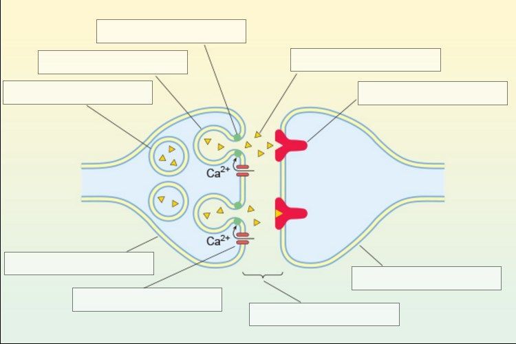
The release of [blank_start]neurotransmitter[blank_end] from a chemical synapse requires that there is first an influx of [blank_start]Ca2+ ions[blank_end] across the [blank_start]presynaptic membrane[blank_end].
After release, the neurotransmitter binds to [blank_start]receptors[blank_end] on the [blank_start]postsynaptic membrane[blank_end] and, directly or indirectly, this leads to the opening of [blank_start]channels[blank_end] so that ions can cross the membrane.
Answer
-
neurotransmitter
-
Ca2+ ions
-
presynaptic membrane
-
receptors
-
postsynaptic membrane
-
channels
Question 16
Question
Depending on the type of [blank_start]channel[blank_end] opened and the specific [blank_start]ion[blank_end] that can cross the [blank_start]postsynaptic membrane[blank_end], the membrane will be [blank_start]depolarised[blank_end] (EPSP) or [blank_start]hyperpolarised[blank_end] (IPSP). The neurotransmitter is [blank_start]actively removed[blank_end] from the [blank_start]synaptic cleft[blank_end].
Neurons [blank_start]integrate[blank_end] afferent inputs using [blank_start]spatial[blank_end] and temporal [blank_start]summation[blank_end].
Answer
-
channel
-
ion
-
postsynaptic membrane
-
depolarised
-
hyperpolarised
-
actively removed
-
synaptic cleft
-
spatial
-
summation
-
integrate
Question 17
Question
There is a great variety of [blank_start]sensory receptor cells[blank_end] but they all have specific [blank_start]receptive fields[blank_end] and are capable of generating a [blank_start]receptor potential[blank_end] which can affect the firing of [blank_start]afferent neurons[blank_end] sending sensory information to the [blank_start]brain[blank_end]. In some cases, the afferent neuron and receptor cell are the same.
Alternatively, where the receptor cell is not equipped to produce an action potential, it will [blank_start]synapse[blank_end] with an afferent [blank_start]neuron[blank_end] capable of doing so.
Answer
-
sensory receptor cells
-
receptive fields
-
receptor potential
-
afferent neurons
-
brain
-
synapse
-
neuron
Question 18
Question
Sensory signals reach the CNS via the [blank_start]spinal[blank_end] and cranial [blank_start]nerves[blank_end].
Once in the CNS, the majority of information crosses the [blank_start]midline[blank_end] to be processed on the [blank_start]contralateral[blank_end] side of the brain.
Answer
-
spinal
-
nerves
-
midline
-
contralateral
Question 19
Question
The majority of information travels to the [blank_start]cortex[blank_end] for processing via the [blank_start]thalamus[blank_end] which acts as a filter for [blank_start]sensory information[blank_end].
The architecture of the cortex includes both [blank_start]horizontal[blank_end] layers and vertical [blank_start]columns[blank_end]. These columns are functional units where incoming [blank_start]stimuli[blank_end] are integrated into information [blank_start]processing streams[blank_end] within the cortex.
There are two main types of [blank_start]neuron[blank_end] within the cortex: the [blank_start]pyramidal[blank_end] cell which projects to other areas and [blank_start]interneurons[blank_end] which remain within the cortex.
Answer
-
cortex
-
thalamus
-
sensory information
-
horizontal
-
columns
-
stimuli
-
processing streams
-
neuron
-
pyramidal
-
interneurons
Question 20
Question
Neurons can be combined in a variety of different types of [blank_start]network[blank_end] which can have implications for [blank_start]perception[blank_end], as illustrated by [blank_start]lateral inhibition[blank_end].
There are two main theories for understanding [blank_start]sensory signals[blank_end]: the [blank_start]labelled[blank_end] line theory and the [blank_start]pattern[blank_end] theory.
Answer
-
network
-
perception
-
lateral inhibition
-
sensory signals
-
labelled
-
pattern
Question 21
Question
Modern imaging techniques are classified as being structural or functional.
[blank_start]Structural[blank_end] imaging techniques (CT, MRI) are essentially static techniques that provide images of the [blank_start]physical structure[blank_end], or anatomy, of different parts of the body.
[blank_start]Functional[blank_end] brain imaging techniques aim to map sensory, motor and cognitive functions to specific regions. In functional imaging, [blank_start]spatial resolution[blank_end] relates to the smallest distance over which distinct and reliable information relating to brain activity can be established. [blank_start]Temporal resolution[blank_end] relates to the smallest timescale over which functional images can be measured.
Answer
-
Structural
-
physical structure
-
Functional
-
spatial resolution
-
Temporal resolution
Question 22
Question
There are four main functional brain imaging techniques: [blank_start]electroencephalography[blank_end] (EEG), [blank_start]magnetoencephalography[blank_end] (MEG), [blank_start]positron emission tomography[blank_end] (PET) and [blank_start]functional magnetic resonance imaging[blank_end] (fMRI).
MEG and EEG are [blank_start]direct[blank_end] techniques since they measure, respectively, [blank_start]magnetic[blank_end] fields and [blank_start]electric[blank_end] currents associated with neuronal activity.
PET and fMRI are [blank_start]indirect[blank_end] techniques since they measure local changes in the [blank_start]metabolism[blank_end] of the brain, for example those associated with increased [blank_start]blood flow[blank_end], which are taken to correlate with [blank_start]activity[blank_end] in the brain.
PET involves a participant being administered a substance that is specifically labelled with a [blank_start]radioisotope[blank_end] that decays by positron emission.
A popular fMRI technique is based on [blank_start]BOLD[blank_end]: blood oxygenation level dependent contrast.
Answer
-
electroencephalography
-
magnetoencephalography
-
positron emission tomography
-
functional magnetic resonance imaging
-
direct
-
magnetic
-
electric
-
indirect
-
metabolism
-
blood flow
-
activity
-
radioisotope
-
BOLD
Question 23
Question
Which of these are structural imaging techniques ONLY?
Answer
-
CT
-
MRI
-
fMRI
-
PET
-
EEG
Question 24
Question
Which of these are DIRECT functional imaging techniques?
Answer
-
EEG
-
MEG
-
PET
-
fMRI
Question 25
Question
Which of these are INDIRECT functional imaging techniques?
Answer
-
fMRI
-
PET
-
EEG
-
MEG
Question 26
Question
[blank_start]Spatial resolution[blank_end] in the order of a few millimetres can be achieved for MEG, PET and fMRI. In PET and fMRI, all parts of the brain are sampled with equal spatial resolution, whereas using MEG spatial resolution degrades for regions [blank_start]deeper[blank_end] inside the brain. The spatial resolution for [blank_start]EEG[blank_end] is not well defined.
MEG and EEG recordings can be taken on a [blank_start]millisecond[blank_end] timescale. For [blank_start]PET[blank_end], temporal resolution is at best in the order of 40 s, whereas for [blank_start]fMRI[blank_end] it can be reduced to the order of a second.
Answer
-
Spatial resolution
-
deeper
-
EEG
-
millisecond
-
PET
-
fMRI
Question 27
Question
Diffusion tensor imaging ([blank_start]DTI[blank_end]) is a relatively new procedure that is able to chart the probabilistic course and distribution of [blank_start]fibre bundles[blank_end] in the white matter of the living brain on the basis of the diffusion of [blank_start]water molecules[blank_end] along [blank_start]axons[blank_end].
Answer
-
DTI
-
fibre bundles
-
water molecules
-
axons
Question 28
Question
Designing suitable stimulus paradigms is an important part of functional imaging and the choice of [blank_start]stimulus[blank_end] itself can be critical. A common stimulus presentation pattern in PET and fMRI is to use regular periods of [blank_start]rest and stimulus.[blank_end] In EEG and MEG studies a single, [blank_start]random event[blank_end] strategy is often used.
Answer
-
stimulus
-
rest and stimulus.
-
random event
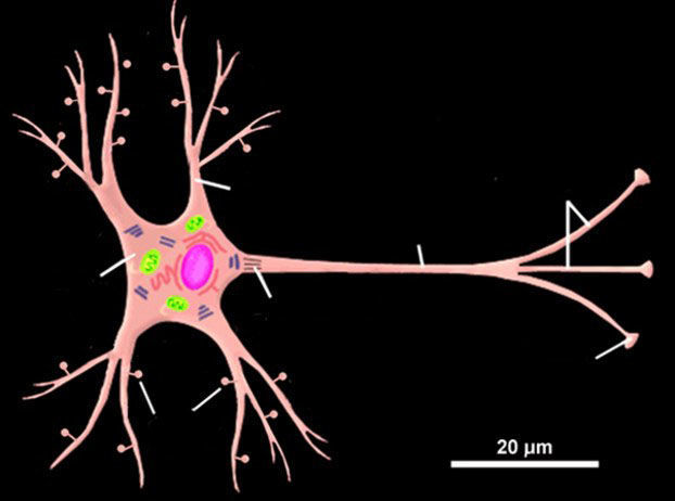
Question 29
Question
Neurons in the CNS have a central [blank_start]cell body[blank_end] containing different [blank_start]organelles[blank_end]. Most neurons have numerous [blank_start]dendrites[blank_end] which radiate outwards from the cell body and can have an abundance of [blank_start]dendritic spines[blank_end] over their surfaces. A neuron also possesses an [blank_start]axon[blank_end] which can transmit electrical signals in the form of [blank_start]action potentials[blank_end]. These signals are generated at the [blank_start]axon hillock[blank_end]. If the axon is [blank_start]myelinated[blank_end] the electrical signals travel via [blank_start]saltatory[blank_end] conduction to the axon [blank_start]terminals[blank_end] where [blank_start]synapses[blank_end] are established with other neurons.
Answer
-
cell body
-
organelles
-
dendrites
-
dendritic spines
-
axon
-
action potentials
-
axon hillock
-
myelinated
-
saltatory
-
terminals
-
synapses
Question 30
Question
Which of these organelles is directly involved protein production in neurons?
Answer
-
nucleus
-
rough endoplasmic reticulum
-
smooth endoplasmic reticulum
-
Golgi apparatus
Question 31
Question
Which of these statements about glial cells are true? Pick 3.
Answer
-
Glial cells have axons that project to different parts of the brain.
-
Glial cells do not communicate with each other.
-
Microglia are sedentary cells located around blood vessels.
-
Astroctyes play no role in the functioning of synapses.
-
In the brain, oligodendrocytes can ensheath the axons of neurons in an insulating material called myelin.
-
Astrocytes supply neurons with oxygen, glucose and other nutrients.
-
Glial cells provide a structural scaffolding for neurons - especially during development.
Question 32
Question
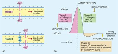
The first phase in the production of an action potential is the opening of [blank_start]voltage gated Na+ channels[blank_end] which cause [blank_start]Na+[blank_end] to enter the cell. This initiates a [blank_start]depolarisation[blank_end] of the potential difference across the cell membrane from its [blank_start]resting potential[blank_end] of [blank_start]-70mV[blank_end] towards a membrane potential of about +30mV.
During the second phase, [blank_start]voltage gated K+ channels[blank_end] open allowing [blank_start]K+[blank_end] to leave the cell. As a result there is a [blank_start]repolarisation[blank_end] of the membrane potential back towards its initial negative value.
The loss of K+ together with the [blank_start]closure[blank_end] of Na+ channels, causes the potential difference across the membrane to become more negative inside the axon than its initial polarised value - this third phase is called [blank_start]hyperpolarisation[blank_end]. The situation is reversed by the action of [blank_start]Na+/K+[blank_end] pumps which gradually return the membrane potential to its resting potential - the time when the neuron is in this state is named the [blank_start]refractory[blank_end] period.
Answer
-
voltage gated Na+ channels
-
Na+
-
depolarisation
-
resting potential
-
-70mV
-
voltage gated K+ channels
-
K+
-
repolarisation
-
closure
-
hyperpolarisation
-
Na+/K+
-
refractory
Question 33
Answer
-
synaptic vesicle
-
fused synaptic vesicle
-
complex of docking proteins
-
neurotransmitter
-
cell surface receptor
-
postsynaptic cell
-
synaptic cleft
-
presynaptic cell
-
presynaptic calcium channel
Question 34
Question
For each of the thalamic nuclei below select the appropriate type of sensory information processed from the drop down menu.
Answer
-
auditory system
-
bodily senses
-
gustatory system
-
visual system
-
auditory system
-
bodily senses
-
gustatory system
-
visual system
-
auditory system
-
bodily senses
-
gustatory system
-
visual system
-
auditory system
-
bodily senses
-
gustatory system
-
visual system
Question 35
Question
Which of the following statements about the cerebral cortex are correct? Select 3
Answer
-
The cerebral cortex can be divided up into four anatomically discrete areas.
-
There are two basic types of neuron in the cortex – pyramidal cells and ganglion cells.
-
Sensory afferents to the cortex are topographically organised.
-
Vertically oriented columns of neuronal cell bodies in the cortex form structurally and functionally isolated units.
-
Afferent input to the cortex from thalamic nuclei processing specific sensory information mainly terminates in layer IV.
-
The cortex is a highly folded laminated sheet with each layer having characteristic types of neurons and neuron densities, as well as specific input and output pathways.
Question 36
Question
This question concerns imaging techniques used to study human brain functions. You should select three choices.
Identify the correct statements below.
Answer
-
Positron Emission Tomography (PET) is a completely safe procedure which can be used repeatedly on the same individual.
-
For both Magnetic Resonance Imaging (MRI) and functional MRI (fMRI) there is a requirement to have a large static magnetic field and a radio frequency (rf) magnetic field.
-
EEG and magnetoencephalography (MEG) both have better temporal resolution than the indirect methods that can be used to image the brain.
-
Diffusion Tensor Imaging (DTI) detects the magnetic behaviour of the hydrogen nuclei in water.
-
Electroencephalography (EEG) uses squids to detect very small electric currents produced in the brain.
-
In MEG all parts of the brain are sampled with equal spatial and temporal resolutions.
Want to create your own Quizzes for free with GoConqr? Learn more.