9110539
Description
Quiz by Narda Carrion-Hernandez, updated more than 1 year ago
|
|
Created by Narda Carrion-Hernandez
over 8 years ago
|
|
Question 1
Answer
-
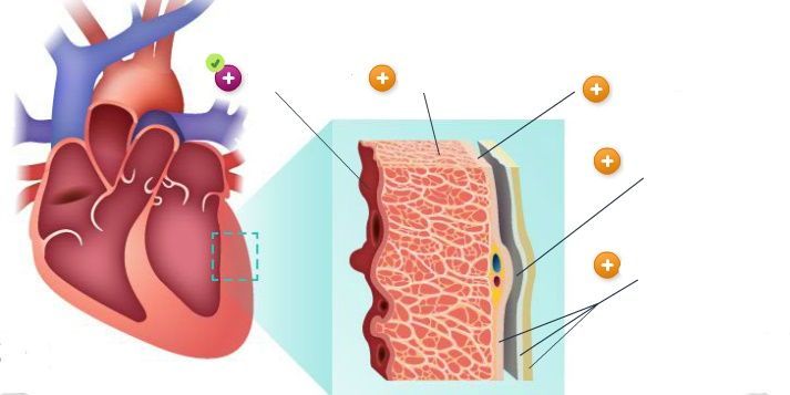
Endocardium
-
Myocardium
-
Epicardium
-
Pericardial cavity
-
Pericardium
Question 2
Question
The [blank_start]endocardium[blank_end] is the third layer, or the inner surface of the heart. It is the delicate tissue that gives rise to the cardiac valves, reduces friction, and deters clot formation. The [blank_start]myocardium[blank_end] is the thick muscle mass composed of cardiac muscle cells. The [blank_start]epicardium[blank_end] is the outer surface of the heart muscle. The [blank_start]pericardial cavity[blank_end] is between the serous layer of the pericardium and the outside of the heart. It contains 10-30 mL of serous fluid to prevent friction and protect from trauma. The [blank_start]pericardium[blank_end] is the fibrous sac surrounding the heart and the roots of the great vessels.
Answer
-
endocardium
-
myocardium
-
epicardium
-
pericardial cavity
-
pericardium
Question 3
Question
The pericardium is composed of two layers: [blank_start]fibrous external[blank_end] layer and [blank_start]smooth serous[blank_end] layer. The latter is composed of two layers, the [blank_start]parietal[blank_end] bonded to the underside of the fibrous pericardium and the [blank_start]visceral[blank_end] lays over the outside of the heart and is also known as the epicardium.
Answer
-
fibrous external
-
smooth serous
-
parietal
-
visceral
-
smooth serous
-
fibrous external
-
parietal
-
visceral
-
parietal
-
visceral
-
fibrous external
-
smooth serous
-
visceral
-
fibrous external
-
smooth serous
-
parietal
Question 4
Question
The RA is a thin walled chamber with low pressures: 2-6 mm Hg, It receives unoxygenated blood from the [blank_start]superior and inferior vena cava[blank_end] and the [blank_start]coronary sinus[blank_end]. It pumps the blood to the RV through the tricuspid valve.
Answer
-
superior and inferior vena cava
-
coronary sinus
Question 5
Question
The LA is a thin walled chamber that receives oxygenated blood from the lung via the [blank_start]pulmonary veins[blank_end]. It pumps blood to the LV through the mitral valve. Its normal pressure is 4-12 mm Hg,
Answer
-
pulmonary vein
Question 6
Question
The LV is a thick, powerful muscle that pumps the blood to the [blank_start]aorta[blank_end] through the [blank_start]aortic valve[blank_end]. The LV must push against the systemic vascular resistance which is 6 times the pressure present in the pulmonary circuit. Normal systolic pressure in LV is 100-130 mm Hg and diastolic is 4-12 mm Hg.
Answer
-
aorta
-
aortic valve
Question 7
Question
The RV is a thin walled chamber, approximately 1/3 the thickness of the LV. About 80% of the venous return from RA passively flows into RV. An additional 20-30% of atrial filling occurs during contraction referred to as the atrial kick- at the end of the diastolic filling of the ventricles. The RV delivers blood to the [blank_start]pulmonary arteries[blank_end] and [blank_start]lung[blank_end] through the [blank_start]pulmonic valve[blank_end]. Normal pressures are 15-25 mm Hg systolic and 0-8 mm Hg diastolic.
Answer
-
pulmonary arteries
-
lung
-
pulmonic valve
Question 8
Question
The [blank_start]chordae tendineae[blank_end] are strong fibrous cords that connect the AV valve leaflets to the papillary muscle. The valve leaflets are attached to a [blank_start]valve annulus[blank_end], a ring of fibrous tissue that provides support to the structure. The [blank_start]papillary muscles[blank_end] are attached to the ventricular wall and contract during systole.
Answer
-
chordae tendineae
-
valve annulus
-
papillary muscle
-
valve annulus
-
chordae tendinaea
-
papillary muscle
-
papillary muscles
-
valve annulus
-
chordae tendinaea
Question 9
Answer
-
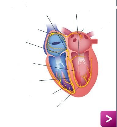
Bachmann's bundle
-
Left posterior fascicle
-
AV node
-
SA node
-
Bundle of His
-
Left bundle branch
-
Right bundle branch
-
Purkinje fibers
-
Left anterior fascicle
Question 10
Answer
-
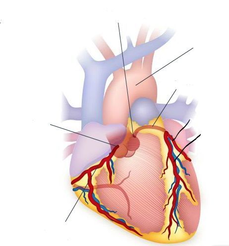
RCA
-
Right sinus of Valsalva
-
Left sinus of Valsalva
-
aorta
-
LCA
-
Circumflex artery
-
LAD
Question 11
Question
The RCA forms the posterior descending artery in 85% of people.
Answer
- True
- False
Question 12
Question
Coronary venous blood flows to the coronary sinus, a large vein on the posterior side of the heart. The coronary sinus then return the blood to the RA. [blank_start]Two thirds[blank_end] of coronary blood flow occurs during diastole.
Answer
-
Two thirds
-
One third
-
Three quarters
Question 13
Question
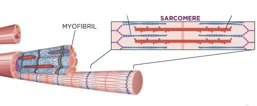
Myosin filaments have cross bridges that reach out, pulling actin in toward the center of the sacromere and causing it to shorten, producing a contraction. Around the actin fibril are interwoven protein rods of troponin and tropomyosin, which inhibit the ability of actin to connect with myosin. At the beginning of a contraction, calcium is released and attaches to troponin, allowing cross bridges on myosin to attach to actin. Relaxation of the cardiac muscle results from calcium uptake. This blocks the actin-myosin cross bridges and allows filaments to slide back to resting length.
Answer
-
actin
-
myosin
Question 14
Answer
-
sodium
-
sodium
-
sodium
-
sodium
-
sodium
-
calcium
-
potassium
-
sodium
-
calcium
-
potassium
-
sodium
-
potassium
Question 15
Question
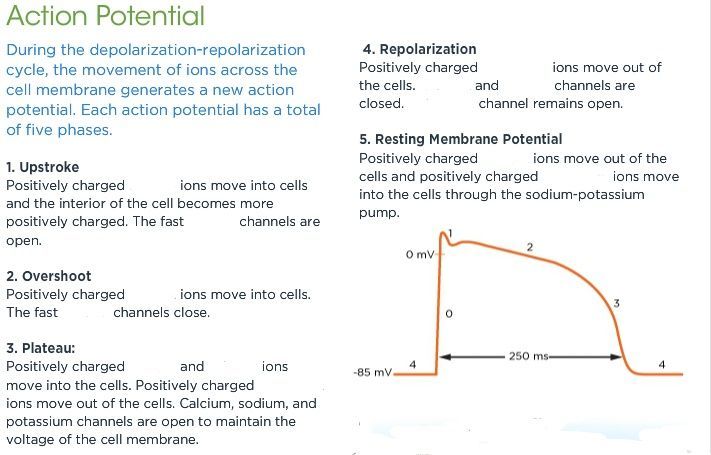
[blank_start]Absolute Refractory Period[blank_end]: The cell will not depolarize no matter how strong the impulse delivered to the cell.
[blank_start]Relative Refractory Period[blank_end]: The cell will depolarize only if it receives a strong stimulus.
[blank_start]Vulnerable Refractory Period[blank_end]: The cell responds to even a very weak stimulus.
Answer
-
Absolute Refractory Period
-
Relative Refractory Period
-
Vulnerable Refractory Period
Question 16
Question
Cardiac output is defined as the volume of blood ejected from the heart over one minute, normal range is [blank_start]4-8 L/min[blank_end]. CO is affected by HR and SV.
Cardiac index relates the CO to the pt's BSA. Normal ranges are [blank_start]2.5-4.3 L/min/m2[blank_end].
SV is the volume of blood ejected from the LV with each ventricular contraction. A healthy heart ejects more than half the total ventricular blood volume. Normal range is [blank_start]50-100 mL/beat[blank_end]. SV is affected by preload, afterload, and contractility.
Answer
-
4-8 L/min
-
2.5-4.3 L/min/m2
-
2.5-4.3 L/min/m2
-
4-8 L/min
-
50-100 mL/beat
-
4-8 L/min
-
2.5-4.3 L/min/m2
-
50-100 mL/beat
-
50-100 mL/beat
Question 17
Answer
-
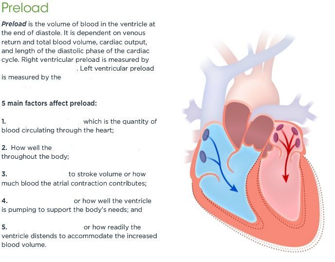
CVP
-
PAOP
-
Absolute blood volume
-
blood volume is distributed
-
Atrial contribution
-
Ventricular function
-
Ventricular compliance
Question 18
Answer
-
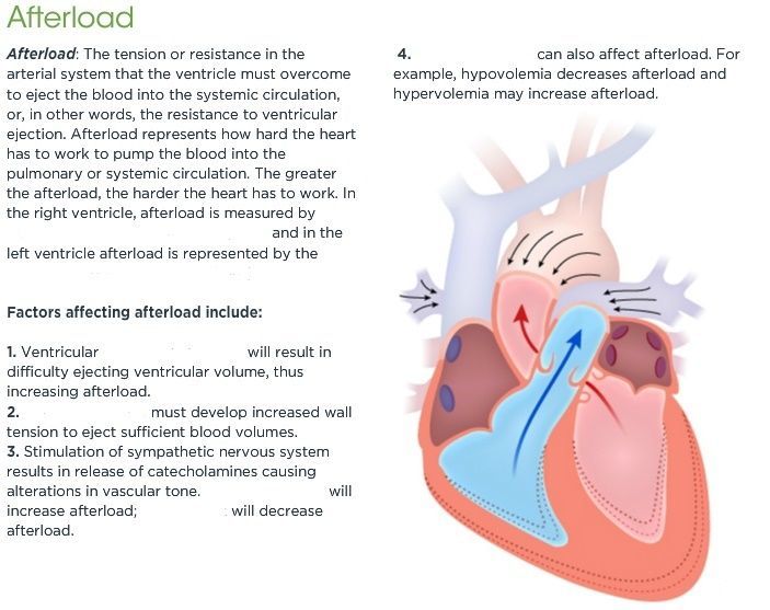
PVR
-
SVR
-
outflow obstruction
-
Dilated ventricles
-
Vasoconstriction
-
Vasodilation
-
Vascular Volume
Question 19
Question
Contractility is described using inotropy and inotropic state. [blank_start]Positive[blank_end] inotropy strengthens the contractile force of the muscle. [blank_start]Negative[blank_end] inotropy lessens the contractility of the muscle.
Answer
-
Positive
-
Negative
-
Negative
-
Positive
Question 20
Question
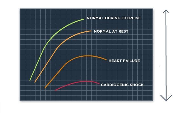
Starling's Law defines the dependent relationship between the ventricular preload, afterload, and cardiac contractility on SV. The greater the stretch or the longer the stretch exerted by EDV, the more forceful the contraction until the the muscle is overloaded. The CO proportionately increases until it peaks.
Image:
Starling Law Li (image/jpeg)
Answer
-
SV
-
EDV
-
Cardiac function
Question 21
Question
The sympathetic nervous system has the greatest effect on the ventricles and blood vessels. The sympathetic fibers are adrenergic and tend to be excitatory though the release of [blank_start]norepinephrine[blank_end]. There are three types of sympathetic receptors: [blank_start]alpha-adrenergic[blank_end], [blank_start]beta-adrenergic[blank_end], and [blank_start]dopaminergic[blank_end]. The cardiac effects tend to be increased CO as a result of increase HR and contractility.
The parasympathetic nervous system originates in the medulla and is mediated by the vagus nerve. Most of the fibers are cholinergic and secrete [blank_start]acetylcholine[blank_end], which tends to be inhibitory in its actions. The cardiac effects include slowing the HR, decreasing speed of conduction through the AV node, and slightly depressing contractility.
Answer
-
norepinephrine
-
alpha-adrenergic
-
beta-adrenergic
-
dopaminergic
-
acetylcholine
Question 22
Answer
-
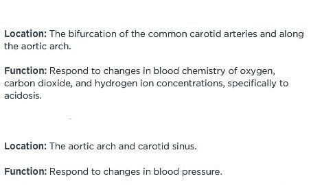
Chemoreceptors
-
Baroreceptors
Question 23
Answer
-
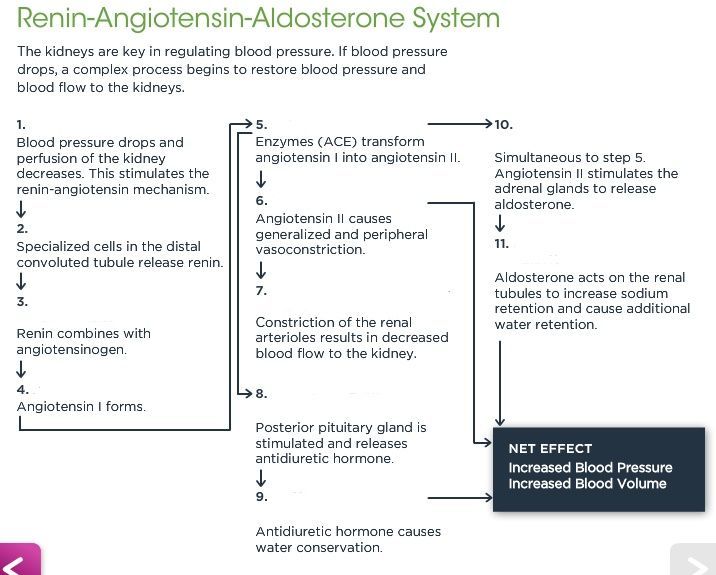
BP DROPS
-
RENIN RELEASED
-
RENIN + ANGIOTENSINOGEN
-
ANGIOTENSIN I
-
ANGIOTENSIN II
-
VASOCONSTRICTION
-
DEC BLOOD FLOW TO KIDNEY
-
RELEASE OF ADH
-
CONSERVATION OF WATER
-
RELEASE OF ALDOSTERONE
-
INC WATER RETENTION
Want to create your own Quizzes for free with GoConqr? Learn more.