
Quiz
Biology 1010 LAB Test
Dieses Quiz hat ein Zeitlimit und öffnet sich deshalb im gewohnten Player.
Fragen in diesem Quiz (55)
1Eukaryotes contain:
- AMembrane-bound nuclei and other organelles
- BNonmembrane-bound nuclei and other organelles
- CTonoplasts and cell walls
- DAmyloplasts and chromoplasts
2What are chloroplasts?
- AElliptical green structures in plant cells. They are the sit of photosynthesis and are green because of the pigment chlorophyll.
- BStructures in plants cells that are blue because of the pigment chlorophyll.
- CComplex carbohydrates used to make the cell wall in plant cells.
- DGreen membrane-bound organelles that dispel water out of the plant cells.
3How do you prepare a wet mount?
- APlace a sample in a drop of water on a microscope slide and cover it with a coverslip.
- BTake a sample of cells and soak them in distilled water and then place them on a microscope slide.
- CYou put lens oil on the microscope slide when it is on 40x power and then look at the sample at 100x power.
4Most plant cells contain a large central vacuole surrounded by a membrane called a tonoplast. As a consequence, the liquid cytoplasm containing the chloroplasts and other organelles is restricted to the edges of the cell.
- ARichtig
- BFalsch
5What is cytoplasmic streaming?
- AA slow, circular movement of chloroplasts at the edges of the cell.
- BThe expulsion of excess water out of the cell.
- CThe storage of molecules in the cell's cytoplasm.
6Chloroplasts are a type of plastid.
- ARichtig
- BFalsch
7What are plastids?
- AOrganelles in plants that are the sites of food manufacture and storage.
- BLight pigments that absorb various wavelengths of light.
- CLarge organelles that help maintain cell shape.
8What are amyloplasts?
- APlastids that store starch.
- BLight pigments that absorb light at 700 nm.
- CPlastids containing pigments other than chlorophyll.
- DCarotenes and xanthophylls.
9Chromoplasts are a type of plastid containing pigments other than chlorophyll, such as carotenes and xanthophylls.
- ARichtig
- BFalsch
10What is a cell wall? Pick the best answer.
- AThe layer that surrounds the cell membrane in plants. It is made of cellulose, a complex carbohydrate formed of glucose molecules attached end to end.
- BA semi-permeable membrane embedded with proteins that allow for the transport of molecules and other substances.
- CA characteristic of plant cells.
- DThe plasma membrane that is used to expel contents out of the cell.
11Select all the organs present in a paramecium and their correct functions.
- AAmyloplasts - plastids that store starch.
- BCilia - propulsion of the organism and sweeping of food into the oral groove.
- COral groove - a canal used to ingest nutrients that leads to a food vacuole.
- DFood vacuole - travels through the cell and the food is digested.
- EContractile vacuole - A clear and round vesicle that slowly builds in size and then fuses with the plasma membrane and contracts to expel water from the cell.
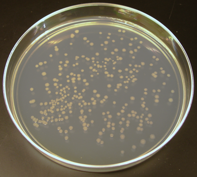
12What process is shown in the image below?
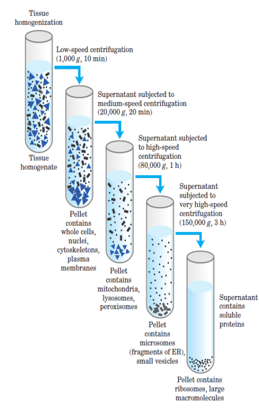
- ACell fractionation
- BInoculation of a streak plate
- CIsolation of bacterial colonies
- DGram staining
13During the first lab, you would have collected epithelial cells from the inside of your cheek and then stained them with methylene blue. Why were the cells stained with methylene blue?
You may have noticed that some of the cells may have had their edges folded over. What does this indicate about the thickness of the cells?- AThe cells were stained with methylene blue in order to be able to see them under the microscope. The folding over of the cells indicates that they are quite thin.
- BThe cells were stained with methylene blue in order to stain the nucleus of each cell. The folding over the cells indicates that they are quite thin.
- CThe cells were stained with methylene blue in order to be able to see them under the microscope. The folding over of the cells indicates that they are quite thick.
- DThe cells were stained with methylene blue to make them appear pretty. The folding of the cells does not indicate anything about their thickness.
14What is differential centrifugation?
- AA technique used by biologists to study cell parts. It is used to isolate parts of a cell creating a homogenate suspension. It uses different amount of centrifugal force to separate cell parts of different densities and sizes.
- BA technique used by biologists to study cell parts. It crushes cells by centrifugal force to expel the contents and stains the organelles based on their sizes and densities.
- CA technique used to put a cell back together after the organelles have been expelled. The centrifugal force forces the cell to create a new cell membrane around all the cell organelles.
- DA technique used by biologists to study cell parts. It is used to isolate parts of a cell creating a homogenate suspension. It separates the smallest cell organelles first and then the bigger cell parts and dissolves the cellular membrane.
15What is a homogenate?
- AA suspension of cell parts and organelles.
- BA pellet at the bottom of a centrifuge tube of large nuclei and intact cells.
- CThe supernatent formed after a round of centrifugation of the remaining organelles that were not separated.
- DAnother name for cell fractionation.
16Identify the definition of:
1) Supernatent
2) Pellet- AA suspension of cell parts and organelles.
- BA collection at the bottom of a centrifuge tube of large nuclei and intact cells.
- CThe liquid formed after a round of centrifugation of the remaining organelles that were not separated.
- DThe mixture created before a round of centrifugation.
17Cell fractionation breaks apart cells and separates the compounds. Cell organelles remain intact and biochemically active so their functions may be studied.
- ARichtig
- BFalsch
18What is the basic process of differential centrifugation?
- AA tissue sample is homogenized to make a homogenate. The tissue sample is crushed in a blender with a buffer then placed in tubes. The tubes are placed in the centrifuge for a certain time at a certain speed. When centrifugation is over, the supernatents can be placed into new tubes and centrifuged again, while the pellet at the bottom can be examined. The process may be repeated a third time.
- BA tissue sample is homogenized to make a homogenate. The tissue sample is crushed in a blender with a buffer then placed in tubes. The tubes are placed in the centrifuge for a very long time so that all the cell parts can be isolated. A sample will contain a pellet at the bottom of the tube with small cellular organelles, and the supernatent on the top will contain large cellular organelles.
- CA sample is placed into a tube and placed into the centrifuge for 5, 10, then 15 minutes. Each time it is placed in the centrifuge, it is centrifuged at a faster speed. One sample will be created that is filled with all of the isolated cell parts.
19What enzyme is an integral protein of the mitochondrial inner membrane and participates in the generation of energy via oxidative phosphorylation?
- ASuccinate dehydrogenase
- BDNA ligase
- CNADH
- DATP
20Label the parts of the microscope numbered 2,3,6,8,9,11,12,13.
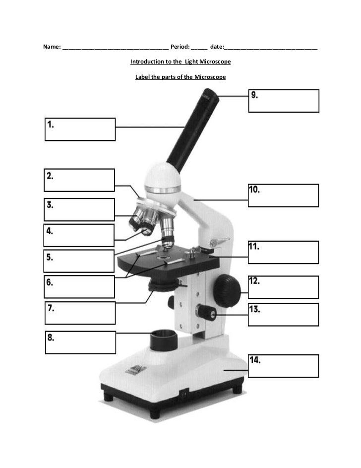
- AEyepiece
- BZoom adjustment
- CStage
- DCoarse adjustment knob
- ECoarse adjustment knob
- FFine adjustment knob
- GMicroscope body
- HEyepiece
- IFine adjustment knob
- JCoarse adjustment knob
- KArm
- LObjective lens
- MLight source
- NDiaphragm
- OBase
- PCoarse adjustment knob
- QStage clips
- RStage
- SStage
- TArm
- UStage clips
- VObjective lens
- WRevolving nosepiece
- XEyepiece
- YRevolving nosepiece
- ZObjective lens
- AAArm
21What is an enzyme assay?
- AA method for measuring enzyme activity.
- BA method used for separating cellular parts based on size.
- CA method used to identify the enzymes used in a chemical reaction.
- DA method used to catalyze a reaction and lower the activation energy.
22What is conducted during an enzyme assay?
- AThe supernatent and pellet are poured in separate tubes to be tested separately. A fixed amount of the enzyme substrate (in the case of our lab, Succinate) is added along with the appropriate indicator. A buffer is added to each solution and the tubes are heated at a temperature that is appropriate for the experiment.
- BAn enzyme substrate (in the case of our lab, Succinate) and an indicator are added to a homogenate and left to sit at room temperature until the indicator changes the colour of the homogenate.
- CA sample is centrifuged for various times and speeds with an indicator present in the sample, and after a certain about of time the sample will change colour to indicate the presence of an enzyme.
23Organelles are often studied by separating them on a sucrose gradient. A centrifuge tube is filled with a sucrose solution of increasing concentration from the top of the tube to the bottom. What would you expect to observe if you put a layer of cell homogenate on top of the sucrose and then subjected the sample to high speed centrifugation?
- AThe cells with the highest density will sink to the bottom of the solution and the organelles with the lowest density will stay at the top. The organelles stop moving in the solution when their densities and concentrations are equal to that of the solution surrounding them.
- BThe cell organelles will equally distribute throughout the solution so that each concentration layer of sucrose has an equal amount of cell constituents.
- CThe homogenate will stay at the top of the sucrose solution because it is less dense.
- DThe homogenate will sink to the bottom of the sucrose solution because it is very dense.
- EThe cells with the lowest density will sink to the bottom of the solution and the organelles with the highest density will stay at the top. The organelles stop moving in the solution when their densities and concentrations are unequal to that of the solution surrounding them.
24Janus Green is blue when in an oxidized state, and as it is reduced, it become colourless. Why would Janus Green be a useful stain to identify mitochondria?
- AMitochondria are the sites of cellular respiration, which involves the transfer of electrons. Janus Green can be used to identify mitochondria because it will be blue when the mitochondria are oxidized and will become colourless as they are reduced.
- BMitochondria are the sites of photosynthesis which involves the transfer of electrons. Janus Green can be used to identify mitochondria because it will be blue when the mitochondria are oxidized and will become colourless as they are reduced.
- CJanus Green is not useful for identifying mitochondria because is would not change colours as mitochondria do not become oxidized or reduced.
25How are prokaryotic cells different from eukaryotic cells?
- AProkaryotic DNA is not packaged into a membrane bound nucleus like eukaryotes, but rather, in a region called the nucleoid. Bacterial DNA is primarily a single, circular chromosome, whereas eukaryotes have DNA organized into linear chromosomes. Prokaryotes also lack membrane bound structures.
- BProkaryotes have membrane bound organelles and eukaryotes do not. Prokaryotic DNA is circular chromosome, whereas eukaryotes have DNA organized into linear chromosomes. Prokaryotes do not have a nucleus.
- CProkaryotic DNA is not packaged into a membrane bound nucleus like eukaryotes, but rather, in a region called the nucleoid. Bacterial DNA is primarily a single, circular chromosome, whereas eukaryotes have DNA organized into linear chromosomes. Prokaryotes also have many membrane bound structures.
- DProkaryotic cells and eukaryotic cells are exactly the same.
- EProkaryotes have membrane bound organelles and eukaryotes do not. Eukaryotes do not have a nucleus, but keep their DNA in a region called the nucleoid, and prokaryotes keep their DNA in a membrane bound nucleus with discrete, linear chromosomes.
26Peptidoglycan is a molecule composed of polymers of amino sugars and is found in bacterial cell walls.
- ARichtig
- BFalsch
27Aseptic technique involves precautionary measures that are taken to ensure personal safety in the lab is maintained. This involves the prevention of contamination of the environment from the cultures used. However, this is optional because pathogenic bacteria are never studied in the lab.
- ARichtig
- BFalsch
28What is the goal of making a streak plate?
- ATo dilute a bacterial culture enough so that single cells are separated and each cell will then give rise to a single colony. Single colonies contain only one type of bacterial cell, so streak plates allow us to separate and isolate organisms in mixed culture.
- BStreak plates allow us to mix different organisms together and their study their interactions under a microscope.
- CTo dilute bacterial culture enough so that singe cells are separated based on their size and density. Single colonies of bacteria are created and then separated based on size.
- DTo create single colonies of bacteria which will then be stained and used in differential centrifugation so that their cell parts can be isolated and studied.
29How is a streak plate prepared?
- AAn inoculation loop is flamed so that it is disinfected. A loop of culture is obtained and then streaks are made on the sterile plate, such as an agar plate. The inoculating loop is flamed again, and a second set of streaks is made, but this time only crossing over the set of initial streaks once. The loop is flamed once more, and a third set of streaks is made, this time only going over the second set of streaks once.
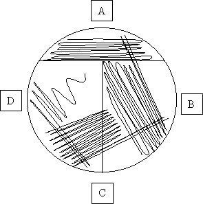
- BAn inoculation loop is flamed so that it i disinfected. A loop of culture is obtained and then streaks are made on the sterile plate, such as an agar plate. The streaks are made all over the plate, and the the inoculation loop should be used to gouge the agar.
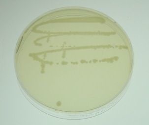
- CA single bacterial cell is obtained from a sample using a disinfected inoculation loop and is transferred to an agar plate. The bacterial cell is incubated for a period of time so that the bacterial cell can multiple and create a new colony.
- AAn inoculation loop is flamed so that it is disinfected. A loop of culture is obtained and then streaks are made on the sterile plate, such as an agar plate. The inoculating loop is flamed again, and a second set of streaks is made, but this time only crossing over the set of initial streaks once. The loop is flamed once more, and a third set of streaks is made, this time only going over the second set of streaks once.
30What is the point of using oil immersion with a light microscope?
- AIt enables you to maximize both the magnifying power and resolution of the light microscope to view very small objects.
- BIt maximizes the magnifying power and resolution of the light microscope so that you can view very small objects at lower powers such as 4x and 10x.
- COil immersion is used as a way to clean the objective lens so that magnifying power and resolution is increased.
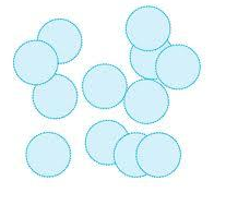
31Identify the coccus shaped bacteria.
- A
- B

- C

- A
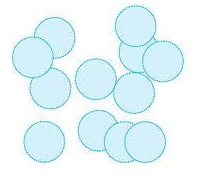
32Identify the bacillus shaped bacteria.
- A

- B

- C
- A
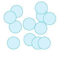
33Identify the spirillum shaped bacteria.
- A

- B
- C

- A
34Some bacteria, but not all, have flagella that help with digestion.
- ARichtig
- BFalsch
35Select the 5 characteristics of a Gram-negative bacteria.
- AMore complex cell wall.
- BHave a thin peptidoglycan layer.
- CHave an outer lipopolysaccharide wall layer.
- DDisplay safranin stain.
- EHave a red/pink cell colour after gram staining.
- FRelatively simple cell wall.
- GHave a thick peptidoglycan layer.
- HDisplay crystal violet stain.
- IDo not have an outer lipopolysaccharide wall layer.
- JHave a dark purple cell colour after gram staining.
36Identify the 5 characteristics of Gram-positive bacteria.
- ARelatively simple cell wall.
- BHave a thick peptidoglycan layer.
- CDo not have an outer lipopolysaccharide wall layer.
- DDisplay crystal violet stain.
- EHave a dark purple cell colour after gram staining.
- FMore complex cell wall.
- GHave a thin peptidoglycan layer.
- HHave an outer lipopolysaccharide wall layer.
- IDisplay safranin stain.
- JHave a red/pink cell colour after gram staining.
37What is a gram stain?
- AA differential staining technique that aids in bacterial identification. It can be used to chemically distinguish between bacterial species. Bacteria are generally divided into gram-positive and gram-negative groups.
- BA differential staining technique that aids in bacterial identification by staining the internal cell parts of the bacteria so that they can be identified. Bacteria are generally divided into gram-positive and gram-negative groups.
- CGram staining identifies the type of cell wall that bacteria by staining them with crystal violet and then uses a decolourizer such as ethanol to wash away the stain so that the cell wall details can be seen.
- DA technique used to identify different bacterial colonies by using different stains. Different types of bacteria show different colours of stains based on what their cells wall and internal cell parts are composed of.
38What is the point of heat fixation?
- AIt kills the cells and then they stick to the microscope slide.
- BIt kills the cells so that they do not move around when being viewed under a microscope slide.
- CIt makes the coverslip stick to the microscope slide so that the cells on the slide do not move around.
39What is the process of gram staining?
- AA culture is smeared on a slide to be about the shape of a dime and then heat fixed. A drop of crystal violet is added to the smear, let to dry, washed with water, and then covered with two drops of Gram's iodine solution. The slide is rinsed with water and the a decolourizer such as ethanol. A drop of safranin is added to the slide and left to dry, and the sample is then observed under a microscope.
- BA culture is smeared on a slide to be about the shape of a dime. It is stained with crystal violet and safranin the heat fixed so that the stains will identify the type of bacteria present.
- CA smear of culture is prepared on a slide, and is the mixed with crystal violet and safranin so they stick to the bacteria. Gram positive bacteria will be stained with crystal violet, and gram negative bacteria will be stained with safranin.
40Biochemical tests are used to determine information about the metabolic properties of an organism.
- ARichtig
- BFalsch
41What is a catalase test and how can it be used to identify types of bacteria?
- AA catalase test catalyzes the reaction of hydrogen peroxide to water and oxygen. Bacteria that the have the enzyme catalase will bubble when hydrogen peroxide is added to the cells because of the production of oxygen.
- BA catalase test is used to distinguish bacteria in the presence of cytochrome c oxidase. When the enzyme is present, is can oxidize a reagent to a compound that is a dark purple colour. If catalase is not present, there is not colour change.
- CA catalase test is used to to test the pH of products that are produced through fermentation. A gas, usually CO2 is produced. Phenol Red is a medium to which lactose is added and will be either red or yellow depending on the pH.
42What is an oxidase test and how can it be used to identify types of bacteria?
- AAn oxidase test is used to distinguish bacteria in the presence of cytochrome c oxidase. When the enzyme is present, is can oxidize a reagent to a compound that is a dark purple colour. If oxidase is not present, there is not colour change.
- BAn oxidase test catalyzes the reaction of hydrogen peroxide to water and oxygen. Bacteria that the have the enzyme oxidase will bubble when hydrogen peroxide is added to the cells because of the production of oxygen.
- CAn oxidase test is used to to test the pH of products that are produced through fermentation. A gas, usually CO2 is produced. Phenol Red is a medium to which lactose is added and will be either red or yellow depending on the pH.
43Disinfectants are substances that can be used on living tissue to reduce the microbial population.
- ARichtig
- BFalsch
44What is an antiseptic?
- AA substance that can be used on living tissue to reduce the microbial population.
- BA substance used to kill or inhibit microorganisms on inanimate objects.
- CAn antibiotic used to kill pathogenic microorganisms.
45What does it mean when a molecule is amphipathic?
- AIt has both hydrophilic and hydrophobic parts.
- BThe entire molecule is hydrophilic.
- CThe entire molecule is hydrophobic.
- DIt is a phospholipid.
46Osmosis is the diffusion of water across a semipermeable membrane, and is another from of passive transport.
- ARichtig
- BFalsch
47What is diffusion?
- AThe movement of a substance from an area of high concentration to an area of low concentration.
- BThe movement of a substance from an area of low concentration to an area of high concentration.
- CThe movement of charged particles through a selectively permeable membrane.
- DA form of active transport.
48Select the definitions of a:
1) Hypertonic solution
2) Hypotonic solution
3) Isotonic solution- AA solution with higher concentrations of solutes compared to the other.
- BA solution with lower concentrations of solutes compared to the other.
- CThe net concentrations of solutes are equal.
- DA solution with a high concentration.
- EA solution with a low concentration.
- FThe movement of a substance from an area of low concentration to an area of high concentration.
49What process is shown in the image?
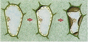
- APlasmolysis
- BOsmosis
- CDiffusion
- DActive transport
- EPassive transport
50What are enzymes and how do they affect the rate of biochemical reactions?
- AEnzymes are catalysts that speed up chemical reactions but are not consumed or changed during the reaction. They speed up chemical reactions that would be too slow to keep up with metabolic needs in organisms.
- BEnzymes are catalysts that slow up chemical reactions but are not consumed or changed during the reaction. They slow down chemical reactions that would be too fast for metabolic needs in organisms.
- CEnzymes are catalysts that speed up chemical reactions and are consumed or changed during the reaction.
- DEnzymes are catalysts that slow down chemical reactions and are consumed or changed during the reaction.
51Enzymes speed up a chemical reaction by raising the activation energy.
- ARichtig
- BFalsch
52Changes in temperature, alterations in pH, the addition of ion or molecules, and the presence of inhibitors can all affect the structure of an enzyme's active site.
- ARichtig
- BFalsch
53What is a respirometer?
- AA device for measuring respiration.
- BA device for measuring the rate of glycolysis.
- CA device used to measure the rate of a chemical reaction.
54Based on the gel electrophoresis test, who is the father of Mary's child?
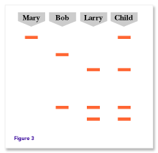
- ABob
- BLarry
- CNeither Bob or Larry are the father.
- DThere is not enough information to determine who is the father.
55What is gel electrophoresis?
- AA method used to separate and analyze macromolecules based on their charge and size.
- BA method used to amplify a single copy of few copies of DNA.
- CA method used to remove the introns and splice the exons together during transcription.
Informationen
- Beschreibung
- Biology 1010 lab practice test
- Autor
- Tiana M
- Sprache
- English
- Aufrufe
- 612
- Schlagwörter
- Bewertung
- Noch keine Bewertungen Bewerte dieses Quiz