
Karteikarten
Week 6
Karten in diesem Stapel (65)
- 1Describe the hip joint-Articulatory surfaces between head of femur and cup-shaped acetabulum of innominate bone
-Synovial joint (highly mobile)
-Multi-axial ball and socket - 2Describe the acetabulum-Where hip bones unite
-Widest and thickest superiorly
-Articulatory surface is deepened by fibrocartilaginous rim (acetabular labrum) which is defective inferioly
-Articulatory surface is horseshoe-shaped which is covered with cartilage
-Non-acticulatory surface is divided into acetabular fossa/notch (ligamentum Teres orginates here) - 3What are the 4 ligaments of the hip joint?-Iliofemoral (thickest and strongest)
-Pubofemoral
-Ischiofemoral (thinnest and weakest)
-Ligamentum Teres (weak) - 4Describe the blood supply to the hip joint-Vessels derived from the external iliac artery enter the head through a large foramina at the base of the head
-Vessels derived from the obturator artery in the Ligamentum Teres enter the head through the small foramina
-Femoral vessels through diaphysis - 5Describe the nerve supply to the hip joint-Femoral nerve (L2-L4)
-Sciatic nerve (L4-L5)
-Obturator nerve (L2-L4) - 6What are the 3 main causes of avascular necrosis of the head of the femur?1) Hip joint dislocations:
-femoral head is displaced superiorly and posterioly behind the acetabular labrum (takes 2 years for avascular necrosis to be detectable)
2) Intracapsular fracture of head of femur (common in elderly post-menopausal osteoporotic population)
3) Slipped epiphysis in some congenital abnormalities - 7What are the joints in the knee?2 condylar joints:
-medial/lateral condyles of femur and plateau of tibia
Gliding joints:
-patella
-patella surface of femur - 8Describe the condylar joints of the knee-Surface of femoral condyle is convex
-Surface of tibial condyle is flat
-To improve fit, meniscii are present inbetween - 9Describe the meniscii-C-shaped cartilagenous pieces
-Acts as a cushion
-Peripherally: thick, convex and attached to joint-capsule
-Centrally: thin, concave, form a free border - 10What are the extracapsular ligaments of the knee joint?-Ligamentum patellae
-Lateral collateral ligament
-Medial collateral ligament
-Oblique popliteal ligament - 11Describe the intracapsular/cruciate ligaments-Cross each other in an X shape
-Main bond between the femur and tibia
-Not part of the synovial cavity - 12What are common injuries of the knee joint?-Medial meniscus is more commonly damaged than the lateral
-Damaged meniscii can be surgically removed entirely or partly without disability - 13What is the important of the ankle?-Region that bears all body weight as it transfers it to the foot
-Centre of gravity of the body passes in front (making the joint unstable)
-Important for locomotion - 14Clinical important of the ankle-Arterial pulses of limb are checked
-Venous and lymph drainages change from horizontal to vertical
-Injuries occur in the form of sprains/fractures - 15Describe the direction of vessels/nerves in the ankle What keeps tendons from bow-stringing?Arteries, motor nerves and tendons enter the food turning forward
Veins, sensory nerves and lymphatics exit the foot turning upwards
Retinacula - 16Describe the articulatory surfaces of the ankle jointTibia has 2:
-superior and medial
Fibrula:
-1 lateral surface
Talus:
-3 surfaces, inferior, infero-medial and infero-lateral - 17What ate the bony prominences of the ankle joint? Importance?-Medial malleolus (distal end of tibia)
-Lateral malleolus (distal end of fibula)
Key landmarks for distal superficial venous drainage of lower limb - 18What are the 3 articulations of the tibia and fibula?Proximal: tibio-fibular articulation
Intermediate: interosseous membrane
Distal: tibio-fibulaar syndesmosis (help by 2 ligaments) - 19What type of joint is the ankle joint?Hinge variety of a synovial joint
Mortise and tenon joint:
-leg bones form a recess (mortise)
-Talus of food offers the tenon - 20What is the subtalar joint?-Articulation of the talus with the calcaneus
-Uniaxial hinge joint
-Allows inversion (30 degrees) and eversion (15 degrees) of foot - 21What does a fractured ankle involve? How does a Pott's fracture occur?Fractures of tibia, fibula or both
Results from forced excessive eversion of the foot - 22Define the spine-Multidirectional mobile column linking legs (for locomotion) with arms and head
-Supports thorax
-Contains wiring for control of arms/legs/systems (gut,renal, hepatic and genital) - 23How is bone strength increased/decreased?Increased: exercise, sunshine on skin (Vit. D), moderate alcohol
Decreased: ageing, tobacco, excess alcohol, inactivity, tablets eg. steroids - 24What type of movements do the cervical/thoracic/lumbar spine do?Cervical-circles
Thoracic- twists
Lumbar- lean forwards - 25Describe what is in each section of the spinal cord: -cord (C1-T12) -conus medullaris (T12-L1) -cauda equina (L2-Sacrum)1) Long fibre tracts- UMN
2) Lower limb anterior horn cells
3) Nerve roots- LMN - 26What are the implications of each level of neural compression? -L5 and below -L2-L5 -T1-L1/2 -C5-T1 -Above C4L5 and below: impaired sphincter and sexual function. Foot and ankle weakness
L2-L5: less likely to walk
T1-L1/2: can't walk
C5-T1: lower the injury, better the upper limb function
Above C4: can't breathe - 27Describe myelopathy-Usually cervical
-Consequence of pressure on spinal cord
-Weakness/sensory changes affects upper limb more
-Person has a shuffling gait (drags feet)
-Accompanied by upper limb radiculopathy
-Increased reflexes
-Rarely has urinary symptoms - 28Difference between radiculitis and radiculopathy?Radiculitis: root pain (sclerotomal), tension and irritation
Radiculopathy: root dysfunction - 29Describe the principal cord blood supply Which regions are particularly vulnerable?Formed of:
-anterior spinal artery
-2 posterior spinal arteries
-Segmental vessels
-Upper thoracic (T1-4)
-Thoraco-lumbar junction - 30What type of spinal deformity is bending... -backwards -forwards -left/right-Lordosis
-Kyphosis
-Scoliosis - 31Physical signs of scoliosis-Shoulder asymmetry
-Scapula prominence
-Waist asymmetry
-Hip prominence
-Visible curve
-Rib/lumbar hump becomes more prominent when bent forwards - 32What are 2 reasons for component abnormality in spinal deformity?-Failure of formation
-Failure of segmentation - 33How can neurally mediated spinal deformities be classified?UMN:
-patient is pulled over
-cerebral palsy
-cord abnormality
LMN:
-patient falls over
-viral myelitis eg. polio
-spinal muscular atropy
-trauma
-muscular dystrophies - 34Describe the femoral stretch test (FST)-Tests the femoral nerve (L2-L4)
-Patient lies prone, the knee is flexed upwards and the hip is extended
-Back pain indicates dural tension
-Leg pain indicates root tension - 35What is stenosis?Abnormal narrowing of vertebral foramen. Spinal nerves are compressed
- 36Symptoms of stenosis-Insidious onset
->50 yrs old
-Numb, aching dysaesthetic leg pain
-Brought on by standing/walking
-Relieved by sitting, leaning forward
-Back pain
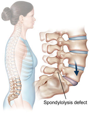
-Weakness/sphincter symptoms - 37What is spondylolysis?-Uni/bilateral break in pars interarticularis
-Occurs in L3-L5 (usually L5)
-Fatigue/stress fracture - 38What is spondylolisthesis? Different causes?Vertebral slip
Causes:
-dysplastic (congenital)
-isthmic (due to spondylolysis)
-degenerative
-traumatic
-pathological ie. cancer - 39How can degenerative spondylolisthesis cause stenosis?-Reduced diameter of vertebral foramen
-Traction and compression of roots - 40What is dissociated sensory loss in neoplasms?Weakness and alteration of pain/temperature precedes loss of fine touch and proprioception in neoplasms
- 41What is discitis?Infection of intervertebral disc space
- 42What is vertebral osteomyelitis? What is spondylodiscitis?Infection of vertebral body. Usually spread by vascular route
Combination of discitis and spondylitis (arthritis of spine. Bones fuse together) - 43What is the difference between the abdominal and pelvic cavity?-2 cavities are continuous with each other
-Lined with the same continuous sheet of parietal peritoneum
-Some viscera straddle both cavities - 44What are the ... boundaries of the abdominal cavity? -superior -anterolateral -posterior -inferiorSuperior: coastal margins and abdominal surface of abdomen
Anterolateral: anterolateral muscles of abdomen
Posterior: vertebral column and erector spinae muscles
Inferior: inguinal ligament and arcuate line - 45Describe the layers of tissue from the outside in-Skin
-Superficial fascia (binds skin to rest of tissues)
-Deep fascia (encloses organs, divides muscles into compartments). Made up of investing layer, intermediate layer
-Muscles
-Deepest layer of deep fascia - 46Describe the muscles of the body wall3 muscle layers:
-external oblique
-internal oblique
-transversus abdominis muscle
-3 muscle layers fuse together in the ventral midline to form the rectus abdominis muscle - 47Describe the external oblique muscle-Arises from 8 digitations of the lower 8 ribs at their anterior angles
-Upper 4 slips interlock with costal fibres of latissimus dorsi
-Lower 4 interlock with digitations of the serratus anterior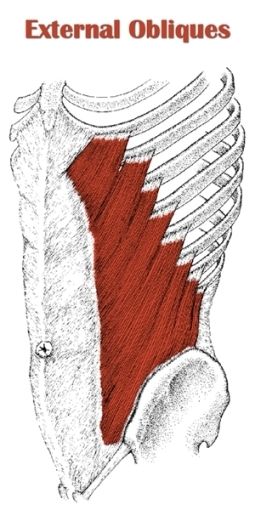
- 48Describe the internal oblique-Fibres run perpendicular to external oblique muscle
-Begin in the thoracolumbar fascia of the lower neck, anterior 2/3 of iliac crest and lateral half of the inguinal ligament
-FIbres run superiomedially
-Inserts into 10, 11, 12th rib and linea alba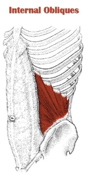
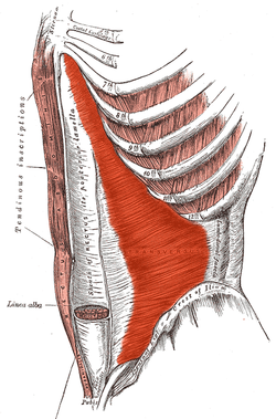
- 49Describe the transversus abdominisMuscle layer of the anterolateral wall which is deep to the internal oblique muscle
- 50Describe the rectus abdominis AKA your 'abs'-Constitutes anterior wall of the abdomen
-2 parallel muscles separated medially by the linea alba
-Separated into 'muscle bellies' by tendinous intersections
-Extends inferiorly from pubic symphysis, pubic crest, pubic tubercle
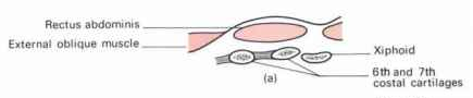
-Inserts on the xiphoid process and costal cartilages of ribs 5-7
-Outlined by linea semilunaris - 51What is the rectus sheath?-Formed by aponeuroses of external oblique, internal oblique and transversus abdominis
-Contains the rectus abdominis muscle and pyramidalis muscle
-Above the sheath is the camper's fascia and the scarpa's fascia (most superficial) - 52Describe the transverse section above the umbilicusThe anterior sheath is made up of the external oblique aponeurosis only which lies anterior to the rectus abdominis and costal cartilages
- 53Describe the transverse section at the level of the umbilicus-Sheath anterior to rectus abdominis is made up of aponeurosis of external oblique blending with anterior lamellae of internal oblique
-Sheath posterior to rectus abdominis is made up of aponeurosis of transversus abdominis blending with posterior lamellae of internal oblique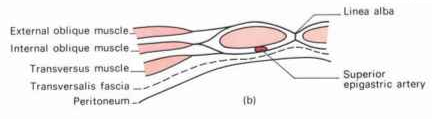
- 54Describe the transverse section below the umbilicusEntirety of the aponeuroses run in front of the rectus abdominis
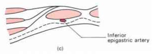
- 55Describe the inguinal canal-Oblique passage
-Lies parallel and above the inguinal ligament
-In a newborn, the deep ring lies almost directly posterior to the superficial ring
Permits passage of:
-testis to the abdomen
-round ligament of the uterus from the uterus to the labium majus
-ilioinguinal nerve - 56What are the boundaries of the inguinal canal?Anterior: aponeurosis of external oblique
Posterior: transversalis fascia
Floor: inguinal ligament
Roof: internal oblique and transversus abdominis
Medial: conjoint tendon - 57What is the clinical importance of the inguinal canal?-Superficial ring is normally palpable, but can become dilated in athletic pubalgia
-Abdominal contents may protude out resulting in a direct inguinal hernia
-Abdominal contents can also protrude through the ring into the scrotal sac which is an indirect hernia - 58What forms the deep and superficial inguinal rings?Deep:
-opening of transversalis fascia
-transmits spermatic cord/round ligament
Superficial:
-opening of the external oblique muscle - 59What is the pelvic floor?-Soft tissue complex that closes-off the inferior pelvic aperture
-Composed of muscles and pelvic fascia
-Separates minor pelvis from perineum - 60Describe the pelvic diaphragm-Sheet of muscle covered by fascia
-Closes of pelvic cavity
-Assists in support of abdominopelvic viscera
-Stretched between the pubis in front and coccyx behind
-Attached along the lateral pelvic wall to a thickened band of obturator fascia - 61What does the pelvic diaphragm consist of?-Superior layer of pelvic fascia
-2 Levator ani (anterior) and 2 coccyegus (posterior) muscles
-Inferior layer of pelvic fascia - 62What is the perineal body?-A thickened mass of connective tissue are the centre of the bones of the pelvis
-All the muscles of the pelvic floor attach here
-The anterior triangle is known as the urogenital triangle
-Posterior triangle is the anal triangle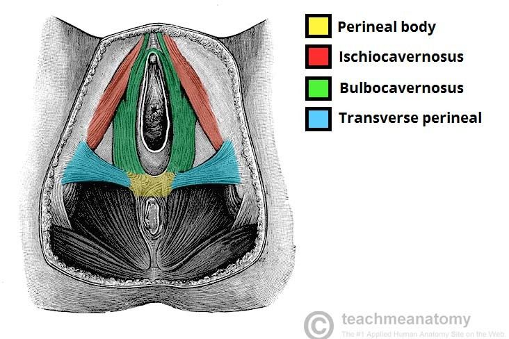
- 63What is the perineum?-External features are the external genitalia
-Serves as a support for the pelvic diaphragm and an anchor for the external genitalia
-Defines opening of urethra and vagina - 64What is a prolapse?Where organs fall down/slip out of place. Caused by weakening of tissues supporting the organ
- 65What is an umbilical hernia? Why might it develop?Fatty tissue/part of bowel pokes through an area near the umbilicus through a weak spot in the surrounding abdominal wall
Caused by: pregnancy (multiple ie. twins), obesity, persistent cough, straining when lifting heavy objects
Informationen
- Beschreibung
- Major joints of lower limb Clinical applications of spinal anatomy Muscles of pelvic floor
- Erstellt von
- Dolu Falowo · Feb 2018
- Aktualisiert
- 8 Mär 2018
- Aufrufe
- 3
- Bewertung