
Karteikarten
BMS10-1018 - Histology of muscle
Karten in diesem Stapel (29)
- 1Skeletal muscle cells are unique becauseThey are multinucleated
- 2Place the following in order of formation: Myofibril Myotube Myoblasts Satellite Cells Myofiber1. Satellite cells (mitotically quiescent)
2. Myoblasts fuse
3. Myotube
4. Myofibril
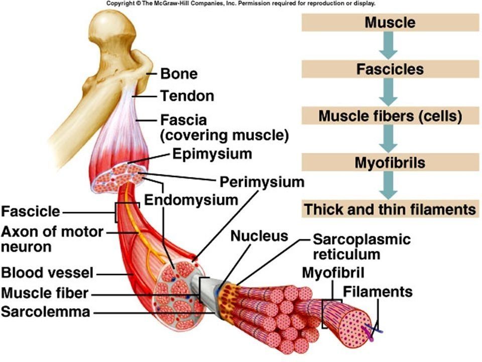
5. Myofiber - 3A muscle is made of ... wrapped together to form a ... cell which then are wrapped to form a ... which then are grouped to form muscle1.myofibrils
2. Muscle fiber
3. fascicle - 4The endomysium wraps around what? What is it made of?- Each muscle fiber cell (containing many myofibrils)
- Blood vessel, nerves, fibroblast - 5The perimysium wraps around what? What is it made of?- Each fascicle made up of many muscle fiber cells
- Connective tissue - 6The epimysium wraps around what? What is it made of?- Around skeletal muscle made of many fascicles
- Fibrous connective tissue (irregular) - 7What is the myotendinous junction?Where muscle fiber merges with dense collagenous connective tissue of the tendon
- 8What do coastameres do? Where are they located?Provide stability to the contractile apparatus
Located on top of the sacrolemma at the Z lines - 9What is a muscle fiber innervated by?Motor neuron
- 10Where do the neuron and the muscle meet? This is the location of the neuromuscular junctionMotor end plate
- 11What is a motor unit made of? What does smaller motor unit indicate?All the muscle fibers a single motor neuron innervates
Finer degree of muscle control - 12What are the four types of muscle cells?1. Skeletal muscle
2. Smooth muscle
3. Cardiac muscle
4. Myo-epithelial cells - 13Are cardiac myocytes multinucleated?Sometimes- they are uni or bi nucleated
The nuclei is centrally located - 14What is the structure of a cardiac myocyte?- Striated with actin and myosin (but shorter than skeletal muscle)
- many mitochondria - 15What joins cardiac myocytes together and allow them to function as a functional syncytium?Intercalated discs
- 16Why do cardiac myocytes have gap junctions in between them?To allow for the transmission of action potentials
- 17Can cardiac myocytes regenerate?No, no satellite cells unlike in skeletal
- 18What are the three junctions in an intercalated disc and their functions?1. Gap junction, to allow for waves of depolarisation to spread through heart
2. Adherans junction (actin-actin) to tether disc
3. Desmosome (IF-IF) between cardiac cells so they do not split during contraction - 19What is contraction of the heart caused by? What modulates it?No neurons, generated involuntarily by SA/AV node
Modulated by PNS/SNS - 20What are Purkinje fibers and what do they do?Conductive myocytes that make sure atria contracts from top-->down/ventricles contract down-->up
- 21Where is smooth muscle found?1. Gut walls
2. Blood vessels
3. Respiratory tract
4. Urinogenital tract - 22Is smooth muscle multinucleated? What is the structure?- No, single nucleus
- Cell surrounded by basal lamina
- Gap junctions to allow for group contraction and Ca2+ to pass through - 23What do pericytes do?Line small blood vessels and divide to form new SM cells
- 24Is SM striated?No, actin and myosin meshwork instead
- 25What controls SM contraction?Hormones
PNS/SNS - 26Which muscle type does not contain troponin? How does it contract instead?SM
Calcium binds to calmodulin in smooth muscle.
- It activates myosin light chain kinase which uses ATP to phosphorylate myosin-cross bridges
- ATP —> ADP + Pi
- Myosin binds to actin and forms cross bridges - 27Which muscle type cannot renew itself?Cardiac myocytes
- 28Which muscle type has no striations?Smooth Muscle
- 29Distinguish muscle types1. Skeletal
2. Smooth muscle
3. Cardiac myocytes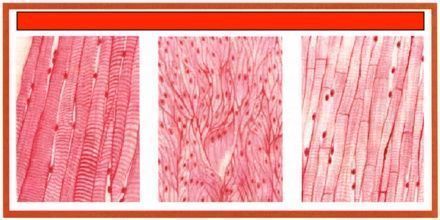
Informationen
- Beschreibung
- BMS08 - Introduction to Anatomy and Physiological Systems Karteikarten am BMS10-1018 - Histology of muscle, erstellt von Evian Chai am 18/04/2020.
- Erstellt von
- Evian Chai
- Aufrufe
- 398
- Tags
- Bewertung