
Mindmap
Heart
Inhalt der Mindmap
Heart
Anatomy
- layers
- heart chamber
- endocardium
- myocardium
- epicardium/visceral layer of serous pericardium
- pericardial cavity
- parietal layer of serous pericardium
- fibrous pericardium
- filled with a watery fluid
- parietal layer of serous pericardium
- pericardial cavity
- epicardium/visceral layer of serous pericardium
- myocardium
- endocardium
- heart chamber
- chambers
- right atrium
- superior and inferior vena cava
- right atrioventricular valve - tricuspid valve
- when valves open, they offer very little resistance to the blood flow so a very small pressure difference across the valves will be enough to produce a large blood flow
- right ventricle
- pulmonary semilunar valve
- pulmonary artery
- lungs
- the systemic circulation to the lungs is the bronchial arteries from the aorta
- lungs
- pulmonary artery
- pulmonary semilunar valve
- chordae tendinae
- papillary muscles
- ventricle wall
- cardiomyopathy and valvular disease are problems with heart mechanics but arrhymias and conducting system disease are problems with heart electricity
- coronary heart disease is a problem with heart pumping
- cardiomyopathy and valvular disease are problems with heart mechanics but arrhymias and conducting system disease are problems with heart electricity
- prevents valve prolapse
- do not open or close the valve but limit the valve's movement
- ventricle wall
- papillary muscles
- left atrium
- 4 pulmonary veins
- left atrioventricular valve - bicuspid/mitral valve
- left ventricle
- aortic semilunar valve
- aorta
- rest of body
- 3 cusps
- aorta
- aortic semilunar valve
- left ventricle
- right atrium
- interventricular septum separates left and right ventricles
- between the atria and ventricles, anteriorly, lies the atrioventricular groove/coronary sulcus
- between the ventricles lies the anterior interventricular artery
- anteriorly only the auricle of the left atrium can be seen
- posteriorly between the ventricles is where the posterior interventricular artery lies
- anteriorly only the auricle of the left atrium can be seen
- between the ventricles lies the anterior interventricular artery
- between the atria and ventricles, anteriorly, lies the atrioventricular groove/coronary sulcus
- layers
Innervation
- rich supply of sympathetic and parasympathetic fibres
- parasympathetic fibres are contained within the vagus nerve
- fibres terminate mainly on the atria and release ACh
- receptors for ACh on the heart are muscarinic
- fibres terminate mainly on the atria and release ACh
- sympathetic fibres innervate the entire heart and mainly arise from T2-T4
- release noradrenaline and cardiac muscle has B-adrenergic receptors that noradrenaline binds to
- parasympathetic fibres are contained within the vagus nerve
- rich supply of sympathetic and parasympathetic fibres
Coronary blood supply
- coronary arteries
- coronary artery flow is diastolic, diastolic flow is a window for coronary flow as coronary arteries cannot perfuse during systole, the heart is the only organ that is perfuse by diastolic pressure
- exits the heart from behind the aortic valve cusps, in the very first part of the aorta
- leads to a branching network of small vessels
- most cardiac veins drain into the coronary sinus which is a single larger vein and this empties directly into the right atrium
- there is variation in the coronary arteries e.g. posterior interventricular artery can arise from the left coronary artery
- the posterior interventricular artery supplies the majority of the left ventricle and interventricular septum, it is important to determine coronary dominance when planning interventions
- branches of the coronary arteries are described as functional end arteries
- there are very few anastomoses between coronary artery branches
- the left coronary system includes the left main stem, the left anterior descending (with its diagonals and septal) and the circumflex artery (with its obtuse marginal and left posterior descending artery)
- the right coronary artery system involves the right coronary artery with its right posterior descending artery and posterior left ventricular branch
- 3 main coronary veins
- the great coronary/cardiac vein
- anterior interventricular vein
- middle coronary/cardiac vein
- small cardiac vein
- right marginal vein
- coronary spasm is a spasm of the smooth muscles that line the coronary vessels
- a person usually does not experience symptoms until an artery 70-80% narrowed
- myocardial ischemia can be typical or atypical, there are two types (type 1 and type 2)
- ejection fraction = % of blood leaving the heart every time it contracts
- myocardial ischemia can be typical or atypical, there are two types (type 1 and type 2)
- a person usually does not experience symptoms until an artery 70-80% narrowed
- the great coronary/cardiac vein
- 3 main coronary veins
- the left anterior descending supplies 50% of the myocardium and is the largest artery of the heart
- the right coronary artery system involves the right coronary artery with its right posterior descending artery and posterior left ventricular branch
- the left coronary system includes the left main stem, the left anterior descending (with its diagonals and septal) and the circumflex artery (with its obtuse marginal and left posterior descending artery)
- there are very few anastomoses between coronary artery branches
- in 67% of people the posterior interventricular artery arises from the right coronary artery which is called right dominance
- in 15% of people the posterior interventricular artery arises from the circumflex branch of the left coronary artery which is called left dominance
- the other 18% of people have the interventricular artery arising from both right and left coronary arteries which is called codominance
- there is variation in the coronary arteries e.g. posterior interventricular artery can arise from the left coronary artery
- most cardiac veins drain into the coronary sinus which is a single larger vein and this empties directly into the right atrium
- leads to a branching network of small vessels
- right and left coronary arteries
- the left circumflex artery goes around to the back of the heart to supply the posterior left ventricle
- coronary arteries
Cellular structure
- cardiac muscle cells are striated, mononucleated cells
- contain intercalated discs which are junctions that separate each muscle cell and allows the cells to contract in unison
- cardiac muscle cells also contain T-tubules and sacroplasmic reticulum which are important for calcium ion release
- the plasma membrane of heart cells contains L-type and T-type calcium channels
- action potentials propagate along the muscle's cells membrane, calcium ions enter the cytosol and the cycling of force-generation cross-bridges is activated
- 1% of cardiac cells do not function in contraction but have specialised features essential for normal heart excitation
- conducting system of the heart
- the system is in electrical contact with the cardiac muscle cells via gap junctions
- the gap junctions interconnect myocardial cells and allows the action potentials to spread from one cell to another
- the initial excitation of one cardiac cell, therefore, eventually results in the excitation of all cardiac cells
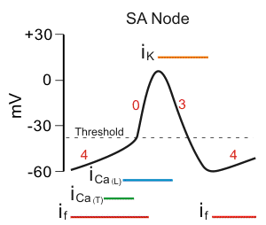
- initial depolarisation usually arises in a small group of conducting system cells called the sinoatrial node
- located in the right atrium near the entrance of the superior vena cava
- SA node is the pacemaker for the entire heart
- discharge rate of SA node determines the heart rate
- depolarisation of the SA node first spreads to the muscles of the atria
- conduction is rapid enough that the right and left atria contract essentially at the same time
- the link between atrial and ventricular depolarisation is another portion of the conducting system called the atrioventricular node
- the AV node is located at the base of the right atrium
- the AV node causes a relatively slow propagation of an action potential that goes through it and this delay produced by the AV node allows atrial contraction to be completed before ventricular excitation occurs
- after the AV node has become excited, the action potential propagates down the interventricular septum
- this pathway has conducting-system fibres called the bundle of His, or atrioventricular bundle
- the AV node and bundle of His constitute the only electrical connection between the atria and the ventricles
- except for this pathway, the atria are completely separated from the ventricles by a layer of nonconducting connective tissue
- within the IVS, the bundle of His divides into the right and left bundle branches
- the bundles separate at the apex of the heart and enter both ventricle walls
- the fibres in turn, make contact with Purkinje fibres which are large-diameter conducting cells that rapidly distribute the impulse throughout the ventricles
- Purkinje fibres are the last part of the conducting system
- depolarisation and contraction begins slightly earlier in the apex of the ventricles and then spreads upwards
- the right and left ventricles contract almost simultaneously due to the rapid conduction of the Purkinje fibres
- ventricle contraction and the process of pumping blood out of the heart is called systole
- atrial contration and the filling of the relaxed ventricles with blood is called diastole
- systole and diastole usually refers to ventricles but can refer to atria
- atrial contration and the filling of the relaxed ventricles with blood is called diastole
- electricity of the heart: membrane potential, action potential, specialised conducting system and excitation-contraction coupling
- the membrane potential is determined by the ion concentration inside the cell vs. outside the cell
- resting potential is usually -80mV, resting potential is when the electrical gradient is equal to the concentration gradient and an action potential is the sequential activation and inactivation of inward and outward currents
- a sodium and calcium inwards flow is usually depolarising while a potassium outward flow is usually repolarising
- the fibres in turn, make contact with Purkinje fibres which are large-diameter conducting cells that rapidly distribute the impulse throughout the ventricles
- the bundles separate at the apex of the heart and enter both ventricle walls
- within the IVS, the bundle of His divides into the right and left bundle branches
- except for this pathway, the atria are completely separated from the ventricles by a layer of nonconducting connective tissue
- the AV node and bundle of His constitute the only electrical connection between the atria and the ventricles
- this pathway has conducting-system fibres called the bundle of His, or atrioventricular bundle
- after the AV node has become excited, the action potential propagates down the interventricular septum
- the AV node causes a relatively slow propagation of an action potential that goes through it and this delay produced by the AV node allows atrial contraction to be completed before ventricular excitation occurs
- the AV node is located at the base of the right atrium
- the link between atrial and ventricular depolarisation is another portion of the conducting system called the atrioventricular node
- conduction is rapid enough that the right and left atria contract essentially at the same time
- depolarisation of the SA node first spreads to the muscles of the atria
- located in the right atrium near the entrance of the superior vena cava
- the gap junctions interconnect myocardial cells and allows the action potentials to spread from one cell to another
- the system is in electrical contact with the cardiac muscle cells via gap junctions
- conducting system of the heart
- 1% of cardiac cells do not function in contraction but have specialised features essential for normal heart excitation
- action potentials propagate along the muscle's cells membrane, calcium ions enter the cytosol and the cycling of force-generation cross-bridges is activated
- the plasma membrane of heart cells contains L-type and T-type calcium channels
- cardiac muscle cells also contain T-tubules and sacroplasmic reticulum which are important for calcium ion release
- the heart has a very limited ability to replace its muscle cells as it has an enormous workload and beats usually once every second
- 1% of all muscle cells are replaced each year
- contain intercalated discs which are junctions that separate each muscle cell and allows the cells to contract in unison
- cardiac muscle cells are striated, mononucleated cells
A dual pump
Cardiovascular tests
- purpose of cardiovascular imaging: - define the structure of the heart, normal and abnormal - valve function - ventricular function - coronary physiology - image of the heart during stress
- electrocardiogram
- when systemically approaching an ECG always ask for clinical context, check the date and time and patent details, make sure to assess the technical quality, like artefact, paper speed (should be 25mm/s) and gain (should be 10mV/mm)
- there are 10 leads in a 12 lead ECG because the 12 leads produce 10 vectors, a lead is an electrical vector not the wire attached to the patient
- 6 chest leads
- V4 - cardiac apex
- V1 - right, 4th intercostal space
- V2 - left, 4th intercostal space
- V3 - between V2 and V4
- V5 - anterior axillary line at the same level as V4
- V6 - mid-axillary line at the same horizontal level as V4 and V5
- unipolar leads
- measures the potential variation at a single point
- limb leads
- lead I
- records the difference between the left arm and the right arm
- RA-LA
- bipolar leads that form Einthoven's triangle
- bipolar leads measure the potential difference between two points
- records the difference between the left arm and the right arm
- lead II
- records the difference between the left foot and the right arm
- RA-LL
- records the difference between the left foot and the right arm
- lead III
- records the difference between the left foot and the left arm
- LA-LL
- records the difference between the left foot and the left arm
- AVR
- records the potential difference between the right arm and zero
- augmented limb leads
- records the potential difference between the right arm and zero
- AVL
- records the potential difference between the left arm and zero
- AVF
- unipolar leads that collect information to a central point which is said to have zero voltage
- records the potential difference between the left foot and zero
- the limb leads can be placed anywhere on the limbs but if the leads are put proximally on the arm then they have to be put proximal on the legs
- lead I
- the chest and limb leads look at the heart through two perpendicular sets of vectors
- ECG electrodes are usual put at the front of the chest
- the heart is closer to the front of the chest
- 6 chest leads
- P wave represents atrial depolarisation
- Q wave represents conduction through the Purkinje fibres
- R wave represents the electrical stimulus as it passes through the main portion of the ventricular walls
- S wave represents ventricular depolarisation
- T wave represents ventricular repolarisation
- if an ECG wave is inverted positively form the baseline then the electrical current is moving towards the electrode
- an ECG is a biological potential made up of a composite of all the action potentials in the heart
- if you have a positive QRS complex then you should have a positive T wave in the ECG for a particular lead and vice versa with negative
- heart rate can be measured by looking at 2 consecutive QRS complexes, to determine heart rate do 300 divided by the number of large squares between each QRS complex
- 1 square = 300/min 2 squares = 150/min 3 squares = 100/min 4 squares = 75/min 5 squares = 60/min 6 squares = 50/min
- another way to determine HR is to times the number of QRS complexes across the 10 second ECG by 6
- ECG normal ranges
- PR interval
- < 1 large square = < 200ms
- QRS complex
- < 3 small squares = < 120ms
- QT interval
- < 11 small squares = < 440ms
- PR interval
- the QRS axis is the direction of average polarisation in the heart which is dominated by left ventricular depolarisation, this is determined by the limb leads
- the angle of the QRS axis is determined relative to lead I, the angle is normally between -30 and +90 degrees and the axis is approximated by finding the lead with the most positive QRS
- depolarisation near limb lead II will result in a positive wave as the flow of depolarisation is moving towards the electrode
- the waves produced in leads I and II have a normal axis if the QRS is positive
- left axis deviation would be from -30 to -90 degrees and this is an abnormal axis, there will be a positive QRS in lead I but a negative QRS in lead II and aVF
- the most common cause of a right axis deviation is due to the heart moving to a more central position (could be due to a smoking related drug disease), a right axis deviation is from +90 to +180 degrees, there would be a negative QRS in lead I and a positive QRS in the aVF lead
- extreme axis deviation is from +180 to -90 degrees, it produces a negative QRS complex in leads I and II and a positive QRS complex in the aVR lead
- in a normal ECG, the P wave is upright in the inferior leads, inferior leads with a negative P wave inversion show that the heart is not in sinus rhythm
- 2 lateral lead sections, an inferior lead section and an anterior/septal section
- P waves are usually referred to as positive, negative or biphasic
- ST segments are usually referred to as isoelectric, elevated or depressed
- T waves are usually referred to as upright, inverted or flat, they can also be referred to as concordant or discordant to the QRS complex
- nomenclature of QRS
- small deflections are given lower case letters
- large deflections are given upper case letters
- the first positive wave is r or R
- any second wave is r' or R'
- a negative wave that follows R or r is s or S
- a negative wave preceding r or R is q or Q
- an entirely negative QRS complex is referred to as qs or QS
- nomenclature of QRS
- T waves are usually referred to as upright, inverted or flat, they can also be referred to as concordant or discordant to the QRS complex
- ST segments are usually referred to as isoelectric, elevated or depressed
- in a normal ECG, the P wave is upright in the inferior leads, inferior leads with a negative P wave inversion show that the heart is not in sinus rhythm
- extreme axis deviation is from +180 to -90 degrees, it produces a negative QRS complex in leads I and II and a positive QRS complex in the aVR lead
- the most common cause of a right axis deviation is due to the heart moving to a more central position (could be due to a smoking related drug disease), a right axis deviation is from +90 to +180 degrees, there would be a negative QRS in lead I and a positive QRS in the aVF lead
- left axis deviation would be from -30 to -90 degrees and this is an abnormal axis, there will be a positive QRS in lead I but a negative QRS in lead II and aVF
- the angle of the QRS axis is determined relative to lead I, the angle is normally between -30 and +90 degrees and the axis is approximated by finding the lead with the most positive QRS
- ECG normal ranges
- heart rate can be measured by looking at 2 consecutive QRS complexes, to determine heart rate do 300 divided by the number of large squares between each QRS complex
- if you have a positive QRS complex then you should have a positive T wave in the ECG for a particular lead and vice versa with negative
- an ECG is a biological potential made up of a composite of all the action potentials in the heart
- if an ECG wave is negative from the baseline then the electrical current is moving away from the electrode
- if an ECG wave has both positive and negative inversions from the baseline then the electrical current is moving parallel to the electrode
- if an ECG wave is inverted positively form the baseline then the electrical current is moving towards the electrode
- end of S wave and before T wave (ST segment) represents a plateau phase of repolarisation
- T wave represents ventricular repolarisation
- S wave represents ventricular depolarisation
- R wave represents the electrical stimulus as it passes through the main portion of the ventricular walls
- from end of P wave to beginning of Q wave represents the delay at the AV node
- after the P wave and the half just before the Q wave represents conduction through the bundle branches
- a bundle branch block is a block of either the left bundle branch or the right bundle branch in the conducting system, for any bundle branch block there should be a prolonged QRS complex, on ECG, if QRS is narrowed then there is no need to worry about bundle branch block
- left bundle branch block: QRSd is > 120ms, septum depolarises from right to left, right ventricle depolarises early, V1 = positive notch and V6 = negative notch, left ventricle depolarises late, V1 = Q/S wave and V6 = dominant R wave
- right bundle branch block: QRSd > 120ms, septum depolarises from left to right, left ventricle depolarises early, V1 = S wave and V6 = dominant R wave, right ventricle depolarises late, V1 = R' and V6 = s
- it is helpful to think of V1 as the right ventricular electrode and V6 as the left ventricular electrode, if a patient develops bundle branch block it will not go away so it will always be present on an ECG
- after the P wave and the half just before the Q wave represents conduction through the bundle branches
- Q wave represents conduction through the Purkinje fibres
- an ECG gives a visual representation of the electrical activity of the heart, ECGs can show abnormalities of rhythm, conduction and repolarisation
- non-invasive as it records electrical activity from the skin of the chest wall
- basic cardiovascular imaging includes ECG and CXR
- a chest X-ray will give a cardiac silhouette and can also show pulmonary vasculature, great vessels, pulmonary oedema and pleural effusions
- advanced cardiovascular imaging includes ultrasound (trans thoracic echocardiography and transoesophageal echocardiography), ionising radiation (nuclear, CT and invasive angiography) and MRI scans
- echo doppler is a procedure that uses doppler ultrasonography to examine the heart
- ejection fraction = % of blood leaving the heart every time it contracts and it is one of the most powerful predictors of prognosis
- echocardiography is used to assess the structure and function of the heart, valves, the pericardium and incurable ischaemia (stress)
- pros: - cheap - available - portable - no radiation
- cons: - requires good acoustic window - user dependant
- exercise, nuclear and echo stress testing are functional stress tests
- nuclear perfusion imaging is used to assess ischaemia and ejection fraction
- pros: - availability cons: - radiation - no structural assessment
- cardiac CTs are 64-slice scanners with high temporal and spacial resolution, they are used to look at coronary artery anatomy, great vessel anatomy and the levels of atherosclerosis or coronary calcification
- pros: - good rule out for CAD - low risk
- cons: - radiation dose - requires low heart rate - no fuctional assessment of ischaemia
- coronary angiography is the gold standard test
- percutaneous coronary intervention
- invasive angiography is used to assess ischaemia, primary PCI (inflating a ballon and leaving a stent in place), valve assessment and ventricular pressure assessment (right and left)
- pros: - gold standar test - option for intervention during procedure - availability cons: - radiation - risks
- MRI is the gold standard test for testing ejection fraction (CMR), gadolinium is the contrast agent usually used in the cardiac MRIs, CMRs are used to assess structure and function, perfusion/stress, the great vessels and tissue characterisation (infiltrative cardiomyopathies and previous infarction)
- pros: - gols standard LV assessment - reproducible - no radiation cons: - cost - availability - clostrophobia - pacemakers
Angina
- angina pectoris is a crushing pain or discomfort felt in the anterior chest, commonly radiating to the left arm and the jaw
- cardiovascular chest pain can be due to: - ischemia - aortic dissection - pulmonary embolism - aortic embolism - pericarditis - myocarditis - takotsubo cardiomyopathy noncardiovascular chest pain can be due to: - pneumonia - pneumothorax - cancer - musculoskeletal -oesophagitis/reflux disease - oesophageal rupture/mediastinitis - gallstones - neuralgia - psychogenic
- angina itself is not a disease, it is just chest pain
- the pain is caused by coronary artery insufficiency leading to intermittent myocardial ischemia
- build-up of metabolites, like adenosine, stimulates sensory nerves and causes referred pain
- referred pain is a phenomenon whereby visceral pain is not felt in the organ but in the dermatomes related to the origin of its efferent (sympathetic) innervation
- internal organs including the heart, lack nociceptors which means that the internal organs are insensitive to touch, cutting and temperature, pain form internal organs is instead elicited by stretching or chemical changes e.g. ischemia
- pain sensation is transmitted by visceral afferent nerves to the spinal cord and then to the brain, the sensory fibres in the heart carry visceral information to the brain through the sympathetic nervous system, the signal ascends through white matter to the thalamus and then to the cerebral cortex, the spinal cord, however, it also receives and transmits somatic sensory information from the body wall and the brain can confuse these signals by assuming that the pain from the internal organs is actually from the area of skin supplied by the same level of the spinal cord (dermatome)
- pain from the heart during ischarmia is not actually experienced in the heart, instead the brain interprets the pain as coming from the skin supplied by the same levels of the spinal cord, T1-T4
- visceral afferents travel to the spinal cord alongside the sympathetic efferent fibres, there is a theory that the somatic and visceral afferents converge on the same ascending pathway
- visceral referred pain is dull and poorly localised whereas somatic pain is sharp and well localised
- referred pain of the liver, gallbladder and duodenum is so high because the phrenic nerve originates from C3,4,5
- build-up of metabolites, like adenosine, stimulates sensory nerves and causes referred pain
- there are 3 types of angina
- stable angina
- stable angina algorithm: 1 - short acting nitrovasodilator (GTN) + beta blocker or calcium channel blocker + drugs for secondary prevention (anti platelet, lipid lowering, antihypertensive etc) 2 - combine beta blocker and calcium channel blocker 3 - consider adding a long acting nitrovasodilator or Ivabradine 4 - consider surgical intervention, stoning or CABG (coronary artery bypass grafting)
- most common
- angina attacks are predictable (e.g. occurs when exercising or stressed)
- myocardial oxygen demand is not met
- involvement of chronic occlusive coronary artery disease
- myocardial oxygen demand is not met
- angina attacks are predictable (e.g. occurs when exercising or stressed)
- unstable angina
- angina attacks are unpredictable
- coronary artery occlusion is due to platelet adhesion to ruptured atherosclerotic plaque
- an acute coronary syndrome that is defined by the absence of biochemical evidence of myocardial damage
- coronary artery occlusion is due to platelet adhesion to ruptured atherosclerotic plaque
- angina attacks are unpredictable
- variant angina
- least common
- angina attacks are unpredictalbe
- coronary occlusion is due to vasospasm
- the dilation of coronary arteries in this type of angina may be useful but it is dangerous in stable and unstable angina as it may cause coronary steal which is where dilation sends more blood to already well perfused areas (reverse Robin Hood effect)
- coronary steal can cause a problem with dipyridamole which is a vasodilator drug that can be used to treat angina
- the dilation of coronary arteries in this type of angina may be useful but it is dangerous in stable and unstable angina as it may cause coronary steal which is where dilation sends more blood to already well perfused areas (reverse Robin Hood effect)
- coronary occlusion is due to vasospasm
- angina attacks are unpredictalbe
- least common
- stable angina
- risk factors: smoking, hypertension, history, diabetes, hyperlipidaemia and obesity
- symptoms: chest pain, dyspnoea and fatigue
- chest pain - tight, crushing, band-like pain across the centre of the chest and it may radiate to the left arm, jaw or throat, relieving factors for the chest pain includes rest ad nitrates
- investigations
- other tests include coronary angiography and cardiac magnetic resonance
- blood tests and the use of biochemical markers makes it possible to identify much smaller areas of heart tissue death, the main biochemical marker used for this is called troponin, levels of troponin can become elevated in the blood within 3 or 4 hours of heart injury and may remain elevated within 10 to 14 days, a high level of troponin may indicate some degree of damage to the heart, in people with angina, an elevated level of troponin may indicate that their condition is worsening
- the biggest chance of diagnosing a heart attack is with the use of troponin, the peak troponin is at around 12 hours and if a highly sensitive troponin is used and if the levels are not elevated after 6 hours then a myocardial infarction can be ruled out
- electrocardiogram
- the ST segment is a key area that can indicate myocardial ischemia
- slight ST segment elevation is normal in both men and women, especially in V2 and V3 chest leads
- in those with angina they will usually have a normal ECG at rest but exercise can provoke ECG changes like an ST segment depression
- in those with no significant ST elevation but have the associated symptoms, it suggests that they have subendocardial ischemia
- in those with ST elevations on their ECG with associated symptoms, it suggests the patient has transmural ischemia (total occlusion in a coronary artery)
- elevated troponins
- STEMI
- ST-elevated MI due to total occlusion of a coronary artery
- STEMI
- normal troponins
- unstable angina (aborted MI)
- mechanical damage after an acute MI can cause a free wall rupture, a ruptured septum or papillary muscle rupture as well as ischemia, pericarditis, thrombus, arrhythmia and aneurysm
- heart failure is either cariogenic shock (acute heart failure) or prolonged heart failure
- syncope is a loss of consciousness and in the vast majority of patients, this is due to blood pressure
- cardiac
- arrhythmia, valvular disease, outflow tract obstruction
- haemodynaminc
- postural hypotension, vasovagal syncope, autonomic dysfuction
- neurological
- seizures
- breathlessness
- cardiac
- angina equivalent, heart failure, valvular disease
- respiratory
- anaemia
- deconditioning
- palpitation
- sinus rhythm
- inappropriate sinus tachycardia, postural orthostatic tachycardia syndrome
- ectopics
- SVEs, VE, bigeminy and trigemini
- true arrhythmia
- atrial fibrillation, atrial flutter, SVT, ventricular tachycardia
- sudden cardiac death
- myocardial infarction
- arrythmia
- cardiomyopathies
- dilated cardiomyopathy, hypertrophic cardiomyopathy, arrythmiogenic right ventricular cardiomyopathy
- pulmonary embolism
- pleuritic chest pain and breathlessness are symptoms of pulmonary embolisms
- aortic dissection
- sinus rhythm
- cardiac
- cardiac
- syncope is a loss of consciousness and in the vast majority of patients, this is due to blood pressure
- heart failure is either cariogenic shock (acute heart failure) or prolonged heart failure
- elevated troponins
- elevated troponins
- NSTEMI
- non-ST elevated MI due to partial coronary block
- NSTEMI
- normal troponins
- unstable angina
- in those with ST elevations on their ECG with associated symptoms, it suggests the patient has transmural ischemia (total occlusion in a coronary artery)
- in those with no significant ST elevation but have the associated symptoms, it suggests that they have subendocardial ischemia
- the ST segment is a key area that can indicate myocardial ischemia
- exercise ECG
- myocardial perfusion imaging
- stress echocardiography
- dobutamine
- multi-slice CT or CT angiography
- angiographs can be used to show occlusions in a major coronary artery
- treatment
- management of risk factors e.g. smoking cessation, weight loss, tightly controlled blood glucose levels for patients with diabetes etc
- if someone is on two anti-anginal drugs and the desired effect is still not being obtained them an angiogram along with a 3rd drug should be considered
- aspirin
- acts to reduce platelet aggregation (atherosclerotic plaques)
- beta-blockers
- has negative inotropic (contraction of heart muscle) and chronotropic (heart rate) effects
- reduced myocardial oxygen demand
- also antihypertensive drugs
- reduced myocardial oxygen demand
- has negative inotropic (contraction of heart muscle) and chronotropic (heart rate) effects
- calcium channel antagonists
- e.g. nifedipine, diltiazem and verapamil (also amlodipine)
- slowing of calcium influx into the myocardial cells causing a negative inotropic response
- can also cause vasodilation in peripheral arteries
- blockage of calcium channels in the AV node will increase the refractory period which slows the heart rate
- verapamil, nisoldipine, diltiazem, amlodipine, flodipine, nicardipine and nifedipine
- L-type calcium channel blockers like verapamil and diltiazem work like a cork in a bottle whereas nifedipine works by allosteric modulation (binds to the allosteric site and reduced the channel opening)
- smooth muscle
- nifedipine > diltiazem > verapamil
- cardiac muscle
- verapamil > diltiazem > nifedipine
- dilate arteries but have little effects on veins
- useful in all forms of angina but may cause coronary steal in stable and unstable angina
- may also cause headaches and constipation
- useful in all forms of angina but may cause coronary steal in stable and unstable angina
- smooth muscle
- L-type calcium channel blockers like verapamil and diltiazem work like a cork in a bottle whereas nifedipine works by allosteric modulation (binds to the allosteric site and reduced the channel opening)
- verapamil, nisoldipine, diltiazem, amlodipine, flodipine, nicardipine and nifedipine
- blockage of calcium channels in the AV node will increase the refractory period which slows the heart rate
- can also cause vasodilation in peripheral arteries
- nitrates
- acts by converting into nitric oxide to dilate the vessel
- can be administered sublingually (glyceryl trinitrate or isosorbide dinitrate), transdermally as a patch or cream or orally as a pill
- GTN, isosorbide dinitrate and isosorbide mononitrate are all nitrates
- GTN is taken sublingually but amyl nitrate is inhaled
- both drugs are rapid on onset but their action is short lived, they are used as prophylaxis in stable angina and provide rapid relief of ongoing anginal attacks
- all nitrovasodilators are prodrugs, they are lipophilic and readily enter smooth muscle cells when reduced to NO which is why they are called NO donors
- prodrugs are drugs that have to be broken down, digested, before they can become active
- NO activates soluble guanulate cyclase (SGC) which is a cytoplasmic soluble enzyme with receptors that contain a iron haem moiety where NO binds to which causes enzyme activation, resulting in the conversion of GTP to cGMP and the increase in cGMP causes vasodilation
- cGMP = cyclic guanine monophosphate
- nitrovasodilators cause more venous dilation than arterial and side effects include: headaches and tolerance developed on prolonged use so there needs to be drug free washout periods to restore drug efficacy
- isosirbides are taken orally and have a slower onset but are more prolonged in duration than GTN
- they are used for prophylaxis (treatment given or action taken to prevent disease) in all forms of angina
- GTN is taken sublingually but amyl nitrate is inhaled
- GTN, isosorbide dinitrate and isosorbide mononitrate are all nitrates
- can be administered sublingually (glyceryl trinitrate or isosorbide dinitrate), transdermally as a patch or cream or orally as a pill
- acts by converting into nitric oxide to dilate the vessel
- potassium channel openers
- e.g. nicoradanil
- acts to precondition the heart against ischemia
- e.g. nicoradanil
- ivabradine has recently been introduced to treat all forms of angina, it blocks If (sodium) funny currents which contribute to SA node depolarisation
- management of risk factors e.g. smoking cessation, weight loss, tightly controlled blood glucose levels for patients with diabetes etc
- angina pectoris is a crushing pain or discomfort felt in the anterior chest, commonly radiating to the left arm and the jaw
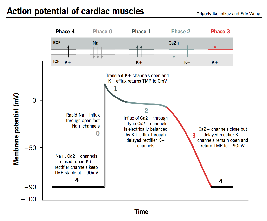
Action potential
- upstroke - phase 0: opening of voltage gated sodium channels allows the inwards flow of sodium and causes depolarisation
- notch - phase 1: activation of outward potassium flow causes repolarisation
- plateau - phase 2: balance of inward calcium and outward potassium flow so that depolarisation = repolarisation
- repolarisation - phase 3: inward currents are inactivated and outward potassium flow predominates which causes repolarisation
- resting - phase 4: resting potential is reached
- repolarisation - phase 3: inward currents are inactivated and outward potassium flow predominates which causes repolarisation
- plateau - phase 2: balance of inward calcium and outward potassium flow so that depolarisation = repolarisation
- notch - phase 1: activation of outward potassium flow causes repolarisation
- upstroke - phase 0: opening of voltage gated sodium channels allows the inwards flow of sodium and causes depolarisation
Heart sounds
- heart sounds can visually be seen through a phonocardiogram
- heart sounds are made at the peak of the R and T waves in an electrocardiogram
- when the atrioventricular valves are closed due to ventricular contraction but the semilunar valves are not open, this state is called isovolemic contraction
- isovolumetric relaxation - as ventricles relax, pressure in the ventricles drop and blood flows back against the cusps of the semilunar valves and forced them closed
- Frank-Starling mechanism
- the contractile strength of the heart depends on what degree the heart is stretched, the more blood put into the heart, the more blood will be pumped out
- we have 2 heart sounds
- S1
- caused by closing of the mitral and tricuspid valves
- S2
- caused by the closing of the aortic and pulmonic valves
- between S1 and S2 is systole and between S2 and S1 is diastole
- a heart murmur is an abnormal or extra sound from a heartbeat
- systolic murmurs occur between S1 and S2 and diastolic murmurs occur between S2 and S1
- systolic murmurs can be due to aortic/pulmonic stenosis or mitral/tricuspid regurgitation
- ejection murmurs and regurgitant murmurs
- diastolic murmurs can be due to aortic/pulmonic regurgitation or mitral/tricuspid stenosis
- continuous murmurs happen throughout the cardiac cycle
- systolic murmurs can be due to aortic/pulmonic stenosis or mitral/tricuspid regurgitation
- the majority are harmless but some signal underlying problems that require medical attention
- innocent heart murmurs are harmless and make up the majority of cases whereas abnormal heart murmurs are caused by a heart condition
- heart murmurs can be cauased by holes in the walls of the heart (atrial or ventricular septal defect), pregnancy, fever or anaemia
- heart murmurs, especially innocent ones, usually do not have any symptoms apart from the murmur itself
- those with abnormal heart murmurs may also experience chest pain, chronic cough, dizziness, sudden weight gain and shortness of breath
- all heart murmurs are analysed for location, pitch, frequency, loudness and duration
- loudness on a scale of 1 to 6
- heart murmurs are first detected when a doctor listens to a patient's heart using a stethoscope during physical exmaination
- aortic valve: right, 2nd intercostal space
- pulmonary valve: left, 2nd intercostal space
- tricuspid valve: left, 4th intercostal space
- mitral valve: left, 5th intercostal space
- a chest X-ray, electrocardiogram, echocardiogram and cardiac catheterisation can also be used to assess a patient's heart that has heart murmurs
- heart murmurs, especially innocent ones, usually do not have any symptoms apart from the murmur itself
- heart murmurs can be cauased by holes in the walls of the heart (atrial or ventricular septal defect), pregnancy, fever or anaemia
- innocent heart murmurs are harmless and make up the majority of cases whereas abnormal heart murmurs are caused by a heart condition
- systolic murmurs occur between S1 and S2 and diastolic murmurs occur between S2 and S1
- a heart murmur is an abnormal or extra sound from a heartbeat
- S1
- when the atrioventricular valves are closed due to ventricular contraction but the semilunar valves are not open, this state is called isovolemic contraction
- heart sounds are made at the peak of the R and T waves in an electrocardiogram
- heart sounds can visually be seen through a phonocardiogram
Response to exercise
- heart rate increases
- stroke volume increases
- cardiac output increases
- systolic blood pressure and oxygen consumption increase
- cardiac output increases
- peripheral resistance decreases
- prevents the blood pressure from becoming too high
- carbon dioxide production increases
- increases tidal volume and respiratory rate
- the autonomic nervous system acts on the SA node to increase its firing rate and this, therefore, increases the amount of times the heart contracts which increases heart rate
- reduced oxygen demand is achieved by...
- dilation of arteries
- reduce after-load, myocardial work and myocardial oxygen demand
- dilation of veins
- reduce pre-load, venous return, strength of the ratio and ventricles, strength of contracition, myocardial work and myocardial oxygen demand
- Preload is the initial stretching of the cardiac myocytes (muscle cells) prior to contraction. It is related to ventricular filling. Afterload is the force or load against which the heart has to contract to eject the blood.
- the Bainbridge (atrial) reflex is a sympathetic reflex initiated by increased blood in the atria, it causes the stimulation of the SA node and it also stimulates the baroreceptors in the atria causing increased sympathetic nerves system stimulation
- reduce pre-load, venous return, strength of the ratio and ventricles, strength of contracition, myocardial work and myocardial oxygen demand
- dilation of arteries
- reduced oxygen demand is achieved by...
- heart rate increases
Heart failure
- a syndrome in which patients have typical symptoms and signs of an abnormality of cardiac structure or function
- prevalence of heart failure increases with age, mean age is 78
- 1% of 25-54 and 4-5% of 65-74
- heart failure is a progressive condition with high morbidity and mortality
- HF is characterised by a progressive decline in cardiac function, interspersed with acute episodes of exacerbation
- the cardinal symptoms and signs of heart failure include breathlessness, fatigue and ankle swelling
- 4 classes of heart failure
- class I - no symptoms, can perform ordinary activities without any limitations
- class II - mild symptoms, occasional swelling, somewhat limited in ability to exercise or do other strenuous activities
- class III - moderate symptoms, noticeable limitations in ability to exercise or participate in mild strenuous activities, comfortable only at rest
- class IV - serve symptoms, unable to any physical activity with our discomfort, some HF symptoms at rest
- signs of HF: sinus tachycardia, arrhythmia, atrial fibrillation, hypo/hyper/normotensive, ankle/sacral oedema, pulmonary crackles, pleural effusion, displaced apex beat, murmurs, S3 and hepatomegaly
- treatment for HF includes digoxin and Ivabradine
- drugs that precipitate or aggravate HF include NSAIDs, calcium antagonists, antiarrhythmics, tricyclic antidepressants and corticosteroids
- patients that die with HF can be due to pump failure or sudden death
- causes of death may be cardiovascular or non cardiovascular
- an implantable cardioverter defibrillatpr (OCD) although useful does not improve HF symptoms or left ventricular function and it is very expensive, if in appropriately shocking a patient then it can be switched off by placing a magnet on top of it
- inotropes are drugs that improve cardiac function but can cause death
- ACE inhibitors have shown to improve cardiac function, symptoms and clinical status and can decrease mortality and hospitalisation
- heart failure -> treat cause (fluid restrictions - diuretics) -> ACE inhibitors -> beta blockers -> aldosterone antagonists -> other treatments
- an intra-aortic balloon pump is useful and is the 1st line treatment for cariogenic shock, it increases coronary perfusion and reduces afterload as well as improving cardiac output
- levotronic VAD is another way to treat HF and it involved using pumps and tubes to circulate the blood outside and around the patient's body
- heart transplantation is a last resort, the mean survival for this procedure is 10 years but it increases the risk for infection, malignancy, renal failure, hypertension and coronary artery disease
- levotronic VAD is another way to treat HF and it involved using pumps and tubes to circulate the blood outside and around the patient's body
- ACE inhibitors have shown to improve cardiac function, symptoms and clinical status and can decrease mortality and hospitalisation
- inotropes are drugs that improve cardiac function but can cause death
- an implantable cardioverter defibrillatpr (OCD) although useful does not improve HF symptoms or left ventricular function and it is very expensive, if in appropriately shocking a patient then it can be switched off by placing a magnet on top of it
- causes of death may be cardiovascular or non cardiovascular
- patients that die with HF can be due to pump failure or sudden death
- drugs that precipitate or aggravate HF include NSAIDs, calcium antagonists, antiarrhythmics, tricyclic antidepressants and corticosteroids
- treatment for HF includes digoxin and Ivabradine
- 4 classes of heart failure
- the cardinal symptoms and signs of heart failure include breathlessness, fatigue and ankle swelling
- HF is characterised by a progressive decline in cardiac function, interspersed with acute episodes of exacerbation
- heart failure is a progressive condition with high morbidity and mortality
- 1% of 25-54 and 4-5% of 65-74
- prevalence of heart failure increases with age, mean age is 78
- a syndrome in which patients have typical symptoms and signs of an abnormality of cardiac structure or function
Bilder und Dateien der Mindmap
Informationen
- Beschreibung
- Year 1 Mindmap am Heart, erstellt von Rana Al-Khayat am 11/04/2018.
- Erstellt von
- Rana Al-Khayat
- Aufrufe
- 3
- Bewertung