
Mindmap
Muscle Contraction
Inhalt der Mindmap
Muscle Contraction
Types of Muscle
- Types of muscle fibre
- Slow Twitch
- slow, less powerful contractions over a longer period of time
- Adapted for aerobic respiration so don't fatigue
- avoid lactic acid build up
- large store myoglobin
- Store Glycogen
- highly vascularised- oxygen and glucose
- lots mitochodria
- Fast Twitch
- fast, more powerful contractions over short period of time
- Adapted
- thick and more numerous myosin filaments
- lots of enzymes involved in anaerobic respiration
- store of phosphocreatine
- Slow Twitch
- cardiac- in the heart
- Smooth- line tubular organs for peristalsis
- skeletal- attached to bone to produce movement and maintain posture- only one under conscious control
- Types of muscle fibre
Skeletal Muscle Structure
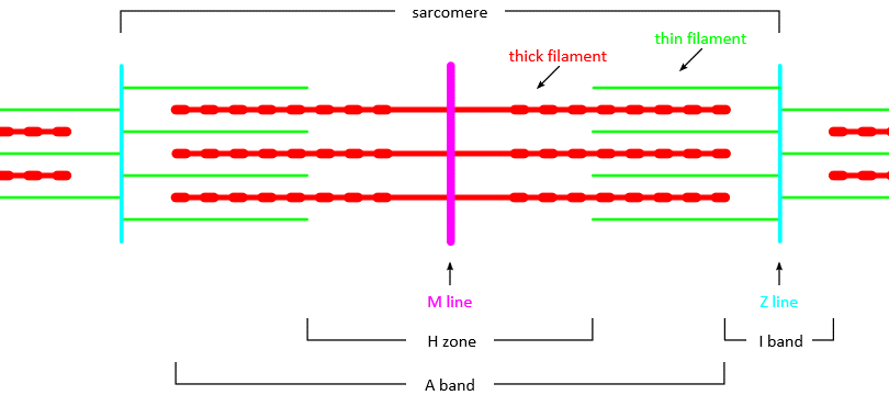
- muscle> mucle fibres> myofibrils> sarcomere
- Diagram
- I band- light just actin
- Z line in middle
- A band- dark overlapping actin and myosin
- H band light just myosin
- M line in middle
Muscle contraction
- I and H band narrower, Z lines move towards one another, a band remains the same
- Proteins in muscle contraction
- myofibrils composed of two
- Actin- thin two strands twisted around each other
- Myosin- rod shaped fibres with bulbous heads
- Tropomyosin- long thin strand wind round actin stops myosin from bonding
- Troponin- moves tropomyosin out of the way so actin can bind to myosin
- myofibrils composed of two
- Sliding Filament theory
- Stimulation
- 1-action potential reaches neuromuscular junction stimulation calcium to move into the synaptic knob
- 2- release acetylcholine into synaptic cleft which binds to receptors on sodium channels on postsynaptic membrane so it depolarises
- Contraction
- 1-action potential moves through T-tubules in sarcoplasm until reaches sarcoplasmic reticulum which opens calcium ion channels on sarcoplasmic reticulum
- 2-calcium ions diffuse into muscle, calcium ions bind to troponin changing its shape, troponin then displaces tropomyosin so actin filament binding sites are free
- 3-ADP attached to myosin head means it can bind to actin and form cross bridge, myosin heads then change their angle pulling actin filament along, in this process they release ADP
- 4-ATP attaches to myosin head causing it to detach from actin filament
- 5-calcium ions activate ATPase which hydrolyses ATP to ADP providing energy to return myosin head to start position
- 6- myosin head is now attached to a ADP and in correct position so it reattaches itself further along actin filament repeating the cycle in a ratchet like mechanism
- Relaxation
- sarcoplasmic reticulum actively transports calcium back into it
- lack of calcium= tropomyosin go back to blocking actin sites
- Stimulation
Energy Supply
- ATP needed for movement of myosin heads and active transport of calcium ions back into the sarcoplasmic reticulum
- ATP from aerobic respiration used up so need anaerobic without building up too much lactate
- Phosphocreatine acts as a supply of phosphate combining with ADP to form ATP
- phosphocreatine store is replenished using ATP when muscle is relaxed
Bilder und Dateien der Mindmap
Informationen
- Beschreibung
- GCSE A2 Biology (Muscle Contraction) Mindmap am Muscle Contraction, erstellt von Clodagh Mullins am 07/04/2016.
- Erstellt von
- Clodagh Mullins
- Aufrufe
- 7
- Bewertung