
Mindmap
Respiratory Tract
Inhalt der Mindmap
Respiratory Tract
UPPER RESPIRATORY TRACT
- air enters the nasal cavity through two openings
- anterior nares (nostrils)
- supported by alar cartilage
- the cartilage is a thin flexible plate situated immediately below the lateral nasal cartilage
- cartilage is bent upon itself in a way that forms the medial and lateral walls of the naris
- the cartilage is a thin flexible plate situated immediately below the lateral nasal cartilage
- the vestibule is a small space inside the nostrils
- covered with a dense network of hair which filters out particles that may be present in inhaled air
- nasal cavity
- receives the Eustachian tube from the inner ear
- walls of nasal cavity are formed by craniofacial skeleton and the septum (that splits the cavity in two) is formed by the vomer bone and an inferior projection of the ethmoid bone
- superior nasal concha
- fold of tissue covering the superior curved plate of bone that hangs down from the ethmoid bone
- space beneath concha in the superior meatus
- fold of tissue covering the superior curved plate of bone that hangs down from the ethmoid bone
- middle nasal concha
- fold of tissue that covers the middle-curved plate of bone which hangs down from the ethmoid bone
- space beneath concha is the middle meatus
- fold of tissue that covers the middle-curved plate of bone which hangs down from the ethmoid bone
- inferior nasal concha
- fold of tissue that covers the inferior bone attached to the ethmoid bone
- space beneath concha is the inferior meatus
- fold of tissue that covers the inferior bone attached to the ethmoid bone
- superior nasal concha
- mucous membrane
- rich blood supply
- mucosa is a pseudostratified ciliated columnar epithelium containing globet cells
- resting on a lamina propria rich in mucous and serous cells
- secrete mucous rich in lysozyme
- resting on a lamina propria rich in mucous and serous cells
- known as the olfactory mucosa
- contains sensory cells that detect odour
- surrounded by and connected to paranasal sinuses
- small irregular spaces filled with air and lined with a mucous membrane, function to lighten the skull and give the voice resonance
- frontal
- ethmoid
- maxillary
- sphenoid
- contains pharyngeal tonsils which protect against inhaled pathogens
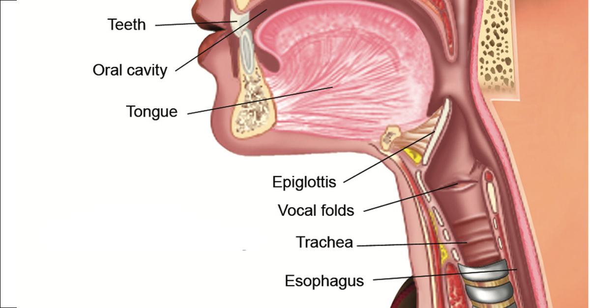
- pharynx
- The pharynx is a muscular tube from the base of the skull to the oesophagus (C6). The inner circular muscles of the pharynx are split into the superior pharyngeal constrictor (in the oropharynx), middle pharyngeal constrictor (in the laryngopharynx) and the inferior pharyngeal constrictor (in the laryngopharynx). The inferior pharyngeal constrictor is further split into the superior thyropharyngeus and the inferior circopharyngeus.
- from the nasal cavity, air passes backwards and downwards to the nasopharynx
- pharynx is a musular tube lined with a mucous membrane which joins the nasal and oral cavities to the oesophagus and larynx
- division between nasal cavity and nasopharynx is marked by posterior nares (choanae)
- oropharynx
- sits between the soft palate and the upper body of the epiglottis
- posterior to the oral cavity and tongue
- laryngopharynx
- lies behind the larynx and terminates at the level of the circuit cartilage by becoming continuous with the oesophagus
- LOWER RESPIRATORY TRACT
- larynx
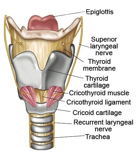
- cartilaginous structure composed of several different cartilage elements
- thyroid cartilage
- largest cartilage of the larynx
- formed by two flat plates that come together anteriorly to join and make the laryngeal prominence (Adam's apple)
- houses and protects the vocal cords
- circoid cartilage
- complete ring of cartilage that forms the inferior border of the larynx
- united with the trachea by the circotracheal ligament
- epiglottis
- thin triangular flap of cartilage located at the entrance of the larynx
- when food is swallowed the epiglottis moves downwards and the larynx moves upwards, blocking off the entrance to the larynx
- food bolus slides down into the oesophagus by passing downwards on either side of the larynx
- food bolus moves into two depressions called the piriform fossae of the laryngopharynx
- food bolus slides down into the oesophagus by passing downwards on either side of the larynx
- two arytanoid cartilages
- two pyramidal cartilages that sit on top of the circoid cartilage and have vocal ligaments attached to them
- the movement of the cartilages tightens or slackens the vocal ligaments which allows us to change the pitch of our voice
- two pyramidal cartilages that sit on top of the circoid cartilage and have vocal ligaments attached to them
- thyroid cartilage
- oesophagus
- accessory muscles helps to hold the cartilage together
- functions
- protecting the airways
- closing and sealing the lower respiratory tract
- supports voice production
- voice box
- trachea
- begins at level C6 below the circuit cartilage of the larynx
- descends through the thorax at divides at T4
- carina
- thick, incomplete cartilaginous ring that runs between the two primary bronchi at the bifurcation of the trachea
- directs air into the principle bronchi during respiration
- primary/principle bronchi
- right primary bronchi
- more vertical, shorter and wider
- more likely to be blocked
- more vertical, shorter and wider
- left primary bronchi
- more horizontal, longer and thinner
- travel obliquely and enters each lung through the hilum
- mucosa
- muscularis layer (submucosa)
- cartilaginous layer
- adventitia
- binds bronchi to the pulmonary artery and lung parenchyma
- hyaline cartilage becomes reduced in size as the bronchial diameter diminishes
- adventitia
- maintains the appropriate diameter of the airway
- submocosa is composed of mucous-secreting glands and adipose tissue is also present in larger bronchi
- cartilaginous layer
- height of epithelial cells and thickness of basal lamina and lamina propria decreases as diameter decreases
- muscularis layer (submucosa)
- secondary bronchi
- one secondary bronchi for each lung lobe
- the right lung has 3 secondary bronchi
- the left lung has 2 secondary bronchi
- tertiary bronchi
- 10 tertiary bronchi in the right lung
- 8 tertiary bronchi in the left lung
- do not have c-shaped cartilages but instead they have cartilaginous plates
- bronchioles
- composed of a fibroelastic membrane and smooth muscle and usually do not contain cartilage
- controls the resistance to airflow and distribution of air in the lungs
- branch in turn repeatedly until they form small branches called terminal bronchioles
- devoid of cartilage and largely composed of smooth muscle
- branch into respiratory bronchioles
- alveolar ducts
- alveolar sacs
- alveoli
- type I pneumocytes
- squamous epithelial cells
- have a flat, dark, oval nucleus and a very thin cytoplasm
- primarily involved in gas exchange
- 40% of the number of cells in the alveoli but make up 95% of the alveolar surface
- squamous epithelial cells
- type 2 pneumocytes
- cubodial cells that bulge into the alveolar space
- produces surfactant
- reduces surface tension of the alveoli and prevents them form collapsing on expiration
- progenitor cells that can proliferative to produce both type I and II pneumocystis after lung injury
- makes up 60% of cells in the alveoli but only 5% of the alveolar surface
- type I pneumocytes
- terminal dilations of the alveolar ducts and connects to at least two alveoli
- alveoli
- present on the distal end of the respiratory bronchioles
- covered in alveoli
- alveolar sacs
- transition zone concerned with both air conduction and gas exchange
- composed of simple cuboidal epithelium with ciliated and Clara cells
- club cells were originally known as Clara cells
- they are dome-shaped cells with short microvilli
- secretes materials like GAGs, lysozyme and conjugation of the secretory portion of IgA antibodies
- protects bronchiolar epithelium
- club cells were originally known as Clara cells
- also contains a lamina propria, separated circumferential smooth muscle fibres, adventitia and scattered, thin-walled outpocketings called alveoli which extend from the lumen
- start of the respiratory zone
- alveolar ducts
- composed of simple cuboidal epithelium
- also contains brush cells, small granule cells and basal cells
- increased number of Clara cells present
- composed of pseudostratified columnar epithelium which gradually transforms into simple ciliated columnar epithelium
- goblet cells are interspersed among the ciliated cells and extend the full thickness of the epithelium
- brush cells, small granule cells and basal cells are also present
- contains a lamina propria, a relatively thick circumferential layer of smooth muscle and an adventitia
- goblet cells are interspersed among the ciliated cells and extend the full thickness of the epithelium
- one secondary bronchi for each lung lobe
- right primary bronchi
- carina
- c-shaped cartilage rings
- united by fibroelastic membrane
- trachealis muscle unites posterior gaps of the cartilages
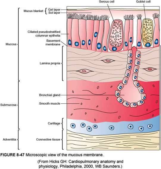
- mucous membrane
- mucosa
- submucosa
- cartilaginous layer
- adventitia
- binds the trachea to the adjacent structures
- composed of loose connective tissue
- hyaline cartilage
- adventitia
- contains mixed sero-mucous glands
- watery secretions humidify inspired air
- mucous as well as mucous from goblet cells trap particles from air
- cartilaginous layer
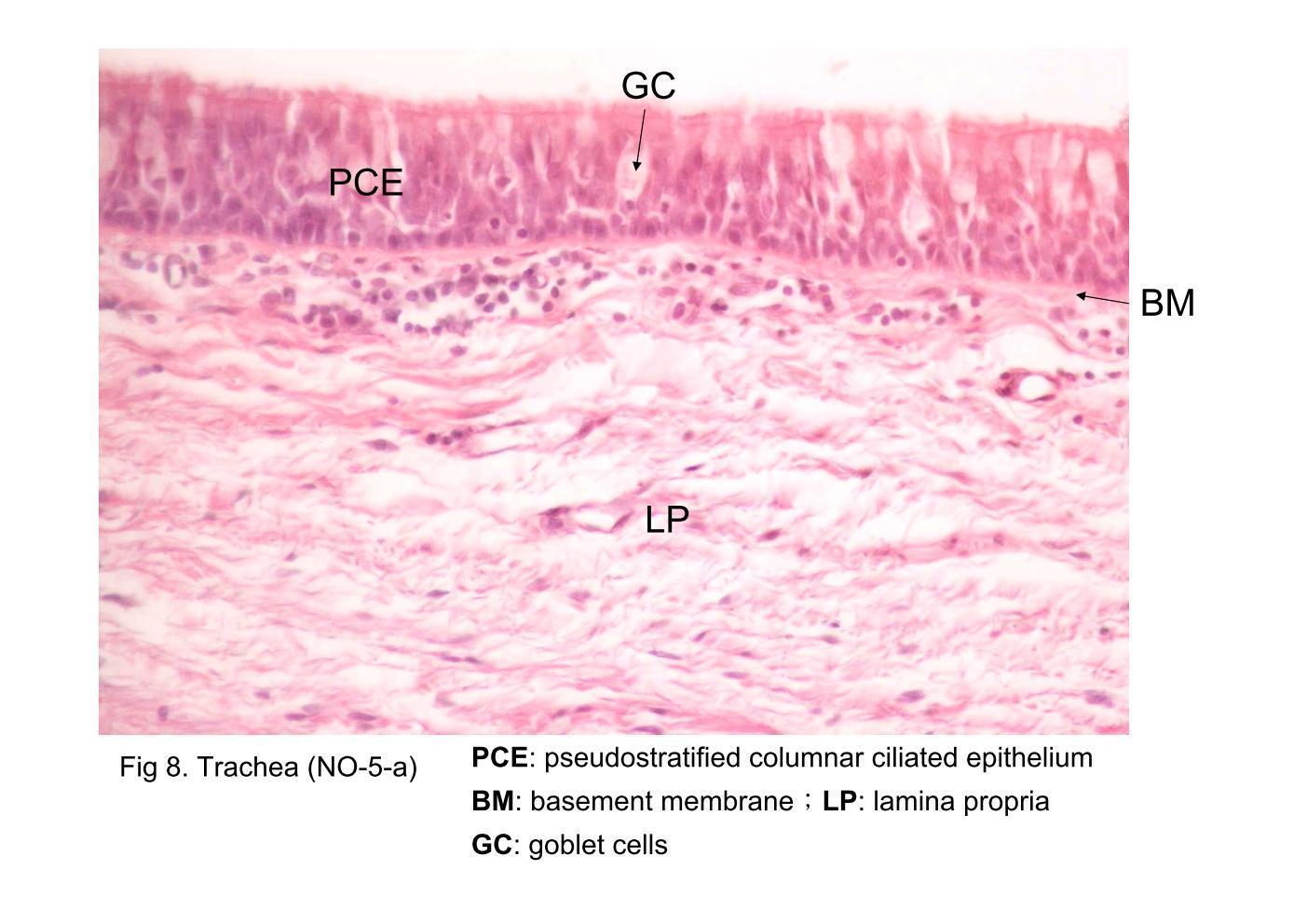
- epithelia
- tall columnar pseudostratified with cilia and goblet cells
- mucocilliary escalator
- brush cells, small granule cells and basal cells
- brush cells - columnar cells that contain microvilli and function as receptor cells for general sensation as their nerve endings penetrate the basal lamina and come together to form the trigeminal nerve basal cells - reserve population by maintaining individual cell replacement in the epithelium
- tall columnar pseudostratified with cilia and goblet cells
- lamina propria
- contains elastin which plays a role in the elastic recoil of the trachea during inspiration and expiration
- submucosa
- mucosa
- start of the conducting zone
- cartilaginous structure composed of several different cartilage elements
- larynx
- also called hypopharynx
- palatine, pharyngeal and lingual tonsils are embedded in the oropharynx
- acute inflammation of the palatine tonsils causes tonsillitis
- formed nodules of lymphatic tissue and function to protect the body against inhaled pathogens
- sits between the soft palate and the upper body of the epiglottis
- separated from the oral cavity by soft palate
- oral and nasal cavities are separated by a hard palate of bone and muscle
- formed by projections of the palatine and maxillae bones
- muscular extension of the hard palate and hangs from the back of it which separates the oral cavity from the nasopharynx
- oral and nasal cavities are separated by a hard palate of bone and muscle
- supported by alar cartilage
- anterior nares (nostrils)
- air enters the nasal cavity through two openings
Lungs
- Blood supplies of the lungs: - pulmonary circulation brings deoxygenated blood from the heart to the lungs and oxygenated blood from the lungs to the heart and then the rest of the body - bronchial circulation (bronchial artery and vein) brings oxygenated blood to the parenchyma, bronchial circulation is part of the systemic circulation
- situated in the thoracic cavity and connected to the mediastinum by a lung root
- lobes
- left
- superior lobe
- splits into 4-5 segments
- oblique fissure
- inferior lobe
- splits into 4-5 segments
- superior lobe
- right
- superior lobe
- splits into 3 segments
- horizontal fissure
- middle lobe
- splits into 2 segments
- oblique fissure
- inferior lobe
- splits into 5 segments
- superior lobe
- further segmented by a connective tissue septum forming pyramid-shaped bronchopulmonary segments
- left
- a primary pulmonary lobule is defined as the lung unit distal to the respiratory bronchioles, it is significantly smaller than an acinus, and is composed of alveolar ducts, alveolar sacs and alveoli
- one arteriole and an accompanying venue supply/drains one pulmonary lobule
- respiratory membrane
- when the capillary wall meets the alveolar wall a respiratory membrane forms
- breathing
- inspiration
- initiated by the neurally induced contraction of the diaphragm and the external intercostal muscles
- activation of the motor neurons in the phrenic nerves that innervate the diaphragm causes it to contract
- dome moves downwards into the abdomen, enlarging the thorax
- simultaneously the activation of the motor neurons of the intercostal nerves to the inspiratory intercostal muscles causes them to contract leading to an upwards and outwards movements of the ribs
- thoracic cavity volume increases
- lungs are stretched and intrapulmonary volume increases
- intrapulmonary pressure drops and air flows down the pressure gradient into the lungs until the intrapulmonary pressure is 0
- expiration
- motor neurons to the diaphragm and inspiratory intercostal muscles decrease their firing rate so that the muscles relax
- diaphragm and intercostal muscles are no longer actively pulled outwards so they start to recoil
- intrapleural pressure becomes less sub atmospheric which decreases the transpulmonary pressure
- the transpulmonary pressure acting to expand the lungs is now smaller than the elastic recoil exerted by the more expanded lungs and the lungs passively recoil to their original dimension
- as the lungs become smaller, the air in the alveoli becomes temporarily compressed so that alveolar pressure exceeds atmospheric pressure
- air flows from the alveoli into the atmosphere passively
- as the lungs become smaller, the air in the alveoli becomes temporarily compressed so that alveolar pressure exceeds atmospheric pressure
- the transpulmonary pressure acting to expand the lungs is now smaller than the elastic recoil exerted by the more expanded lungs and the lungs passively recoil to their original dimension
- intrapleural pressure becomes less sub atmospheric which decreases the transpulmonary pressure
- diaphragm and intercostal muscles are no longer actively pulled outwards so they start to recoil
- motor neurons to the diaphragm and inspiratory intercostal muscles decrease their firing rate so that the muscles relax
- expiration
- intrapulmonary pressure drops and air flows down the pressure gradient into the lungs until the intrapulmonary pressure is 0
- lungs are stretched and intrapulmonary volume increases
- thoracic cavity volume increases
- simultaneously the activation of the motor neurons of the intercostal nerves to the inspiratory intercostal muscles causes them to contract leading to an upwards and outwards movements of the ribs
- dome moves downwards into the abdomen, enlarging the thorax
- activation of the motor neurons in the phrenic nerves that innervate the diaphragm causes it to contract
- initiated by the neurally induced contraction of the diaphragm and the external intercostal muscles
- control of respiration
- medulla oblongata and pons in the brainstem
- medullary rhythmicity area (in the medullar oblongata), pneumotaxic and apneustic areas (in the pons)
- mechanosensroy signals are essential for adapting and adjusting breathing - the receptors are located in the smooth muscle of the airway and are stimulated by rapid lung inflation or deflation or oedema and their reflex is to cause a sigh or shortened expiration
- medullary rhythmicity area: regulates breathing rhythm
- can be further divided into the inspiratory area (DRG) and expiratory area (VRG)
- pneumotaxic area: increased firing rate of this area will increase the breathing rate
- apneustic area: transmits stimulatory nerve impulses to the inspiratory area of the MRA, which would cause prolonged deep inhalations if left alone
- cortical control
- voluntary control of the respiratory centre is exerted prdeominantly via the cortical connections that the brain has with the respiratory centre
- nerve impulses from the hypothalamus and limbic system
- voluntary control of the respiratory centre is exerted prdeominantly via the cortical connections that the brain has with the respiratory centre
- chemoreceptors
- central
- clusters of neurones located in the medulla oblongata
- responds to changes in the concentrations of H+ and CO2 in cerebrospinal fluid
- major role in the moment-to moment control of breathing
- peripheral
- located in the aortic arch and carotid bodies
- respond to changes in O2, CO2 and H+ concentrations in the blood by sending nerve impulses to the brain via the vagus (aortic bodies) and glossopharyngeal (carotid bodies) nerves
- signals are sent to the Nucleus Tractus Solitarius (NTS)
- receives information from mechanoreceptors and peripheral chemoceptors, this information is processed in the brainstem by respiratory neurones and generates the rhythm of breathing - the rhythmic signal is sent to the respiratory muscles
- inspiratory and expiratory neurons
- e.g. phrenic nerve exits the spinal cord at C3-5 and goes on to innervate the diaphragm and other nerves go on to innervate the intercostal muscles
- dorsal respiratory group - other respiratory groups are the pontine and ventral respiratory group
- ventral group generates rhythm
- receives information from mechanoreceptors and peripheral chemoceptors, this information is processed in the brainstem by respiratory neurones and generates the rhythm of breathing - the rhythmic signal is sent to the respiratory muscles
- signals are sent to the Nucleus Tractus Solitarius (NTS)
- central
- stretch receptors
- located in the lung and chest wall and responds to distension
- Hering-breuer reflex prevents further inspiration
- prevents over-inflation of lungs
- proprioceptors
- monitors the angle of the structure that they innervate
- gives the body means of increasing or decreasing the rate of breathing in response to increased muscle and joint movement
- monitors the angle of the structure that they innervate
- medullary rhythmicity area (in the medullar oblongata), pneumotaxic and apneustic areas (in the pons)
- respiratory compensation: links blood pH and breathing rate
- medulla oblongata and pons in the brainstem
- inspiration
- fissures are narrow depressions
- pleura
- the right and left pleura are separated by the mediastinum
- a serous membrane that surrounds the lung
- visceral pleura
- the layer that is superficial to the lungs
- pleural cavity
- the space between the visceral and parietal pleura
- parietal pleura
- outer layer that connects to the thoracic wall, the mediastinum and the diaphragm
- produces pleural fluid and creates cavities that separates the major organs
- pleural fluid is secreted by mesothelial cells from both pleural layers
- acts to lubricate their surfaces
- reduces friction between the two layers
- creates surface tension that helps maintain the position of the lungs against the thoracic wall
- acts to lubricate their surfaces
- prevents the interference of other organs during organ movement
- prevents the spread of infection
- pleural fluid is secreted by mesothelial cells from both pleural layers
- recesses
- costomediastinal recesses
- costodiaphragmatic recess
- lung function tests
- spirometry
- measures the volume of air that the patient is able to expel from the lungs after maximal inspiration
- measures forced vital capacity
- the maximal volume of gas that can be expired from the lungs during a forced expiration from full inspiration
- measures forced expiratory volume in 1 second
- maximum volume of gas that can be expired from the lungs during the first second of a forced expiration from full inspiration
- FEV1 decline graph shows that those that quit smoking can/will end up with the same FEV1 levels as those that do not smoke
- maximum volume of gas that can be expired from the lungs during the first second of a forced expiration from full inspiration
- also measures forced mid-expiratory flow (FEF), maximal voluntary ventilation (MVV), inspiratory reserve volume (IRV), expiratory reserve volume (ERV), residual volume (RV), inspiratory capacity (IC), functional residual capacity (FRC), vital capacity (VC) and total lung capacity (TLC)
- peak expiratory flow rate
- subject breathes as hard and as fast as they can into a meter which measures the maximum expiratory flow that can be sustained for a minimum of 10 milliseconds
- total capacity of the lungs
- determined using either gas dilution techniques or body plethysmography
- diffusing capacity of the lungs for carbon monoxide (transfer factor)
- single breath test that measures the overall gas-exhcange function of the lung
- diffusion capacity is a measure of of the ability of gas to diffuse from the alveolus
- arterial blood gas measure
- measures the arterial oxygen tension (PaO2) and arterial carbon dioxide tension (PaCO2)
- cardiopulmonary exercise testing
- respiratory muscle function
- spirometry
Cough reflex
- comes from the autonomic nervous system
- pulmonary irritant mechanoreceptors in the epithelium of the respiratory tract is sensitive to both mechanical and chemical stimuli
- receptors in the cough reflex are nestled in the epithelial layer and are called "rapidly adapting pulmonary stretch receptors" (RAR). RARs are afferents of the vagus nerve and are activated by dust, smoke, ammonia, oedema etc.
- bronchi and trachea are sensitive to light touch
- a cough is used to remove foreign matter from the respiratory tract before it reaches the lungs
- a cough is a protective reflex that rids the airway of any offending material
- cough receptors are located mainly on the posterior wall of the trachea, pharynx and carina
- when triggered, impulses travel via the internal laryngeal nerve, which stems from the vagus nerve, and goes to the medulla of the brain
- the efferent neural pathway transmits signals to the glottis, diaphragm and other major inspiratory/expiratory muscles
- diaphragm and external intercostal muscles contract, air rushes into the lung, epiglottis closes, vocal cords contract to shut the larynx, abdominal muscles contract to accentuate the action of the relaxing diaphragm, vocal cords relax, epiglottis opens (releasing air at over 100mph), bronchi and non-cartilaginous portions of the trachea collapse to form slits through which air is forced
- the efferent neural pathway transmits signals to the glottis, diaphragm and other major inspiratory/expiratory muscles
- when triggered, impulses travel via the internal laryngeal nerve, which stems from the vagus nerve, and goes to the medulla of the brain
Functions of the respiratory system
- ventilation and gas exchange, regulation of blood pH, air preparation, vocalisation, olfaction, protection and defence
Bilder und Dateien der Mindmap
Informationen
- Beschreibung
- Year 1 Mindmap am Respiratory Tract, erstellt von Rana Al-Khayat am 04/04/2018.
- Erstellt von
- Rana Al-Khayat
- Aufrufe
- 0
- Bewertung