
Mindmap
Embryology
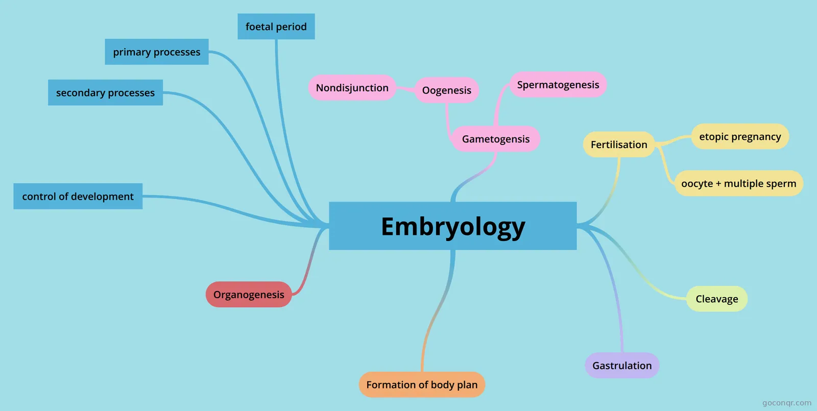
Mind map: Embryology
Inhalt der Mindmap
Embryology
- weel 0-3 conceptus/embryo weeks 3-8 embryonic period weeks 9-40 foetal period
Gametogensis
- formation of a gamete a gamete is is a cell with a chromosome complement of 23 chromosomes
- Oogenesis
- starts in development ends at menopause primary oocytes have begun meiosis by week 30 but arrest in propane until puberty reproductive lifespan determined in fetal life
- Nondisjunction
- 75% of nondisjunction cases occur during oocyte formation nondisjunction is the failure of sister chromatids to separate during cell division. in meiosis this can lead to trisomy.
- Spermatogenesis
- occurs at puberty continues throughout life each cycle tales 64-74 days production rate decreases with age 6.5 mill sperm per gram of testicular tissue/day
Fertilisation
- the union of gametes fusion of sperm and oocyte formation of a zygote typically occurs in ampulla of uterine tubea chemical attracts sperm to egg sperm can survive 5 days in the reproductive tract the zone pellucida is formed by secretions from the oocyte sperm binds to glycoprotein ZP3 in ZP and acrosomal enzymes are released from the sperm head. sperm digests its way into the egg. egg and sperm fuse and sperm contents enter the egg. this triggers completion of meiosis 2 and releases cortical granules from oocyte. release of cortical granules helps prevent fusion with more than one sperm by making ZP impermeable to other sperm
- etopic pregnancy
- topic pregnancy is when implantation is located outside the uterus. dangerous, potential rupture and haemorrhage. abnormal sites are ovary uterine tube peritoneum internal OS of uterus placenta previa condition is implantation towards the cervix. placenta covering cervical loop.
- oocyte + multiple sperm
- if multiple sperm fertilises the egg. there will be three sets of chromosomes. too much gentic material embryo won't make it
Cleavage
- period of rapid cell division rapid mitotic cell division with no increase in size. subdivides fertilised egg into many identical daughter cells called blastomeres. 8+ cell blastomeres = totipotent (can become all cell types including embryonic and placental)mitotic cell division of zygote forms morula formation of morula then blastocyst (days 1-4)mitotic non disjunction =mosacism some might not some all features of trisomy 21 morula is at least 16 blastomere cells. cells become more compact. maximise space they get closer and form cell gap junctions. allow fluid to enter through zona pellucida forming blastocyst cavity. known as blastocyst now as there's an obvious cavity. outer layer of blastocyst thins becomes one cell thickness and trophoblast forms. inner mass cells move to embryonic pole eventually forming embryo itself. blastocyst fully formed around day 4/5. reach uterus for implantation. blastocyst hatches out of ZP and adheres to uterine lining for implantation. implantation only occurs once zona pellucida has started to degenerate and blastocysts has hatched and enlarged. trophoblast attaches to endometrial epithelium on posterior uterine wall. blastocysts has to invade endometrial epithelium and connective tissue to attach and implant. corpus leuteum is important for maintace of a uterine environment that allows implantation and pregnancy. every ovulation = corpus letup developed. corpus leuteum secretes progesterone. progesterone is important in maintaining lining of endothelium which must be maintained so its ready for implanation and further growth of the embryo. trophoblast differentiates and splits into cytotrophoblast (inner part of trophoblast becomes single cell layer) and synctiotrophoblast (more extensive layer, invasive layer, that invades endometrium to help implantation if embryo) embryo organises itself into bilaminar discs. hypoblast and epiblast. cavity forms within epiblast (amniotic cavity)if fertilisation and implantation have occurred the cellsnof syncytiotrophoblast start to secrete hug which stimulates corpus letup to secrete progesterone. bilaminaer embryo has made a defect in wall of uterine cavity. blood clot-fibrin coagulum. vacuoles in syncytiotrophoblast and unite to form lacunae. provides nutrition to embryo via maternal blood = uteroplacental circulation. cells from hypoblast cover lining of ctrotrophoblast layer, forming heursers membrane which lines primitive yolk sac. new cells appears between heusers membrane and cytrophoblast from the extra embryonic mesoderm which degenerates into the chronic cavity which surrounds primary yolk sac. presence of connective stalk from embryo to extra embryonic mesoderm. connective stalk become umbilical cord. second wave of hypoblasts migrate to form secondary definitive yolk sac. embryo implanted within endometrium. endometrium circulation. uteroplacental circulation forms as synctiotrophoblast extends and contacts maternal blood supply.
Gastrulation
- formation of the germ layers - day 15 body axes established by formation of primitive streak primitive streak appears in midline at caudal end of epiblast. primitive pit and node is at cranial end of streak primitive grove cells migrate through primitive streak between the epiblast and and definitive endoderm. mesoderm does not spread out to oropharyngeal membrane or the clocual membrane notochord is a midline structure that forms early on the the trilmainaor embryo. it doesn't exist in adults and it secrets FSH and binds to target cells to produce a signal that helps tissues differentiate and develop. fate of endoderm epidermis of skin (hair nails associated glands) nervous system (brain spinal cord and peripheral nerves) fate if mesoderm paraxial - axial Skelton, voluntary skeletal muscles, parts of dermis (back) intermediate - urogenital systems (kidneys ureter gonads and associated structures) lateral plate somatic - most of dermis, lining of body wall, parts of limbs later plate visceral part- cardiovascular system, mesothelial covering of organs, smooth muscle endoderm - lining of gut tube, lining of respiratory tract lining of bladder and urethra
Formation of body plan
- formation of body plan embryonic folding
Organogenesis
- Formation of organs and organ systems. basis of these systems in place by the end of embryonic period. will continue to develop through foetal period
foetal period
- weeks 9-40 growth and weight gain mainly grows in length during the second trimester (14-26 weeks) and most weight is added in the 3rd trimester (27-40 weeks) tissues mature and become functional overt sexual differentiation bone laid down, connections made in CNS
primary processes
- cell division differentiation cell attachment apoptosis induction cell margination
secondary processes
- Axis formation/ polarity folding/ rotation
control of development
- genetic gene expressed tightly regulated in time and space epigenetic preferential expression of maternal/paternal copy of gene deleted region on chromosme 15 contains 2 paternally expressed genes and 1 maternally expressed gene Angleman's syndrome - deletion on maternal chromosome 15 Prader-Willi syndrome- deletion on paternal chromosome 15
Informationen
- Beschreibung
- Year 1 Anatomy Mindmap am Embryology, erstellt von ashley sharp am 21/11/2020.
- Erstellt von
- ashley sharp
- Aufrufe
- 1
- Bewertung