
null
null
17923181
7. Specialised Cells: CD4 T Cells
Descripción
Sin etiquetas

Diapositivas por Chloe Cavarretta, actualizado hace más de 1 año
Más
Menos

|
Creado por Chloe Cavarretta
hace más de 7 años
|
|
null
Resumen del Recurso
Diapositiva 1
Learning Outcomes: L15-16
What distinguishes the main subsets of CD4 T cell?
What is “signal 3”?
And what is signal 3 for Th1 + Th2 induction?
What intracellular signalling pathways cause Th1 + Th2 induction?
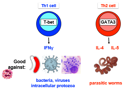
What pathogens are Th1 + Th2 cells effective against?
How do Th1 + Th2 “help” B cell class switching?
How do Th1 cells “help” macrophage activation?
How do CD4 T cells “help” CD8 T cell responses?
Which pathogens are killed by Th17 cells?
What is “Signal 3” for Th17 differentiation and what are the downstream signalling pathways?
What are Th17 effector mechanisms?
Why do we need regulatory T cells?
What are natural and induced Tregs?
What is the relationship between Tregs and Th17 cells?
How do Tregs suppress immune responses?
Diapositiva 2
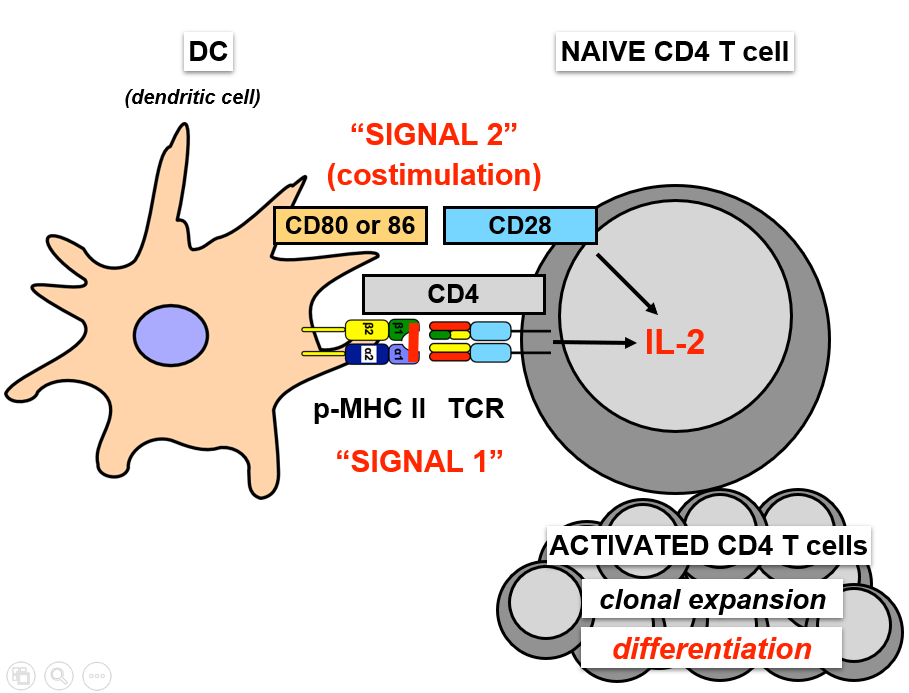
Recap: T Cell Activation
Diapositiva 3
CD4 T Cell Subsets
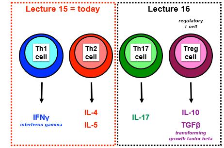
Pie de foto: : CD4 T cell subsets make signature cytokines
Diapositiva 4
CD4 T Cell Subsets
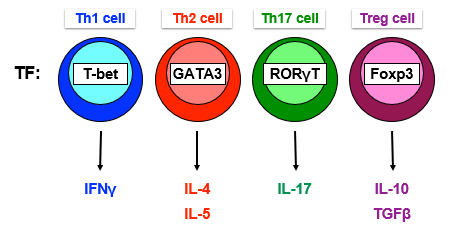
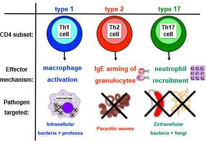
Distinct subsets of CD4 T cells coordinate distinct immune responses to target different pathogens, subsets are distinguished by signature cytokines
Cytokines= proteins secreted by immune and non-immune cells that influence immune function: Interleukins, interferons, tumour necrosis factors, transforming growth factor
CD4 T cell differentiation events:
Production of polarising cytokines - signal 3
Cytokine signalling events in CD4 T cells
Effector functions of CD4 T cell subsets
Diapositiva 5
1. Produce Polarising Cytokines: Th1
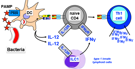
Signal 3-Th1 differentiation, supported by ILC1-derived IFNϒ
Summary: PAMPs stimulate DCs to produce IL12-> main driver of Th1 differentiation (signal 3)
DC recognises bacterial PAMP- presents peptide on MHC-II
PAMP stimulates DC to produce cytokine IL-12
IL-12 binds IL-12R on naive T cells and elicits signal in naive CD4 T cell
ILC1 aLso express IL12R and will bind IL12 and produce IFNϒ
IFNϒ binds to IFNϒR on naive CD4 T cell
Sum of signals (1 +2 ) + 3= cytokines (IL12 and IFNϒ) = cause naive T cell to be activated to Th1 cell that produces signature cytokine IFNϒ
Diapositiva 6
1. Produce Polarising Cytokines: Th1
Diapositiva 7
1. Produce Polarising Cytokines: Th2
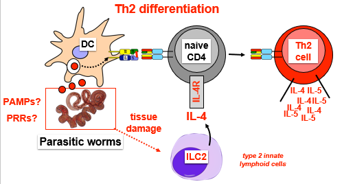
Signal 3-Th2 differentiation
Th2 is response induced by parasitic worms
DC present peptide on MHC-11 (dont know PAMP or PRR)
Parasitic worms cause tissue damage which activates ILC2 to produce IL-4 (signal 3)
IL-4 binds IL-4R on naive CD4 T cell
Sum of signals (1 +2 ) + 3= cytokines (IL4 = cause naive T cell to be activated to Th2 cell that produces signature cytokines IL4 and IL5
Diapositiva 8
1. Produce Polarising Cytokines: Th2
Diapositiva 9
2. Cytokine Signalling in CD4 T Cells
How does Il-12 induce Th1 differentiation and how does IL-4 induce Th2 differentiation
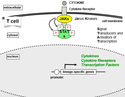
T cell signalling: JAK-STAT pathway GENERAL
JAK is associated with cytokine receptor
When cytokine binds receptor, JAKs activated, phosphorylate cytokine receptor
Phosphorylation acts as docking site for STAT proteins and recruit them to receptor
STATs close to JAKs and get phosphorylated, then translocate to nucleus
STATs bind to promoter of and transcribe lineage specific genes: cytokines, cytokine receptors, TFs
Lineage specific TFs promote the production of signature cytokines by different CD4 T cell subsets
Diapositiva 10
2. Cytokine Signalling in CD4 T Cells
Pie de foto: : JAK-STAT pathway
Pie de foto: : Lineage specific TFs induce signature cytokines
Diapositiva 11
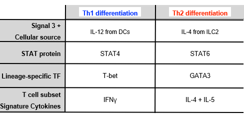
2. Cytokine Signalling in CD4 T Cells: Th1
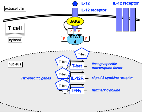
IL-12 signal cytokine binds IL-12R, phosphorylation through JAKs of intracellular portion of IL12R
Recruit STAT4, phosphorylated and translocates to nucleus
Activated STAT4 binds to TH1-specific genes:
T-bet= lineage specific TF- promotes more response to IL12
IL12R- signal 3 cytokine receptor- promotes cell to be more responsive to IL12
IFNϒ= hallmark cytokine of Th1 cells
T-bet binds to all 3 gene loci, activates, reinforces and stabilises Th1 differentiation, promoting expression of all 3
Diapositiva 12
2. Cytokine Signalling in CD4 T Cells: Th1
Diapositiva 13
2. Cytokine Signalling in CD4 T Cells: Th2
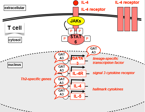
IL-4 signal cytokine binds IL-4R, phosphorylation through JAKs of intracellular portion of IL12R
Recruit STAT6, phosphorylated and translocates to nucleus
Activated STAT6 binds to TH2-specific genes:
GATA3= lineage specific TF- promotes more response to IL-4
IL4R- signal 3 cytokine receptor- promotes cell to be more responsive to IL-4
IL4 +IL5= hallmark cytokine of Th2 cells
GATA binds to all 3 gene loci, activates, reinforces and stabilises Th1 differentiation, promoting expression of all 3
Diapositiva 14
2. Cytokine Signalling in CD4 T Cells: Th2
Diapositiva 15
Summary of Differentiation so far
Diapositiva 16
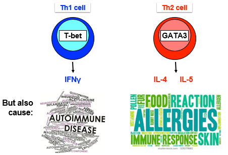
Overview of Th1 + Th2 Effector Function
Diapositiva 17
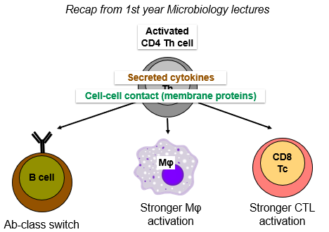
3. Effector Functions of CD4 T cells
Diapositiva 18
3. Effector Functions of CD4 T cells
1. Activated CD4 T cells help Ab class switch
Ab's have different isotypes-> starts as IgM and IgD but class switch into IgG, IgE, IgA
Isotypes have different heavy chain constant regions:
Bind different Fc receptors
Differing ability to activate complement
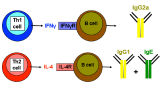
CD4 T cell signature cytokines direct Ab class switching:
Th1 cell produced IFNϒ and B cells express the IFNϒR- Th1 cell IFNϒ induces B cells to class switch to IgG2a
Th2 cell IL-4 induces B cell to class switch to IgG1 + IgE
Diapositiva 19
Activated CD4 T cells help Ab class switch
Diapositiva 20
Activated CD4 T cells help Ab class switch
Eg of Importance of class switching:
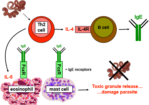
Parasitic worms induce Th2 cell to produce IL4, induces class switch to IgE
Innate immune cells (granulocytes) express receptor for IgE (FcεR)
Immune cells bind IgE, armed with IgE (receptor)
May see pathogen molecules that IgE recognises and bind-> activate immune cells
Th2 also produces IL5 cytokine= survival factor and differentiation factor for eosinophils
Produces lots of eosinophils in blood all armed with IgE
Mast cells will degranulate- release toxic granules onto parasite to attack
Diapositiva 21
Activated CD4 T cells help Ab class switch
Diapositiva 22
3. Effector Functions of CD4 T cells
2. Activated CD4 T cells help macrophages
CD4 T cell activated by DC in LN, macrophage far away in peripheral tissue fighting infection
Activated T cell needs to migrate from LN to peripheral tissue
Diapositiva 23
Activation-induced changes in CD4 T cell surface proteins
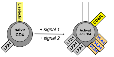
Naive CD4 T cells have lots of L-selectin (to enter HEV) and integrin LFA1 that gets them into LN
Following activation:
Loss of L-selectin
Increase integrins (more LFA1 and new VLA-4)
Induction of CD40L ligand
Activated CD4 T cells help macrophages
Diapositiva 24
Activated CD4 T cells help macrophages
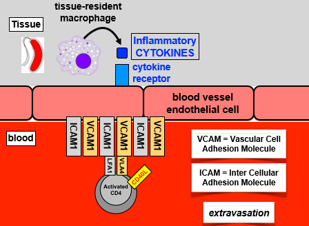
T cells migrate from blood into inflamed tissues:
Tissue resident macrophage produces inflammatory cytokines recognised by receptor on endothelial cell
Endothelium activated, expressed high levels of adhesion molecules: ICAM1 (binds LFA1), VCAM1 (binds VLA4)
Binding of adhesion molecules to integrins arrests activated T cell on endothelial cell and migration into tissue controlled by chemokine gradient
Diapositiva 25
Activated CD4 T cells help macrophages
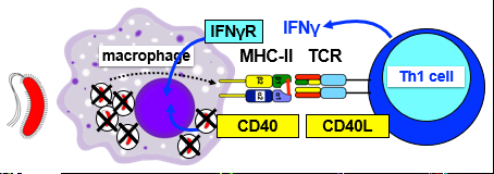
Macrophages present antigens, re-stimulate activated CD4 T cell
Present antigens on MHC-II
T cell with correct antigen specificity
TCR signals delivered by MHC-II:
Th1 produce IFNγ + express high levels of CD40L
CD40L binds CD40 on mac
Activate macrophage to kill intracellular pathogens
Diapositiva 26
3. Effector Functions of CD4 T cells
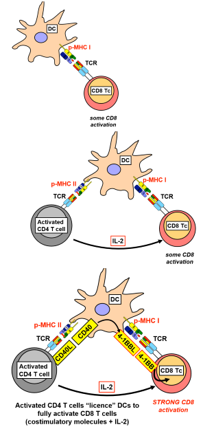
3. CD4 T cells help CD8 CTL activation:
Activated CD4 T cells licence DCs to fully activate CD8 T cells:
DC presents peptide on MHC-I and binds TCR on CD8 T cell = weak activation
Simultaneous presentation of MHC-II:peptide to CD4 T cell (different peptide derived from same pathogen), activated CD4 T cell produces IL2= further activation CD8
CD40L (induced by cross-talk between CD4 + DC) binds CD40 on DC and causes upregulation of other co-stimulatory molecules on DC for CD8 = strong activation of CD8
Diapositiva 27
CD4 T cells help CD8 CTL activation
Diapositiva 28
Type 1 vs Type 2 Immunity
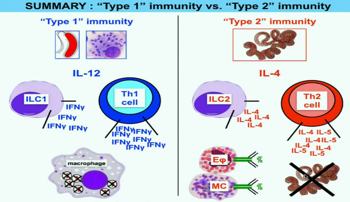
Th1 and Th2 cells are coordinators- directors of broader immune responses (Type 1 and Type 2 immunity)
Type 1 immunity:
Bacteria activates DC and macrophages to produce IL12
ILC1 produce IFNϒ, supports differentiation into Th1
Lots of production of IFNϒ- Th1 and ILC1
Activation of macrophages to kill intracellular pathogens
Type 2 immunity:
Worm activates IL4 production
ILC2 produces IL4 which supports differentiation of Th2 cells
Th2 produce IL4 and IL5 cytokines- lead to activation of B cells to produce IgE class switch Ab
When eosinophils and mast cells recognise parasite they degranulate and kill pathogen
Diapositiva 29
Type 1 vs Type 2 Immunity
Diapositiva 30
Th17 Cells
Make large amounts of cytokine IL-17
Initiate effector immune responses against pathogens not dealt with by Th1 and Th2 cells: Extracellular bacteria and fungi
Diapositiva 31
How are Th17 Cells Induced?
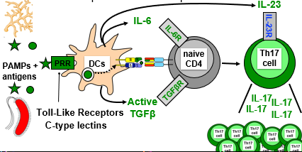
PAMPs bind DC PRR, uptake antigens present on MHC-II
Trigger DCs to produce signal 3= active TGFβ and IL-6 (inflammatory cytokine)
Initial Th17 differentiation
DCs present peptides to CD4 T cell, which binds as well as cytokines to receptors on CD4 T cell triggering initial differentiation into Th17
Th17 characterised by production of IL-17
Need additional signal to keep alive- DCs produce IL-23 cytokine as part of signal 3
Diapositiva 32
How are Th17 Cells Induced?
Diapositiva 33
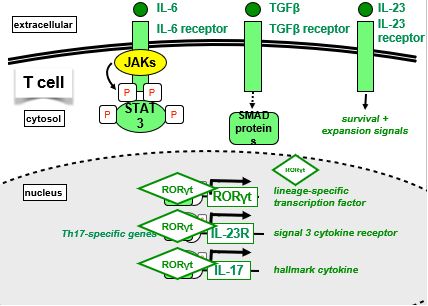
Cytokine-induced Signalling in Th17 Cells
IL-6 binds IL-6R on T cell, activates JAKs to phsophorylate intracellular portion of IL-6R
Phosphotyrosine acts as docking site for STAT3, phosphorylated by JAKs, translocates to nucleus and bind Th17 specfic genes
STAT3 alone not sufficient to induce gene expression
TGFβ binds to receptor on T cell and activates SMAD proteins
SMADs and STAT3 bind and induce expression of Th17 specific genes
IL23 (not there upon initial activation) binds to its receptor on T cell and signals for survival and expansion
RORγT TF protein is expressed and will take over to stabilise expression of all 3 genes (RORγT TF , IL-23R, IL-17)
Diapositiva 34
Cytokine-induced Signalling in Th17 Cells
Diapositiva 35
Th17 Effector Functions
Recruited to initial sites of inflammation:
Infection of extracellular pathogen causes DC to mature and present, DC migrate down afferent lymphatic to draining LN
Interacts with T cell with correct specificity, differentiation into Th17
Some Th17 cells leave via efferent lymphatic, end up in blood and recruited back to inflamed tissue
Tissue resident macrophage induced by pathogen to secrete inflammatory cytokines, act on endothelial wall of blood vessel, activate expression of adhesion molecules, chemokine gradient pulls Th17 cells into tissue
Diapositiva 36
Th17 Effector Functions: Neutrophils
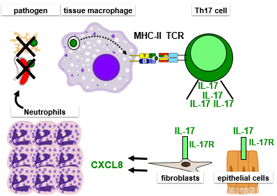
Once at site of inflammation, recruit neutrophils:
Interaction between mac and specific Th17 cell via MHC-II:peptide- reactivates Th-17 (first in LN), produces IL-17
IL-17 binds to receptor (broadly expressed by non-immune cells (stromal: fibroblast, eptihelial cells)
Triggers non-immune cells to produce lots of chemokine CXCL8
CXCL8 recruits neutrophils from blood to site to kill pathogen
Diapositiva 37
Th17 Effector Functions: Neutrophils
Diapositiva 38
Summary: Th17
DC produce active TGFβ, IL-6, IL-23 in response to fungi or extracellular bacteria = Signal 3 for Th17 differentiation
TGFβ + IL-6 induce initial Th17 differentiation
IL-23 promotes expansion + survival of Th17 cells
Lineage-specific TF for Th17 cells is RORγT
Th17 cells produce IL-17
This induces non-immune cells at sites of infection to produce CXCL8 = neutrophil chemoattractant
Recruited neutrophils kill pathogen
Diapositiva 39
Summary of T Cell Subset so far
Diapositiva 40
Regulatory T Cells
Immune responses are inflammatory and potentially pathogenic... autoimmunity and immunopathology (damages tissue even after clearance)
Central tolerance: deletion of self-reactive T cells in thymus
Some self-reactive T cells escape this negative selection, so need peripheral tolerance
Peripheral tolerance: stop activation of mature self-reactive T cells
Via co-stimulation- in absence of infection APCs have low level of CD80/CD86
Not always efficient, need specialised cells to turn off immune responses.. Regulatory T cells
Diapositiva 41
2 Types of Regulatory T Cell (Treg)
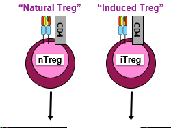
Still have TCR and express CD4 like normal T cells
Difference=repressive, turn off effector T cells, APCs etc
Diapositiva 42
Natural Tregs
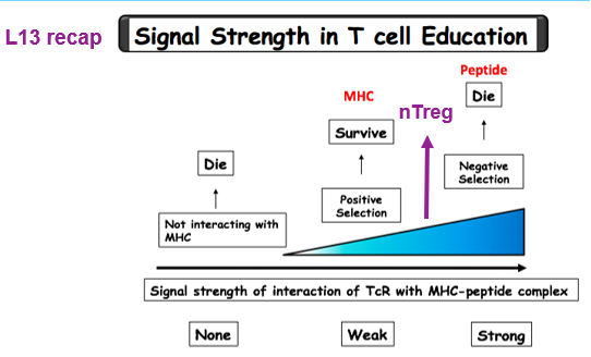
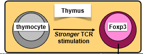
nTregs differentiate in thymus in response to stronger than normal TCR stimulation
TCR binds to self-peptide:MHC stronger than normal but not strong enough to be deleted
Induces expression of Treg lineage-specific TF: Foxp3
T cells exit thymus and circulate through LN and spleen
Function: they recognise self-peptides and have stronger than normal self-reactivity, recognise T cells that escaped negative selection and recirculating-> directly inhibit autoreactive T cell
Diapositiva 43
Natural Tregs
Diapositiva 44
Induced Tregs
iTregs differentiate in the periphery
Signal 3 for iTreg is TGFB (in absence of inflammatory signal)
DC take up antigen, produce active TGFB which binds receptor on naive CD4 and upregulates foxp3 positive iTreg
iTreg potentially specific for any antigen: pathogen, self-antigen, commensal
Diapositiva 45
Differentiation of iTreg vs Th17 Cells
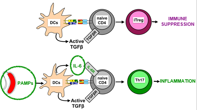
Pathogens induce production of inflammatory cytokines (eg IL-6) that switch immune suppression to Th17 inflammatory response
Diapositiva 46
iTregs Supress Immune Response
Via cell surface molecules:
Treg express high levels of CTLA-4 (co-inhibitory molecule)
CTLA-4 binds and sequesters DC CD80/86 from naive cells - no signal 2
CTLA-4 can physically rip CD80/86 from APCs= no ability co-stimulate
Tregs express high levels of IL-2R
Act as sink for IL-2, take it away from activated cells so they cant proliferate
Outcompete activated T cells for IL-2
Diapositiva 47
Via cell surface molecules
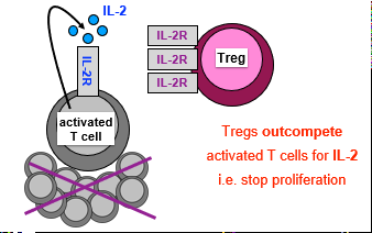
Pie de foto: : Express IL-2R: Outcompete activated T cells for IL-2
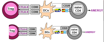
Pie de foto: : Express CTLA-4: no co-stimulation (no signal 2)
Diapositiva 48
iTregs Supress Immune Response
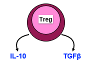
Via secretion of immunosuppressive cytokines:
Inhibit Il-12 production by DCs (no Th1 response)
Reduce MHC-II + CD80/86 levels on DCs
Directly inhibit T cell proliferation
Generate more Tregs (TGFβ)
Diapositiva 49
Mutations in Tregs
Mutations to Foxp3 gene of X chromosome = IPEX syndrome
Cant make functional Foxp3 protein, lack Tregs, die by autoimmune diseases
Fatal early (childhood) onset autoimmune disease in males:
Diabetes, anemia, splenomegaly, skin inflammation, cytokine storm
Diapositiva 50
Tregs Summary
Tregs are essential for peripheral tolerance
nTregs differentiate in thymus + turn-off self-reactive T cells
In absence of inflammatory cytokines, TGFβ induces iTregs
iTregs suppress immune responses through surface + secreted molecules
¿Quieres crear tus propias Diapositivas gratis con GoConqr? Más información.
Similar
null