
Fichas
Week 1
Fichas del conjunto (41)
- 1What happens in the mouth and oropharynx?-Chewing/mastication: increases SA:V of food increasing the rate of absorption
-Initiating carbohydrate and fat metabolism using salivary amylases and lipases (sensory function. More effective when secreted in stomach/pancreas than in saliva)
-Lubrication of bolus - 2Describe the structure of salivary glands?-Made up of parotid, submandibular and sublingual glands
-Exocrine glands that secrete amylase and lipase
-Secretory products include mucous, fluid and antibacterial products (IgA, lysozyme and lactoferrin
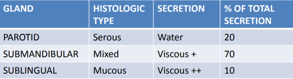
-Acinar cells produce and secrete enzymes. Ductal cells produce mucous and secrete water/electrolytes (modifies protein) - 3Describe the 3 salivary gland in terms of: -histological type -secretion (what is it like?) -% of total secretion What is the total volume secreted in 24 hrs?1.5L
- 4Functions of saliva?-Lubrication
-Hydration: keeps oral mucosa moist, prevents cell death
-Cytoprotection: protects oral/oesophageal mucosa from damage - 5Describe the immune functons of: -IgA -Lactoferrin -Lysozyme-IgA antibodies bind to pathogenic antigens to: neutralise toxin, block viral attachment to cells, opsonize bacteria, activate NK cells and are then taken up by M cells
-Lactoferrin binds to iron and is bactericidal (iron needed for bacterial colonisation)
-Causes cell lysis - 6Salivary duct cells modify the secretion of acinar cells. How/why does this happen?-Extracts Na+/Cl-
-Secretes K+/HCO3-
-Saliva is no hypotonic and alkaline
-This protects teeth from bacterial acid/neutralises gastric acid that refluxes into oesophagus - 7What is Sjoren's syndrome?-Autoimmune attack of salivary/tear glands
-Results in xerostomia (dry mouth/eyes)
-Commonly affects women
-Associated with rheumatoid arthritis - 8What does the mumps virus do?-Parotitis (inflammation of parotid gland)
-Prodrome of headache and fever
-Associated with orchitis (inflammation of one/both testicles)
-Prevention: MMR vaccine - 9Describe the innervation of the salivary glandsANS controls secretion mostly via ParaNS (causes secretion)
-CN VII innervates submandibular/sublingual glands
-CN IX innervates parotid gland
-SymNS also innervates glands via thoracic sympathetics causing vasoconstriction/thick mucous secretion - 10Describe 4 phases to swallowing1) Oral preparatory phase: food is masticated so it can be swallowed
2) Oral phase: tongue propels food posteriorly until pharyngeal swallow is triggered
3) Pharyngeal phase: bolus is transported through pharynx, glottis is closed, breathing is stopped and upper oesophageal sphincter is relaxed
4) Oesophageal phase: oesophageal peristalsis carries bolus from cricopharyngeal juncture down towards lower oesophageal sphincter - 11Where is the swallowing (neural) centre? What nerves are present?In the medulla
CN V, IX, X, XII - 12Difference between pseudobulbar and bulbar palsies? Signs/symptoms:Pseudobulbar- upper motor neurone affected
Bulbar- lower motor neurone affected
Difficulty in swallowing, altered speech, loss of gag reflex leading to aspiration of food/fluid into trachea - 13What type of muscle is present in the oesophagus?Striated and smooth
- 14How does the trachea develop?-Develops as lung buds from the oesophagus
-Trachea matures and lies anterior to oesophagus - 15What is GORD? (Gastro-oesophageal reflex disease)-Chronic symptoms/mucosal damage produced by the abnormal reflux in the oesophagus
ie. chronic heartburn (repeated episodes daily) - 16Causes of GORD?-Obesity (increased intra-abdominal pressure)
-Hiatus hernia
-Drugs that lower tone of lower oesophageal sphincter (loosened)
-Pregnancy
-Zollinger Ellison syndrome (gastrin secreting tumours)
-Supine position after eating meal - 17Symptoms of GORD?-Heartburn
-Regurgitation
-Dysphagia
-Cough, hoarsness and chronic earache (due to laryngopharyngeal reflux) - 18Effects of GORD?-Oesophagitis
-Stricture
-Barrett's metaplasia
-Oesophageal adenocarcinoma - 19How is GORD diagnosed?-24 hr pH monitoring (probe in oesophagus)
-Contrast swallow - 20Treatment for GORD?Conservative: weight loss, avoid eating close to bedtime, decrease alcohol, raise head of bed
Medical: decrease acid production using proton pump inhibitors, antacids, alginates (coat mucosa)
Surgical: anti-reflux surgery, repair hiatus hernia - 21What is Barrett's metaplasia?-Metaplasia: change of epithelial type in response to environmental stress
-Musoca is replaced
-Acid from stomach destroys stratified squamous epithelium
-Exposed stem cells give rise to columnar cells instead
-Pre-malignant condition for oesophageal adenocarcinoma - 22How s Barrett's metaplasia managed?-Only mechanism is surveillance endoscopy
-Expensive and labour intensive
-Controversial as only 0.5% go on to progress to adenocarcinomas - 23Difference between squamous cell carcinomas/adenocarcinomas?Squamous cell- top 2/3 of oesophagus
Adenocarcinoma: bottom 1/3 of oesophagus - 24What is achalasia?-Motor disorder of oesophagus
-Loss of myenteric plexus at lower oesophageal sphincter (remains constricted) - 25Symptoms?-Dysphagia (of BOTH solids and liquids. Oesophageal cancer patients have no difficulty swallowing fluids)
-Regurgitation
-Chest discomfort
-Halitosis (bad breath) - 26Treatment?-Botox injection at lower oesophageal sphincter
-Oesophageal dilation
-Surgery (hellers myotomy) - 27Purpose of the digestive system?Supply the body with energy which is needed for:
-maintenance of deep body temp
-repair + renewal of the body's tissues
-growth, development and maturation of the young
-reproduction
-survival! - 28What are the 5 processes of digestion?1) Ingestion
2) Digestion (breakdown of complex food substances into smaller soluble ones)
3) Absorption
4) Assimilation (absorbed food used to provide energy
5) Egestion - 29What are the key components of the digestive system?-Digestive muscular tube
-Digestive viscera
-Valves
-Lymphatics (incl. MALT organs and Peyer's patches)
-Colonies of bacteria - 30Where does the oesophagus enter the abdomen?-At T10 where it goes through the diaphragm at the oesophageal hiatus
- 31How are the abdominal and pelvic cavity related?-They are continuous with each other
-Have same continuous sheet to parietal peritoneum
-Some viscera are in both cavities - 32Describe the boundaries of the abdominopelvic cavitySuperior border: diaphragm
Inferior anterior border: pubic tubercle
Posterior border: ribs/vertebral column
Inferior border: pelvic floor/peritoneum - 33Describe how the abdomen can be divided into 9 compartments-2 vertical planes (each joining the mid-clavicular point to the mid-lingual)
-2 horizontal planes:
*subcostal (between lowermost bony part of rib cage (10th costal cartilage)
*transtubercular (joining 2 tubercles of the iliac crests) - 34How can the abdominal cavity be divided?-Lesser sac: behind stomach/in front of vertebral column. Size is determined by fullness of stomach
-Greater sac: rest of peritoneal cavity - 35Difference between visceral and parietal peritoneum?Visceral: lines visceral organs as a single continuous sheet of epithelium
Parietal: lines the internal surface of the abdominal wall - 36Describe the innervation of the 2 peritoneum. Implications for pain?Parietal: innervated by sensory division of the somatic nervous system. We are aware of pain and can pin-point it
Visceral: innervated by sensory division of autonomic nervous system. Just sense a general feeling of discomfort, not pain - 37What are retroperitoneal organs?-Organs found lying in relation to the posterior abdominal wall
-Peritoneum only lines 1 side of the organ
-Includes: kidneys, bladder and rectum - 38What is a ligament (of the abdomen)?Any double fold of visceral peritoneum that:
-joins any 2 abdominal organs
-attaches a visceral organ to the posterior abdominal wall - 39What is omentum?-Double fold of peritoneum that starts on the curvature of the stomach, drops, does a u-turn back up and connects it to other abdominal viscera
-Between the layers is fat and peritoneal fluid containing white blood cells
-Adhesions can form here - 40What is a mesentry?-Any double fold of peritoneum that starts and ends on the intestines
-Contains neurovascular structures - 41What are the colic compartments?Supracolic: abdominal arteries
Infracolic: viscera under the intestines
Dividing line is determined by the attachments of the transverse mesocolon
Información
- Descripción
- Functions of the mouth and oesophagus and associated diseases Gross anatomy of the digestive system
- Creado por
- Dolu Falowo
- Visitas
- 0
- Valoración