
Fichas
LL
Fichas del conjunto (164)
- 1flexed knee/ unlock kneeLateral rotation of popliteus
-popliteal contraction pulls lateral meniscus backwards over the slope of tibial plateau
- lateral meniscus moved posteriorly from posteriorly moving lateral femoral condyle, medial meniscus no change
-locking of knee, popliteus relaxes and lateral meniscus resumes position due to elastic recoil - 2extension of knee/ lock kneeFemur rotates medially on tibia
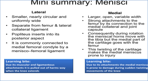
-medial rotation of knee, knee extension - 3medial/ lateral meniscus more easily damaged?medial meniscus, cause it is part of the capsule [cannot move freely]
lateral meniscus, free to move and popliteus is attached to move it out of harms way - 4medial/ lateral meniscus characteristics:
- 5function of meniscus:1) Load spreading, lubrication distribution, proprioceptive feedback, joint stability
2) Flexion and extension between femoral condyles and menisci - 6popliteus attachments:Origin: tibia above soleal line
Insertion: capsule, lateral femoral condyle, posterior aspect of lateral meniscus
function: unlock knee extension - 7Anterior cruciate attachments:APEX:
anterior cruciate from posterior to external (lateral) femoral condyle
Function:
Medial rotation of femur during extension
Stops femur sliding backwards off tibia - 8Posterior cruciate ligaments:Attachments: PAIN
- posterior cruciate from anterior to internal (medial) femoral condyle
function:
Stops femur from sliding forward off the tibia when knee is semiflexed and weight bearing
(ie. upper leg whilst walking downstairs) - 9medial collateral ligements:Insertions:
capsule, medial surface of shaft of tibia, posteriorly to medial meniscus - 10Lateral collateral ligamentInserts: fibula head
- 11Collateral ligaments function:Tighten: knee straightens and rotates in full extension
- 12Extra synovial fat padSeparate synovial membrane from capsule
Function:
Maintains synovial fluid on articular surfaces irrespective of knee position - 13hip and knee referred pain:same innervation:
sciatic, obturator and femoral nerve - 14popliteal artery:Starts: femoral artery exiting adductor canal, closely adherent to joint capsule
End: soleal fibrous arch - 15CapsuleAttachments:
1) Articular margins of femur, tibial plateau and fibula head,
2) Menisci- loose lateral, firm medial meniscus
3) Posteriorly by capsule is perforated by popliteus
Reinforcements:
-medial collateral ligaments
-iliotibial tract
-extensor apparatus
-arcuate and oblique ligaments - 16Bursa:1) Suprapatellar: lower end of quads and femur
2) infra patellar (parson's knee): either side of patella deep/ superficial tendon
3) prepatellar (housemaid's knee): deep to skin overlying patella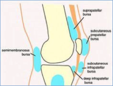
- 17Fibrous capsule function:contain collagen fibres
-thicken lines form ligaments- restricts movement, protect joint from damage - 18synovial membranesecretes synovial fluid- lubricant
- 19articular cartilagehyaline cartilage, frictionless surface
poor blood supply (Avascular) - 20knee joint:Complex hinge joint
- 21Ligaments:Pubofemoral ligament
Iliofemoral ligament- Y shaped
Ischiofemoral ligament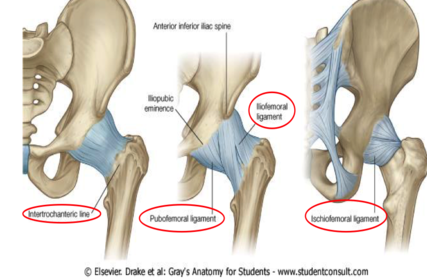
- 22sciatic nerve position at pelvisposterior to piriforms
- 23Acetabulumdeep socket, weight bearing, stable
lined by articular, hyaline cartilage, except acetabular fossa
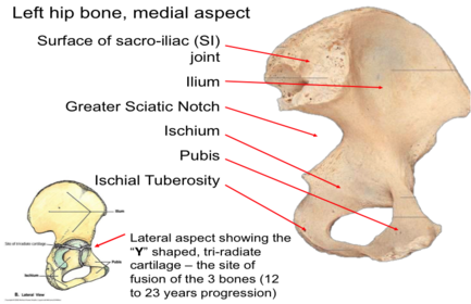
makes space for ligament of head of femur carries some blood supply to femoral head - 24medial hip bone anatomy:
- 25blood supply head of femur from neck:retinacular arteries
- 26Gateways:Sciatic nerve- greater sciatic foramen
Femoral nerve – Gap between inguinal ligament and pelvic bone
Obturator nerve- obturator canal - 27Pas anserinus (goose foot):3 tendons (Sartorius, gracilis, semitendinosus) inserting into upper tibia, with bursa between bone and medial collateral ligament of knee- bursitis
- 283 muscular components: anterior, posterior and medialAnterior- quads and sartorius
[femoral nerve]
Posterior- hamstrings
[sciatic]
Medial- adductors
[obturator] - 29position of fascia lata:3.5 cm below and lateral to pubic tubercle is the saphenous opening for great saphenous and 4 other veins
laterally- Iliotibial tract and gluteus maximus - 30Flexion of hip
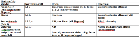
- 31Extension of knee

- 32Adduction of hip:
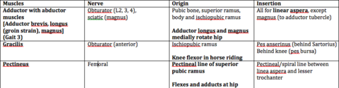
- 33hip lateral rotation:
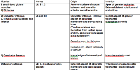
- 34Hip abduction:
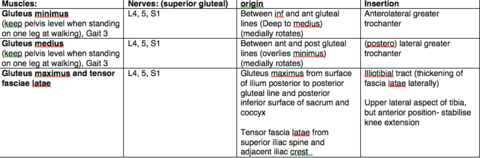
- 35Knee flexion:
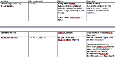
- 36innervation of gluteal muscles:
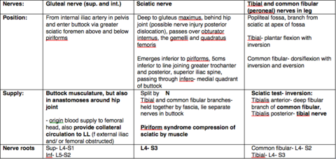
- 37Adductor/Hunter's canal muscles and nervesBetween vastus medialis and adductor longus then magnus, overlain by Sartorius and fascia
[femoral artery, vein, saphenous nerve-supplies sartorius and gracilis
and vastus medialis] - 38Adductor hiatuspassageway from adductor canal to popliteal fossa
[inferior to adductor canal]
[Gap located between adductor magnus and femur]
femoral artery and vein passes through - 39Gait:Gait 1: ‘swing phase’- lift LL, Flex hip and knee
Gait 2: ‘swing phase’- flex hip and extend knee
Gait 3: ‘stance phase’ keep the pelvis level, adductors and abductors
Gait 4: ‘push off phase’- extend hip and knee
Gait 5: 'swing phase'- foot forward
-lateral rotation of hip - 40Femoral triangle: [Lateral to medial] inferior to inguinal ligamentNAVEY
Nerve
Artery
Vein
Canal
Contain lymph vessels and Cloquet’s nodes- receives directly from glands of penis/ clitoris (site of hernia) - 41Femoral ring: base of femoral canalBoundaries:
Anterior- inguinal ligament,
posterior- pectineal ligament,
medial- lacunar ligament,
lateral- femoral vein - 42Femoral sheathExtension of transversalis fascia,
forms sheath around artery and vein,
contains lymph nodes medially in femoral canal ‘’space’’
Femoral nerve lies laterally outside the sheath - 43Femoral triangle:superior: inguinal ligament
Medial: adductor longus
lateral: sartorius
roof: fascia lata
floor: pectineus, illiopsoas, adductor longus - 44Herniaa protrusion of a viscus through the wall of its containing cavity [passes from abdomen through femoral canal]
in relation to pubic tubercle- posterior and lateral - 45Politeal fossa: [behind knee] diamond shaped depressionRoof- fascia lata
Floor- femur, capsule and popliteus
- 46medial compartment (adductors)obturator nerve
From pelvis through obturator foramen
To the medial thigh on each side of adductor brevis - 47Anterior compartment (Extensor)Femoral nerve and vessels passes behind inguinal ligament
- 48femoral and obrturaotr nerves:
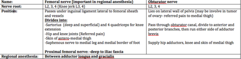
- 49small/ great saphenous veinsSmall saphenous vein:
Start: behind lateral malleolus and enters popliteal [moves along with sural nerve] - 50Great/ long saphenous veinfrom 2.5cm anterior to medial malleolus
to enter femoral 3.5 cm below and lateral to pubic tubercle
(Joins femoral vein in groin)
4 tributaries ligated during a varicaose vein surgery to prevent reoccurance
Passes through the cribiform fascia in the saphenous opening in the fascia lata and enter the femoral vein - 51Venous flow- superficial to deep- superficial to deep (via perforators and muscle pump, arterial pulsation and negative intra-thoracic pressure help venous return to heart
If blood escape backwards, superficial veins become dilated- varicose veins
Compressed by muscle contraction within fascia compartment, forcing blood back to heart - 52Lymph drainage:Inguinal lymph nodes ‘’T shaped’’- parallel to inguinal ligament, alongside great saphenous vein, drain to deep inguinal and external iliac nodes
superficial nodes:
Abdol wall to horizontal, superolateral group
Perineum to horizontal, superomedial group
Lower limb to inferior, vertical group
Deep nodes:
Medial to femoral vein [in triangle]
drainage to
Femoral canal to external iliac nodes - 53Dermatomesstand on S1, squat on S2, Sit on S3 (knee)
-L4, 5, S1- both plantar and dorsal surfaces - 54safe injection region:Injection for intramuscular- upper outer quadrant of buttock
- 55Lateral femoral cutaneous /lateral cutaneous nerve of thigh Femoral branch of genitofemoral Saphenous nerve
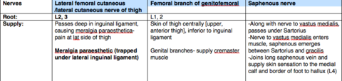
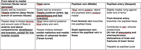
- 56popliteal fossa innervation
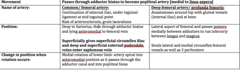
- 57Femoral artery
- 58Sciatic nerve(L4, 5, S1,2)
Midway between ischial tuberosity and greater trochanter
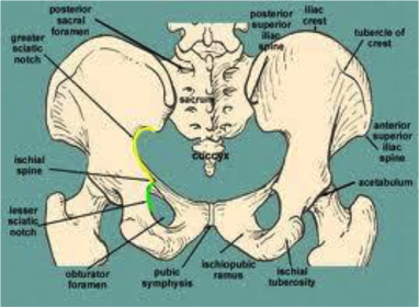
Gateways- greater sciatic foremen, obturator canal (abduction of hips), femoral - 59Hip anatomy
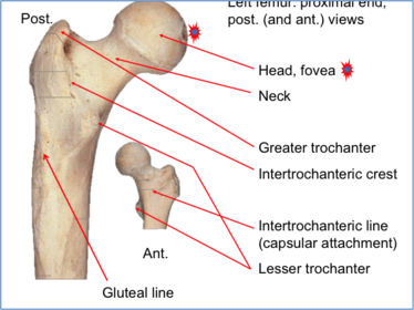
- 60Femur Proximal end (posterior and anterior views)
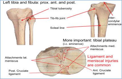
- 61Tibia and fibula (proximal superior part)
- 62Efficacy:max response of tissue obtained by occupying less than 100% receptors
[ability of drug to generate stimulus once bound to receptor] - 63Affinity:concentration of drug needed to occupy 50% of receptors at equilibrium
- 64Efficacy in drug receptor interactions:max response: f [DR]/ [RT]
efficient drugs- occupy fewer receptors to give response than less efficient - 65partial agonist:decrease response, some receptors occupied by partial agonist, smaller response achieved than all receptors occupied
- 66competitive agonist:drug binds reversibly with receptor to form complex, not evoke response
[D] + [R] + [A] = [DR] + [AR] - 67full and partial agonist:
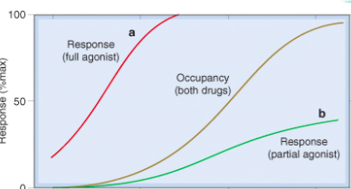
- 68competitive antagonist:max agonist response never change
pA2 = -log10(KA)
The pA2 - comparing the affinity of an antagonist (similar to the pD2 of an agonist)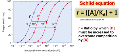
- 69irreversible competitive antagonism:slope decreases
Max response to agonist decreased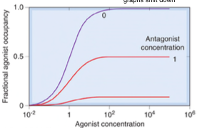
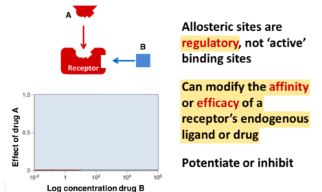
- 70Allosteric binding sites
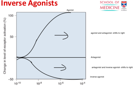
- 71Inverse agonist:
- 72Drug Receptor InteractionsG ligand protein channel
direct control on ion channel
direct control of effector enzyme
control of DNA transcription - 73drug receptor theory:1. Enzymes
2. Carrier molecules
3. Ion channels
4. Neurotransmitter, hormone or local hormone receptors - 74receptor theory:lock key analogy
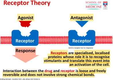
- 75antagonist and agonist definitionsboth bind to same receptor- similar chemical properties
One activates receptor [agonist] and the other does not [antagonist] - 76law of mass action to drug receptorrate of a chemical reaction is proportional to the product of the concentration of the reactants
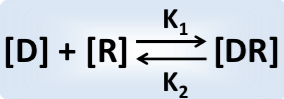
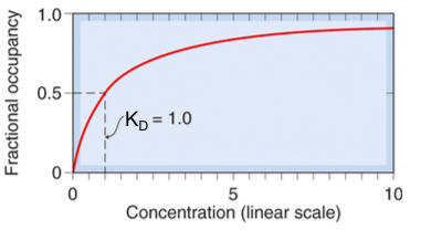
- 77equilibrium dissociation constant (KD):Represents the concentration of drug required to occupy 50% of the receptors at equilibrium.
KD is:
1. different for every drug
2. a measure of the affinity of any one drug for a receptor - 78dissociation constant in log scale
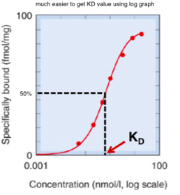
- 79occupancy theory:[D] is the concentration of drug
KD is the equilibrium dissociation constant of the drug
- 80dissociation constant in linear scale
- 81receptor theory:drugs are graded
drug concentration VS the magnitude of the response obtained - 82occupancy theory:
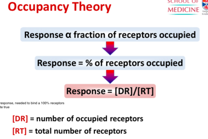
- 83bones and ligaments of gluteal region:Bones
Postero- lateral to hip bone and proximal femur
Passes through pelvis, via greater sciatic foramen to gluteal region, then thigh/ via lesser sciatic foramen to perineum
Ligaments- Sacrotuberous and sacrospinous ligament - 84Hip extension:Gluteus maximus
(Gait 4)
Nerves (Inferior gluteal)
L5, S1, 2
Origin
Ilium posterior and sacrum and coccyx
(Laterally rotates and abducts)
Action of paradox- although extensor, can act like a hip flexor by relaxing against gravity, same as biceps
Insertion
Deep fibres- gluteal tuberosity (extending from upper end of linea aspera)
Superficial fibres- iliotibial tract - 85Trendelenburg gait (Aided by iliotibial tract)Nerve/ muscle injury- shortened femoral neck, congenitally dislocated hip, pelvis dips to opposite side
- 86anterior leg
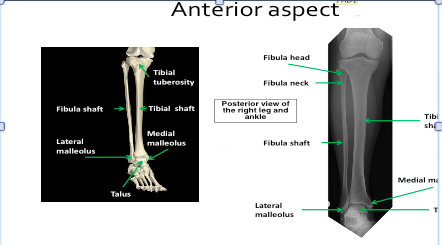
- 87Tibia shaft fractures:Middle and distal third [tibia narrowest]
Poor blood supply [upper third most]-
compound, non union fractures take 6 weeks to unite [tearing of posterior tibial artery] - 88Fibula fracturesNeck of fibula- common peroneal nerve affected- direct trauma
Indirect trauma- Maisonneuve fracture- spiral break in upper fibula, result of excessive ankle inversion - 89Non- healing fractures of fibula shaft rare:1) Surrounded by muscles
2) Supply distally by branches of fibula artery - 90Anterior compartment:
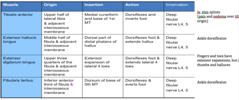
- 91Extend toes if ankle dorsiflexed?2 short toe extensor muscle arise distal to ankle- extensor digitorum brevis [EDB]
and extensor hallucis brevis [EHB]- not affected - 92lateral compartment of muscles:
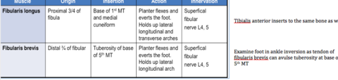
- 93toes flexion and extension
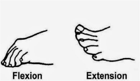
- 94dorsiflex/ plantarflex
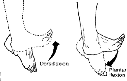
- 95Extensor retinacularSuperior extensor retinaculum
-lies just above ankle joint, runs from tibia to fibula
Inferior extensor retinaculum
-Y shaped
arises from calcaneus laterally and inserts into medial malleolus and fascia overlying sole of foot - 96Fibular retinacularSuperior
Between lateral malleolus and calcaneus
Inferior
From fibular trochlea on lateral calcaneus
Upper: around fibularis brevis, inserts to calcaneus and inferior extensor retinaculum
Lower: around fibularis longus to insert into calcaneus - 97Synovial sheathTendon from extensors and fibular components when passing under retinacula
-Only tibialis anterior has a synovial sheath under superior extensor retinaculum
Overuse trauma- inflammation
Pain and swelling - 98common fibular nerve injury:neck of fibula (Direct trauma. Compression)
impairs ankle dorsiflexion- patient developed high stepping gait to allow toes to clear ground in walking
results in foot drop- paralysis of anterior muscles
- foot laterally rotates - 99Common fibular nervefrom sciatic nerve →between butt and popliteal fossa
from popliteal fossa, run through fibularis longus (fibula neck), divides into superficial and deep fibular nerves - 100Deep fibular nerveFrom CFN, curves around fibula neck within fibularis longus,
Course: pierces anterior intermuscular septum and EDL, descends on tibialis anterior and interosseous membrane, terminates at ankle
All leg’s anterior group of muscles
Short toes extensors, under joints and skin of 1st and 2nd toes [1st web space] - 101Superficial fibular nerveCFN runs in fibularis longus
Course: from fibular compartment pierces the anterior intermuscular fascia
Subcutaneously over the front leg and dorsum of foot [cutaneous sensation] - 102Anterior tibial arteryFrom popliteal artery
descends medial to deep fibular nerve, supply compartment muscles and link anastomosis with knee and ankle - 103Fibular arteryruns laterally within deep posterior compartment
- 104Dorsum of foot:Skin: thin, subcutaneous fat
Dorsal venous arch:
-great saphenous vein, medially and anterior to medial malleolus
-short saphenous vein, laterally and posterior to lateral malleolus
Cutaneous nerves:
Sural lateral skin, saphenous medial skin, deep fibular 1st cleft dorsum of foot
Deep fascia continuous with inferior extensor retinaculum
Anterior tibial artery:
-passes deep to extensor retinacular and continues was dorsalis pedis lateral to extensor hallicus longus
-passes between 2 heads of 1st dorsal interosseous to form deep plantar arch - 105posterior leg
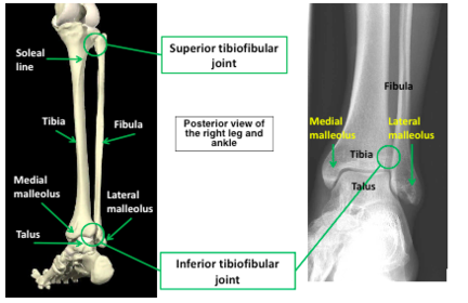
- 106medial aspect:
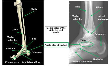
- 107Foot (Dorsal view)
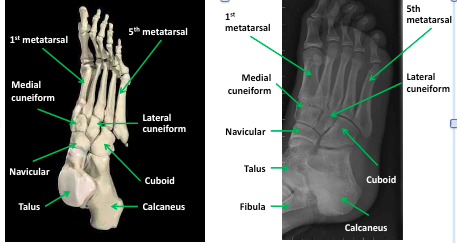
- 108Interosseous membraneTough ligament between tibia and fibula
Function:
Keeps both bones aligned throughout ankle movement
Attachment extensor and flexor muscles of ankle
Border of osteofascial compartments - 109Tibiofibular linksTypes:
Interosseous membrane
Superior tibiofibular joint
Inferior tibiofibular joint
Movement: limited gliding - 110Deep posterior
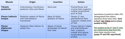
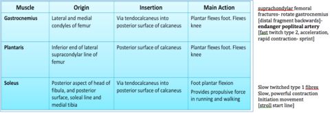
- 111superficial muscle
- 112Achilles tendon bursaAction:
Reduce friction during flexion and extension of ankle
Clinical:
Inflammation following excessive movement
Overlying pressure from large shoe’s heal tab - 113Achilles tendon Pathology:Strains and tears: common,
Gastrocnemius (exercise)
Rupture due to poor blood supply: quick start, sudden pain in posterior calf. Diff weight bear [Simmond’s test]
Mid section:- site of rupture
least vascular area and poor anastomatic linkage both proximally and distally
ability for self repair is reduced leading to progressive degeneration - 114Tarsal tunnel: Tom, Dick And Naughty Harry, A- achilles tendonFriction reduced by synovial sheath, roof- flexor retinacular
Flexor retinacula: From medial malleolus to calcaneus
-prevents the tendons bowstringing when ankle plantar flexes and extends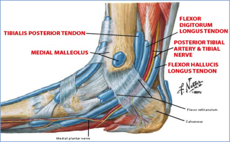
- 115Compartment syndrome:Cause: boundaries provide little stretch, ie. from bleeding, inflammation/ infection [cause intra- compartmental pressure]
Effect: ischaemia, nerve symptoms and pain during stretching
Site at risk: anterior and posterior leg compartments
Deep compartment most likely affected- transverse fascia strong
[pulseless late sign, 2 posterior compartments] - 116structures of posterior leg are risk of damage?medial soleus, tibial nerve, gastronimius, deep compartment- TP, FDL
- 117Posterior nerves:
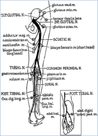
- 118somites
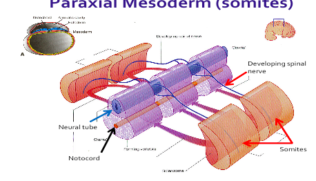
- 119myotomes and dermatomesFormation of myotomes:
- somites develop muscles
- nerve develop from muscle developing adjacent somite
- muscles receive motor nerves from adjacent segment of spinal cord
ie. abduction supplied by nerves from C5,6- MYOTOMES
Formation of dermatome:
Skin develops from ectoderm adjacent to a specific somite innervated by sensory neurons arising from adjacent spinal level ie. skin from lateral side C6- DERMATOMES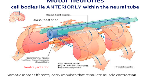
- 120Intramembranous ossificationFormation of bone in fibrous connective tissue (formed from condensed mesenchyme cells)
Formation of flat bones ie. mandible and flat bones of skull
Mesoderm- germ layers: ectoderm, mesoderm, endoderm
Mesenchyme- connective tissues derived from mesoderm - 121Rotation of limbsForelimbs rotate 90 degree laterally, hind limbs rotate 90 degrees medially
Flexor compartment anterior UL and posterior in LL
Sole of foot = palm of hand
Big toe (hallux) = thumb (pollux) - 122Limb development:UL appear approx. 24 days between C5-T1
LL appear approx. 28 days between L1-5/S1/2
Week 8- all maj components of limbs present
Medial rotation of LL complete - 123HOX genes in limb development:Determine body axis and position of limbs
Variation in HOX:
UL (TBX5),
LL (TBX4)- determine shape of bones
HOX B8 controls position of UL - 124Limb growth:
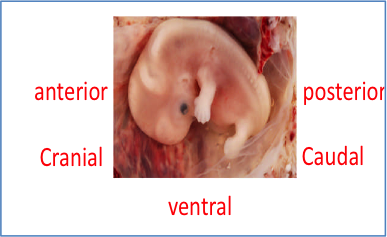
- 1251) proximo- distal axis 2) antero- posterior axis 3) dorso- ventral axis
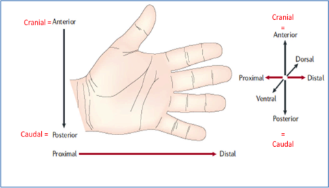
- 126Proximo- distal axisEctoderm form outer layer of limb epidermis, thickened at apex of developing limb to form Apical Ectodermal Ridge (AER)
Limb bud:
Core of mesenchyme derived from parietal layer of lateral plate mesoderm
Undifferentiated, proliferating cells- Progress zone in tissue
Move further away from AER to cartilage and muscle
1) HOX 8 control position of limb on long axis of body
2) Initial outgrowth of forelimb is controlled by TBX5 gene and FGF- - 127ZPA controls cranio- claudial axis (anterior and posterior axis)ZPA-zone of polarizing activity
1) ZPA initiates expression of protein sonic hedgehog (SHH)
2) SHH causes production of retinoic acid
3) Cluster of cells near posterior border of limb produced retinoic acid (vit A)
-Regulates AP axis
4) Ensures thumb grows on (Cranial) anterior side of limb bud - 128Dorso- ventral axisBMP in ventral ectoderm induce EN1,
EN1 represses WNT7a restricting expression to dorsal limb ectoderm - 129limb defects:UL abnormalities more common than LL
Amelia- complete absence of limbs
Meromelia- partial absence of limbs
Phocomelia- absence of long bones
Micromelia- segments are abnormally short - 130Thalidomidesleeping pill and babies with flipper like arms/ legs with cardiac abnormalities and atresia, phocomelia/ Amelia
- 131Holt Oram syndromeTBX 5 mutation leads to failure of limb bud development
UL deformities, heart defects - 132Defect limb development:Brachydactyl- short digits
Syndactyly- fused digits- failure of apoptosis (1 in 2000 births)
Polydactyl, extra digits
Cleft foot- lobster claw deformity - 133Talar shape:Bones: malleoli and inferior tibia, talus
pressure on lateral malleolus in dorsiflexion
-less stability in plantar flexion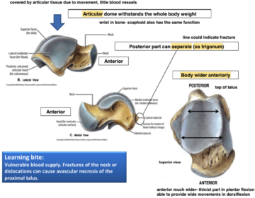
- 134Ankle: superior stabilizing structures-anterior and posterior tibiofibular ligaments
-interosseous ligament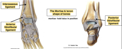
- 135Ankle: medial stabilizing structures Medial (deltoid) ligament -superficial and deep, stronger than sustentaculum talitibiocalcaneal
- strong, doesn’t allow movement [eversion/ inversion]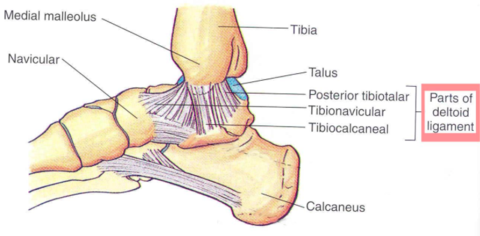
- 136Foot osteology:
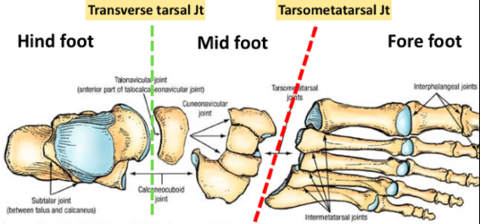
- 137Ankle ligament injury:Stability- max dorsiflexion
Injury most likely: plantarflexion, inversion injuries, lateral ligaments
Severe: many fibres partly/ completely, blood tracks under deep fascia, instability of ankle joint, associated fractures - 138Subtalar joints:2 joints act as 1- clinical subtalar joint
posterior- talus body articulates with posterior calcaneus
Anterior- part of tibionavicular joint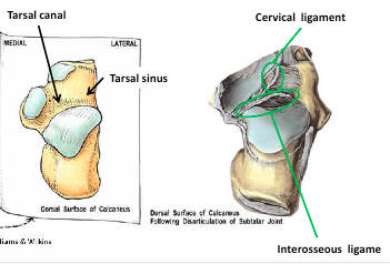
- 139lateral ligaments:Calcaneofibular ligament
[weak]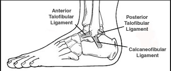
- 140Transverse tarsal joints:Talo- calcaneal- navicular joint:
Ball- talar head,
socket- navicular (Anteriorly),
sustentaculum tali and anterior calcaneus (inferiorly)
(between) spring and bifurcate ligaments
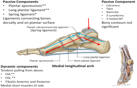
Spring ligament (Calcaneo-navicular):
-sustentaculum tali and posterior nevicular
-supports tala head
-maintains medial longitudinal arch
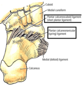
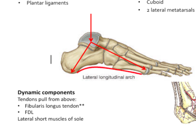
Short plantar- (calcaneo-cuboid):
Overlies spring ligament
Anterior calcaneous and cuboid
-maintain longitudinal arches
Long plantar (long calcaneo- cuboid):
Overlies short plantar
Posterior calcaneous to 2-4 MT heads
Bridges fibularis longus
Maintains longitudinal arches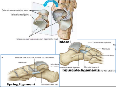
- 141Calcaneo- cuboid joint: Planar, little movement possible, strengthen by bifurcate ligaments medially and long/ short plantar ligaments
- 142Inversion and eversion:1) intial movements around longtudinal axis occur at transverse joint (limited ROM)
2) force transferred passively to subtalar joint, much more mobile
3) reaches limit, talus rotates and cause movement of fibula
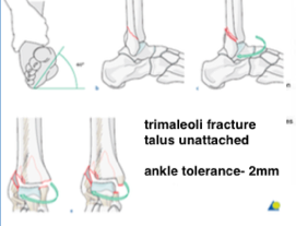
walking on uneven ground- foot remains pointing straight forward due to recipocal rotation at knee, hip/ pelvis mediated by extrapyramidal control - 143injury:Talus rotation:
Impact/ rotation fracture
Malleoli fractire- inversion/ eversion injuries- direct force through talus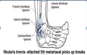
- 144Suppination and pronation: Ankle axis: posterior- plantarflex anterior- dorsiflex subtalar axis- long plantar ligament[not directly on axis]/ Longitudinal axis: medial- inversion and adduction of forefoot lateral- eversion and abduction of forefootAnkle axis:
posterior- plantarflex
anterior- dorsiflex
subtalar axis- long plantar ligament
[not directly on axis]/ Longitudinal axis:
medial- inversion and adduction of forefoot
lateral- eversion and abduction of forefoot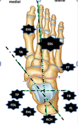
- 145Tarsometatarsal:Cuneiform- 1,2,3rd MT
Cuboid- 4 and 5th MT
Difference from carpometaparcpal:
-little movement/ medial cuneiform 1st MT/ 2nd MT base - 1462nd tarsometatarsal joint:metatarsal base fixed between medial and lateral cuneiforms
Clinical: 2nd MT stress fracture, marker of high energy transfer
March fracture- in army and dancers, 2nd MT fracture
Lizfranc dislocation: dorsal pedis artery affected - 147MTP and IP joint difference:Both have collateral ligaments
Less movement,
risk of Hallux Valgus/ bunions as long tendons only held in position by fibrous bands - 148aponeurotic and muscular layers of the sole and compare them to the plam
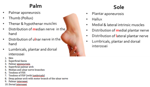
- 149Skin and subcutaneous:Thick and fibrous,
Exposed position: lacerations, embedded foreign bodies
Clinical significance: -trap infection, impede distribution of local anesthetic, specialized skin over weight bearing areas - 150Plantar Aponeurosis: [plantar fasciitisLocation: from calcaneus to 5 fibrous flexor sheaths
Vertical fibrous bands to skin 1st and 5th MT
Function: longitudinal arches support, grip
Difficult and for local anaesthetic - 1511st muscle layer of sole:Flexor digitorum brevis (medial plantar nerve)
FDS [same attachments with soleus]
Lateral 4 toes can flexed with ankle dorsiflexion
Abductor halluces [medial plantar nerve]
-abducts hallucis
Abductor digiti minimi [lateral plantar nerve]
-abducts little toe - 152Superficial neurovascular planeBetween 1st and 2nd muscle layer
Medial plantar nerve= median nerve [more muscle bundle than lateral n. - FHB,FDB]
-Runs with medial plantar nerve
-Abd H, FHB, FDB, and 1st lumbrical
Skin- medial 3.5 toes, plantar surface and nail beds
Lateral plantar nerve= ulnar nerve
Runs with lateral plantar artery
[nerve link over plantar surface of 4th MT/P joint- metatarsalgia could occur] - 1532nd muscle layer of sole:Flexor hallicus longus [TIBIAL n.]
-propulsion, medial arch support
Flexor digitorum longus [TIBIAL n.]
=FDP, connected to FHL, 4 lumbricals and 4 lateral toes [1 medial and 3 lateral plantar n]
Quadratus plantae [lateral plantar n.]
Straighthen the oblique pull of FDL
Lateral 4 toes flexed with ankle dorsiflexion - 1543rd muscle layer of sole:Flexor hallucis brevis [medial plantar n.]
-insertion each contains a sesemoid bone [FHL passage]
-flex 1st MTP joint
Adductor halluces [lateral plantar n.]= adductor pollicis
Flexor digiti minimi [lateral plantar n.] - 155Deep neurovascular planeLateral plantar nerve- supplies intrinsic muscles
Lateral plantar artery- runs with nerve, only 1 plantar arch, same function as hand, joins dorsalis pedis artery - 1564th muscle layer of sole:Interossei (lateral plantar nerve)
Flex MTP and extension of IP joint
Secondary: Abduction and adduction related to axis through 2nd MT and toe
Long tendons: (tibial nerve)
Fibularis longus, tibialis posterior tendons - 157Cutaneous supply:Tibial: Medial calcaneal nerve, medial plantar nerve (median nerve), lateral plantar nerve (ulnar nerve)
Sural nerve: lateral skin
Saphenous nerve: medial skin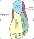
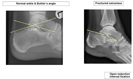
- 158Calcaneal injury:
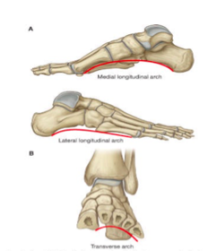
- 159Foot arches functions:Absorb and distribute weight bearing forces,
Adapt to uneven surfaces
[terrain adaptation- standing/ reduce energy expenditure]- standing short period- ligaments, long period- muscles - 160Types of foot arch:
- 161Medial longitudinal arch: Passive/ dynamic structures
- 162Lateral longitudinal arch: Passive/ dynamic structures
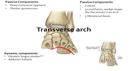
- 163Transverse arch: Passive/ dynamic structures
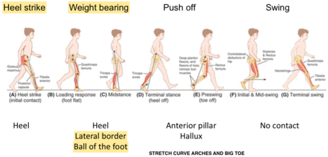
- 164Gait cycle: Heel strike and weight bearing- heel and lateral border and ball of foot Heel rising EHL and EDB extends toes -FHL and FDL tendons stretch, subsequent contraction increase -heightens medial longitudinal arch -increase force of push off by transmitting force through medial toes
Información
- Descripción
- Fichas sobre LL, creado por denise tan1844 el 23/04/2015.
- Creado por
- denise tan1844
- Visitas
- 1
- Valoración