
Fichas
Gross Anatomy of the Heart
Fichas del conjunto (26)
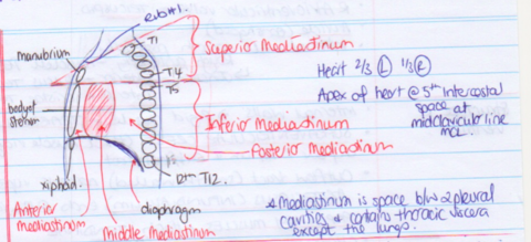
- 1Draw a sagittal plane & label mediastinums & key landmarks
- 2Location of the heart* Middle Mediastinum
* 2/3 Left of midline
* Anterior & Deep to sternum & 3-5 Costal cartilage
* Apex @ MCL 5th Intercostal space - 3Name Pericardium & heart layersPericardium (1) Fibrous, (2) Parietal Serous Pericardium (3) Visceral Serous Pericardium.
Cardiac: (1) Epicardium = Visceral serous, (2) Myocardium, (3) Endocardium.
Then Fibrous skeleton - 4Draw coronal Heart plane
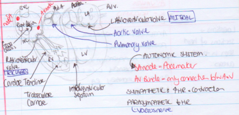
- 5Describe Right Atrium* Veins -> SVC, IVC, Coronary Sinus
* Intra-atrial Septum
* Fossa Ovalis
* Auricle
* Internal wall - Smooth posteriorly; Rough anteriorly = Musculi Pectinati. Junction b/w smooth & rough = SULCUS TERMINALIS exteriorly & CRISTAE TERMINALIS internally - 6Describe Right Ventricle* TRABECULAE CARNAE
* SUPRAVENTRICULAR CREST
* CONUS ARTERIES (INFUNDIBULUM)
* PAPILLARY MUSCLES
* CHORDAE TENDINAE
* Tricuspid valve & Pulmonary valve
* SEPTOMARGINAL TRABECULA - 7What are TRABECULAE CARNAEInternal ridged walls in the Ventricles
- 8Where & What is the SUPRAVENTRICULAR CRESTR Ventricle. thick ridge that separates inflow (rough) & outflow tract (smooth)
- 9What & where: CONUS ARTERIES (INFUNDIBULUM)R Ventricle outflow tract. Narrows superiorly, and ends at the entrance to Pulmonary Trunk. Lined with Semilunar Pulmonary valve
- 10What & where: PAPILLARY MUSCLES. Which one is clinically importantattach to Tendinous cords, which attach to the AV valve. Also attach to the Trabeculae Carnae.
Anterior Papillary Muscle - carries part of the conduction system - 11What & where: SEPTOMARGINAL TRABECULAModerator band. Curved muscle bundle from inferior inter ventricular septum to the base of the anterior papillary muscle - carries part of the conduction system
- 12Structure: Left AtriumMostly smooth walled (except L auricle - Pectinate muscle) fossa ovals depression. 4 Pulm. veins (Superior/Inferior: L/R) enter on the smooth posterior wall.
- 13Diff: Atriums L/RLA wall slightly thicker
- 14Structure: L VentricleThickest wall (x3). Trabecular more & finer. Large papillary muscles. Aortic Vestibule. Bicuspid valve. Has outflow not as obvious.
- 15Where & names: Heart Valves & CuspsSemilunar (1) Pulmonary - Anterior, R, L
(2) Aortic - Posterior, R, L
AV Valves: (R) Tricuspid - Septal, Anterior, Posterior.
(L) Mitral - Anterior, Posterior - 16Coronary Arteries (Left)LCA -> AIB (aka LAD) + circumflex ->
Variation: LAD -> lateral branch (aka diagonal)
Circumflex -> Left Marginal Branch -> (40%) SA nodal branch
(10%) Circumflex -> PIA - 17LCA suppliesLA, Most LV, Part RV, most IVS, AV bundle, RBB & LBB [ & SA node (40%)]
- 18Coronary Arteries (Right)RCA -> Acute (aka Marginal) + PIA + AV nodal branch
(60% - RCA -> SA nodal branch)
(80% RCA -> PIA - posterior descending) = RCA dominant
May supply Apex if LAD is short - 19RCA suppliesRA, Most RV, Diaphragmatic surface LV, 60% SA node, 80% AV node
- 20Cardiac Veins* Great CV (w AIA)
* Middle CV (w PIA)
* Small CV (w R marginal branch)
Terminate @ coronary sinus
Anterior drain directly into heart. - 21LymphFollows blood vessels -> lymph nodes around trachea -> R lymphatic duct OR Thoracic duct
- 22Where & What: Fibrous skeletonDense CT, fibrous ring around each valve, connects valves & forms part of interatrial & interventrical wall.
Function (1) Anchor (2) prevents electrical discord
MYOCARDIUM attaches to it. - 23Conduction System(1) SA
(2) AV
(3) AV Bundle (Bundle of His)
(4) Bundle Branches (L&R)
(5) Purkinje fibres - 24Sympathetic NS on HeartSympathetic: >HR, >force, >contractility, > coronary artery flow.
Preganglionic cells in lateral horn T1-5.
Primarily act on SA node, but also on AV node - 25Parasympathetic NS on HeartTravel in Vagus nerve (CNX). Synapse @ cardiac plexus or near SA & AV nodes.
Vagal tone = 60-70bpm - 26Heart pain: How & WhereDermatomes T1-5. chest, upper limb, & other areas. Neck & Jaw (? from Vagus nerve).
Información
- Descripción
- Week 1 AMW
- Creado por
- Fiona Beer · feb 2016
- Actualizado
- 24 ene 2017
- Visitas
- 83
- Etiquetas
- Valoración