
Fichas
UA
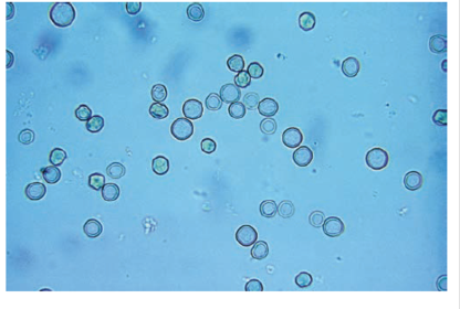
Fichas del conjunto (94)
- 1—Normal RBCs
- 2—Microcytic and crenated RBCs
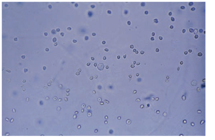
- 3—Budding Yeast
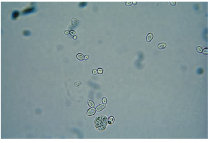
- 4—Air Bubble
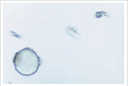
- 5—Squamous epithelial (with oil droplets and NOT RBCs)
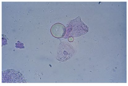
- 6—Dysmorphic RBCs
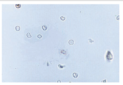
- 7—RBCs and one WBC
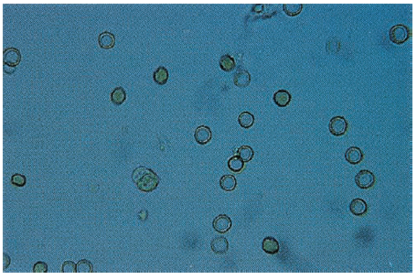
- 8—WBCs (notice the multi-lobed nuclei)
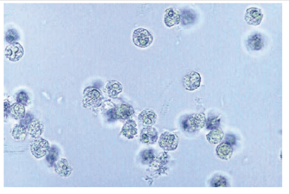
- 9—WBC clump
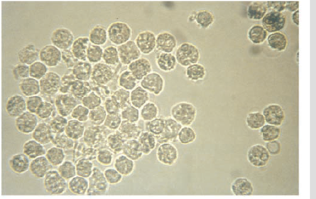
- 10—Eosinophils (Hansel stained)
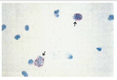
- 11—WBCs (with acetic acid nuclear enhancement)
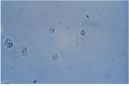
- 12—Squamous epithelial
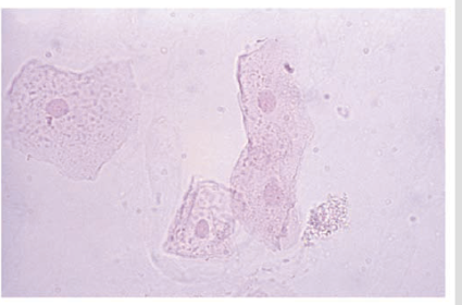
- 13—Squamous, RTE, WBC, caudate transitional, RBC
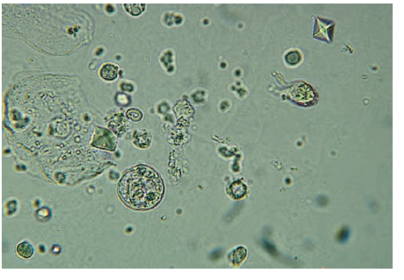
- 14—Squamous epithelial and phenazopyridine crystals after refridgeration
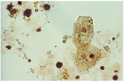
- 15—Squamous epithelial (under low power)
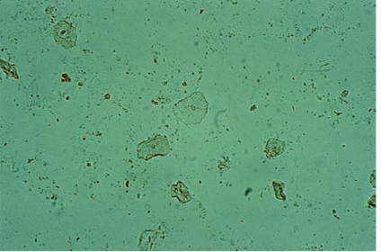
- 16—Clump of squamous epithelial cells
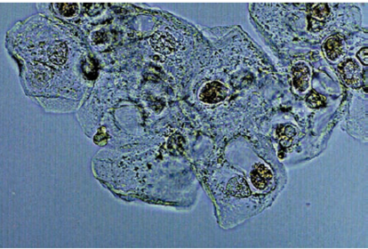
- 17—Clump of squamous epithelial cells with folded forms
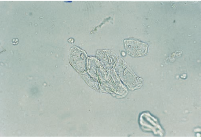
- 18—Spherical transitional epithelial cells
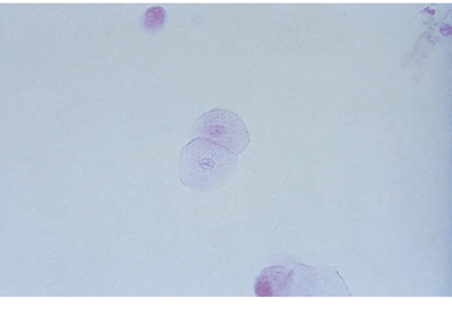
- 19—Caudate transitional epithelial cells
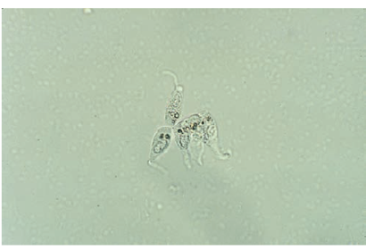
- 20—Syncytia of transitional epithelial cells
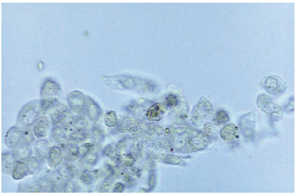
- 21—RTE cells. Oval distal convoluted tubule cells (notice the eccentrically placed nuclei).
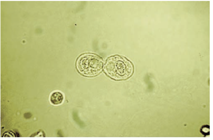
- 22—RTE cells (cuboidal, from collecting duct)
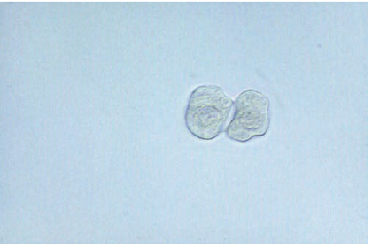
- 23—RTE cell (proximal convoluted tubule cell with granules and attached fat globules).
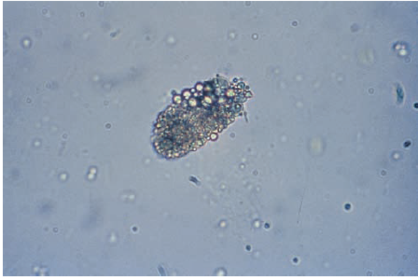
- 24—Fragment of RTE cells from collecting duct
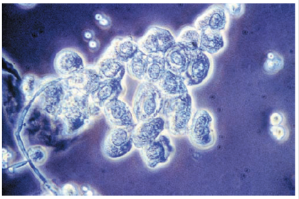
- 25—Oval fat body
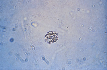
- 26—Oval fat body (sudan III stained)
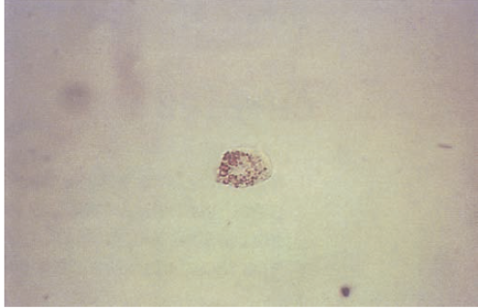
- 27—Oval fat body (bright field on right and polarized on left (notice the Maltese cross))
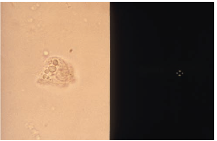
- 28—Hemosiderin granules
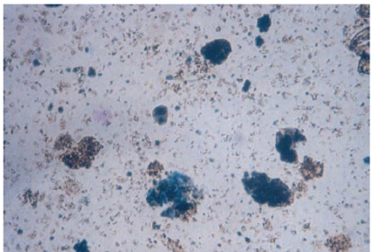
- 29—Bacteria
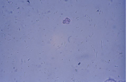
- 30—Yeast (mycelial forms)
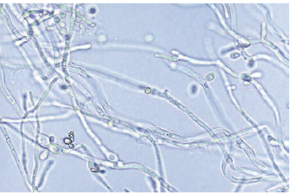
- 31—Mucous threads
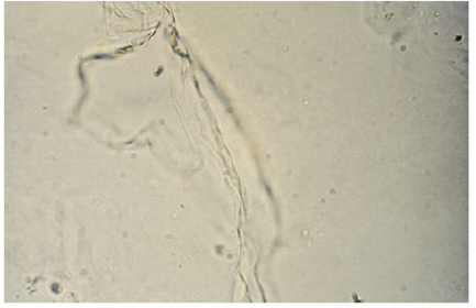
- 32—Hyaline casts
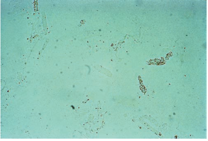
- 33—Hyaline cast (under phase microscopy)
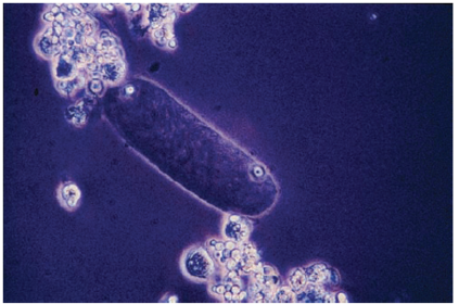
- 34—Convoluted hyaline cast
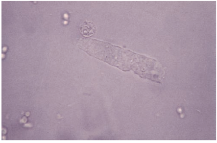
- 35—Hyaline cast (top - with amorphous urates and bottom - attached to mucus pseudocast)
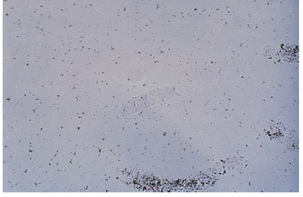
- 36—Hyaline cast
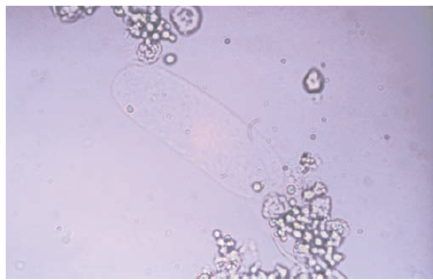
- 37—Hyaline cast containing the occasional granules
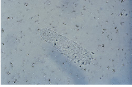
- 38—RBC cast
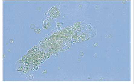
- 39—Disintegrating RBC cast (notice the presence of free RBCs)
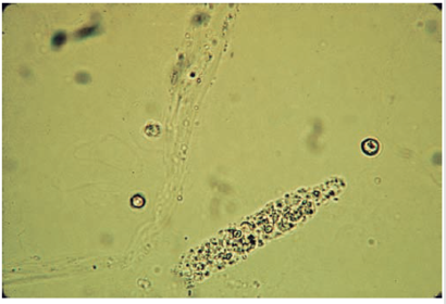
- 40—RBC cast (under phase microscopy)
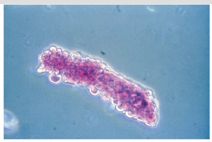
- 41—Cast containing hemoglobin (also RBCs and yeast are present)
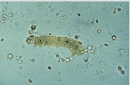
- 42—Granular dirty brown cast
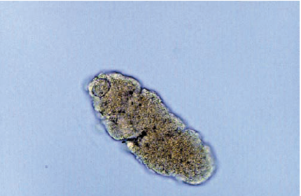
- 43—WBC cast
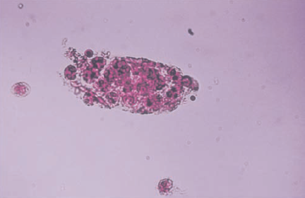
- 44—Disintegrating WBC cast
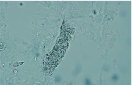
- 45—WBC clump (notice the absence of a cast matrix)
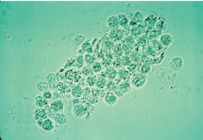
- 46—RTE cell cast
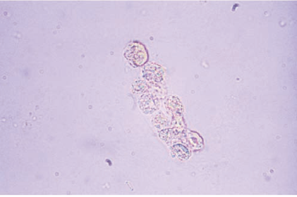
- 47—RTE cell cast
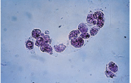
- 48—RTE cell cast
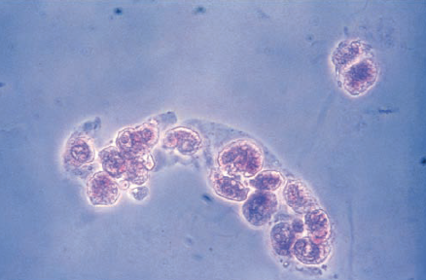
- 49—Fatty cast showing adherence of fat droplets to cast matrix
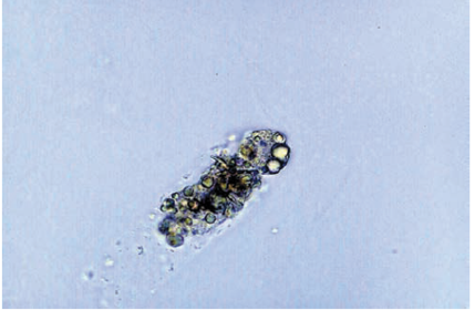
- 50—RTE cast with bilirubin stained cells
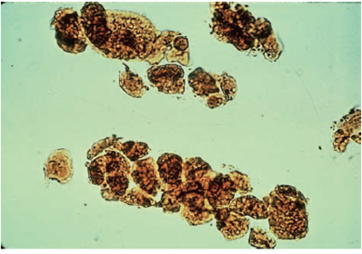
- 51—Fatty cast
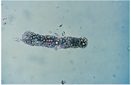
- 52—Finely granular cast and uric acid crystals
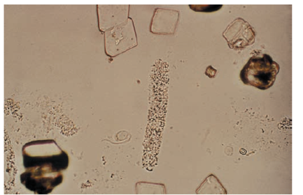
- 53—Granular cast formed at a tubular bend
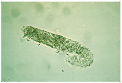
- 54—Granular disitenigrating cellular cast
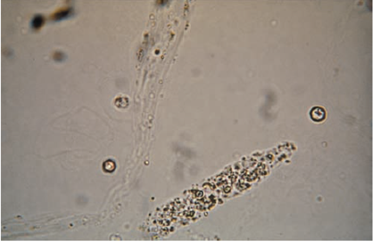
- 55—Fatty cast under phase micriscopy
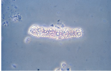
- 56—Coarsely granular cast, squamous epithelial cell, and mucous
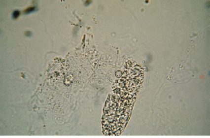
- 57—Waxy casts
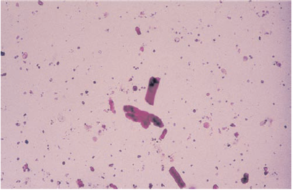
- 58—Granular cast degenerating into waxy cast
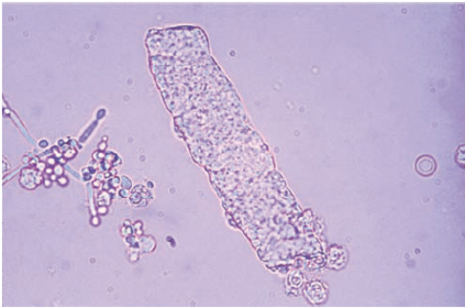
- 59—Waxy cast
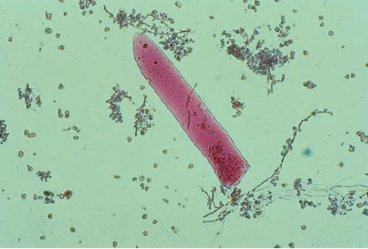
- 60—Waxy cast
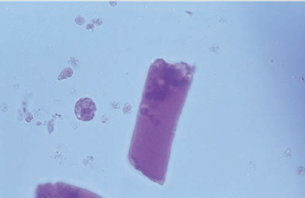
- 61—Broad granular cast becoming waxy
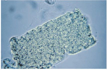
- 62—Broad waxy cast
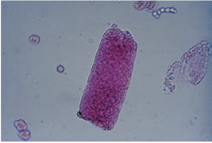
- 63—Broad bile stained waxy cast
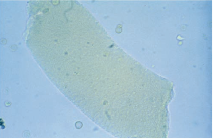
- 64—Amorphous urates
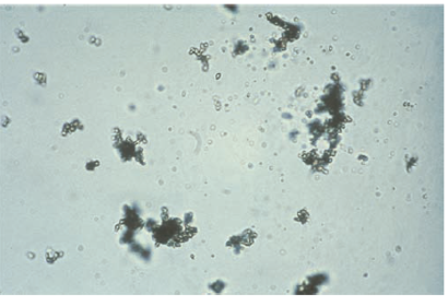
- 65—Uric acid cryststals
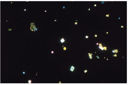
- 66—Uric acid crytals
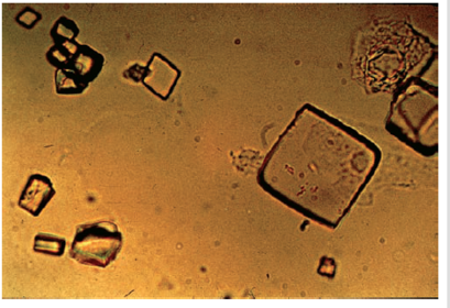
- 67—Uric acid crystals under polarized light
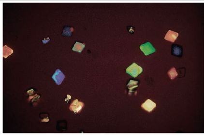
- 68—Clumps of uric acid crystals ( notice the whetstone, not the hexagonal shape that differentiates uric acid crystals from cysteine crystals)
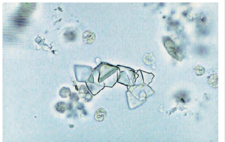
- 69—Dihydrate calcium oxalate crystals
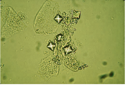
- 70—Dihydrate calcium oxalate crystals under phase microscopy
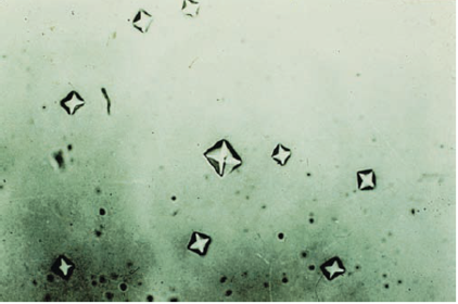
- 71—Amorphous phosphates
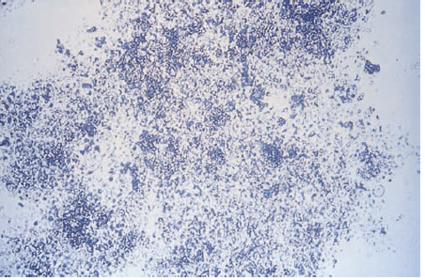
- 72—Triple phosphate crystal
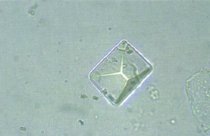
- 73—"coffin lid" and other forms of triple phosphate crystals
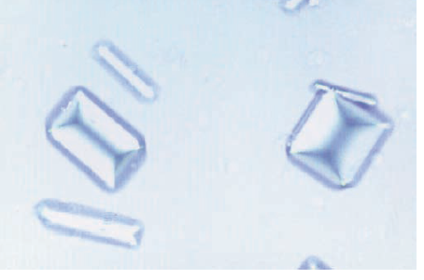
- 74—monohydrate calcium oxalate crystals
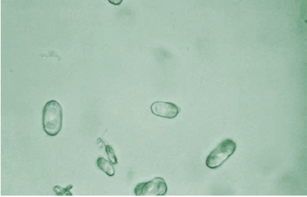
- 75—triple phosphate crystal sand amorphous phosphates
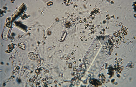
- 76—Ammonium biurate crystals
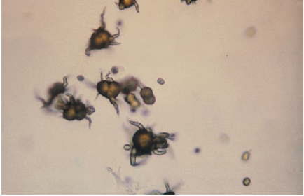
- 77—Calcium carbonate crystals
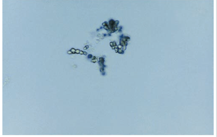
- 78—Cystine crystals
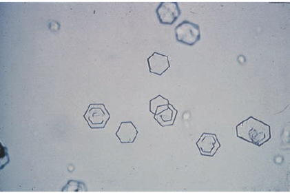
- 79—Clump of cystine crystals
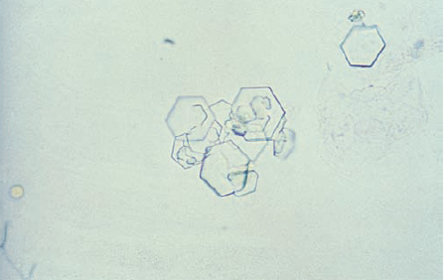
- 80—Cholesterol crystals
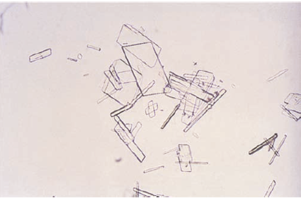
- 81—Tyrosine crystal sin fine needle clumps
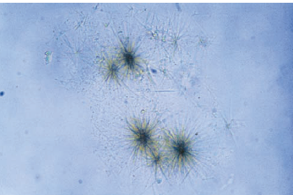
- 82—Tyrosine crystals in rosette form
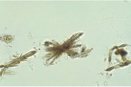
- 83—Leucine crystals
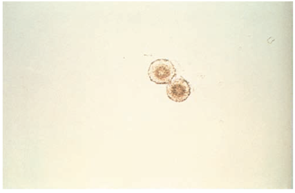
- 84—Cholesterol crystals under polarized light
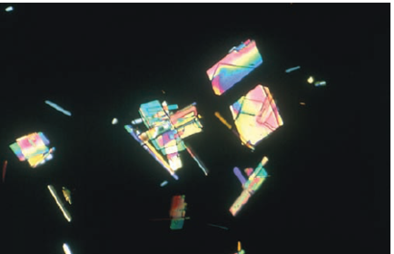
- 85—Bilirubin crystals ( notice the bright yellow color)
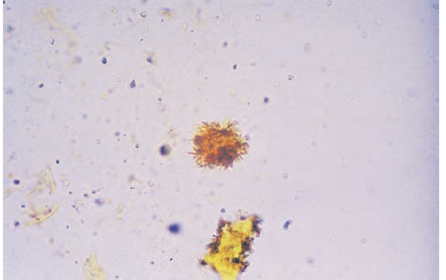
- 86—Ampicillin crystals, non refrigerated
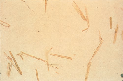
- 87—Sulfa crystals and bacteria seen in UTI
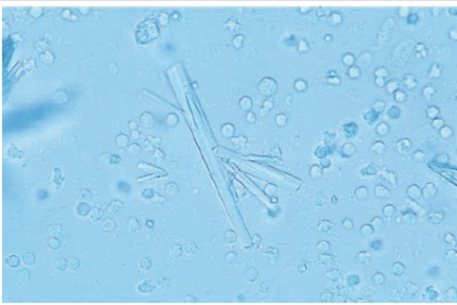
- 88—Ampicilan crystals after refridgeration
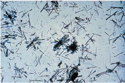
- 89—Sulfa crystals in rossette form
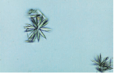
- 90—Starch granules ( notice the dimpled center)
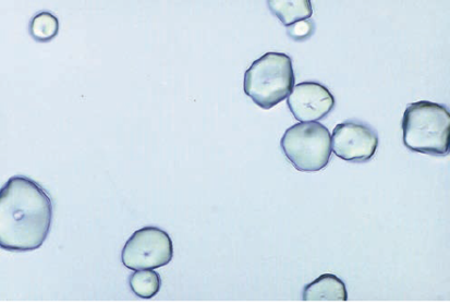
- 91—Pollen granule ( notice the concentric circles)
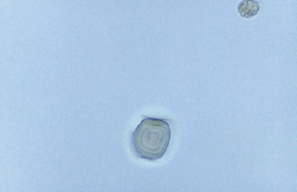
- 92—Fiber resembling a cast
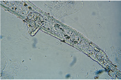
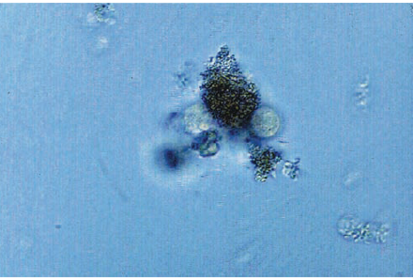
- 93—Fecal material and oil artifacts
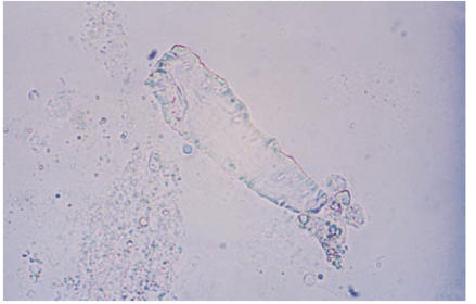
- 94—Vegetable fiber resembling a waxy cast
Información
- Descripción
- 2 Urine and Body Fuid analysis Fichas sobre UA, creado por Fritz Hull el 28/03/2016.
- Creado por
- Fritz Hull
- Visitas
- 2.533
- Valoración