
Mapa mental
Venous Anatomy
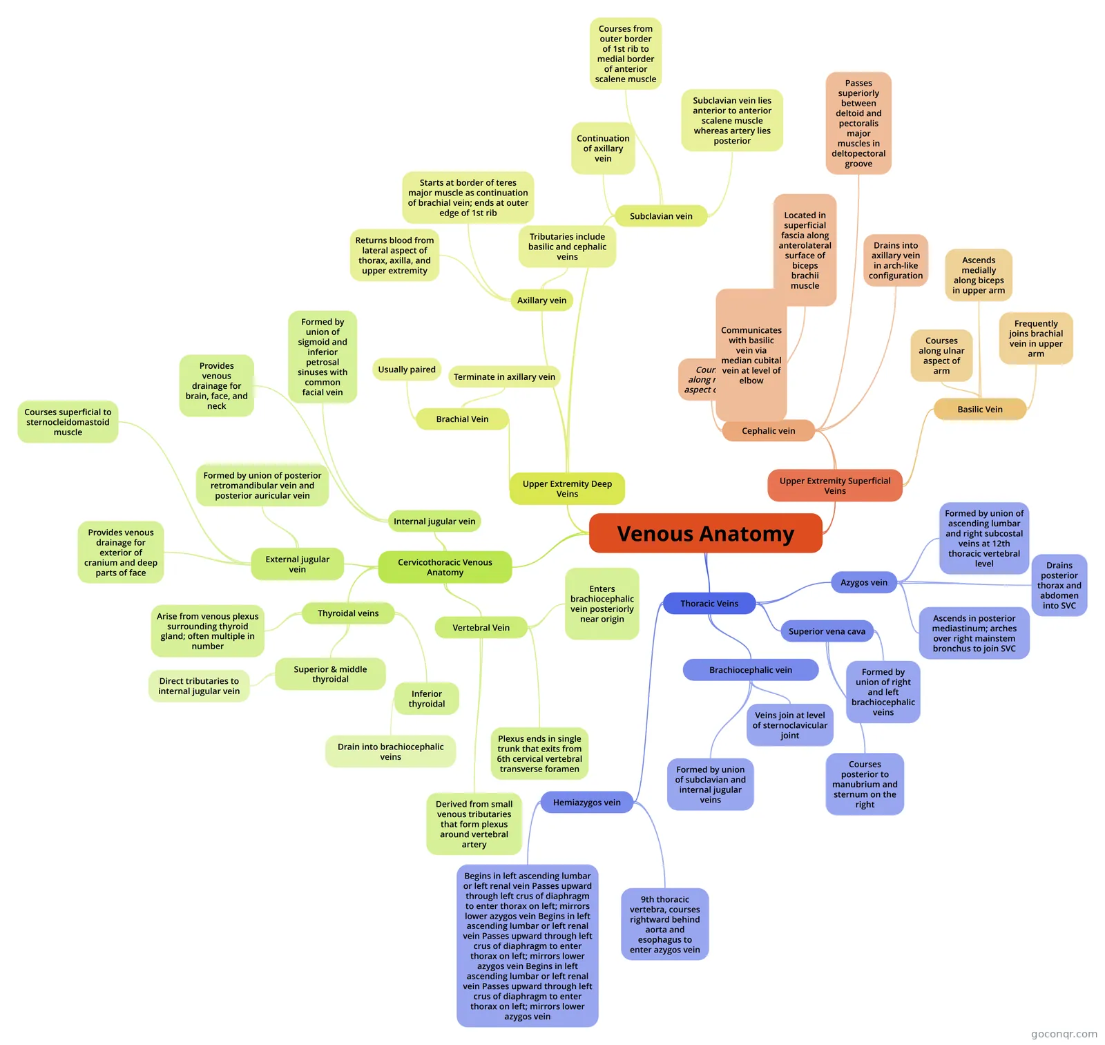
Mind map: Venous anatomy
Contenido del mapa
Venous Anatomy
Upper Extremity Superficial Veins
- Cephalic vein
- Courses along radial aspect of arm
- Communicates with basilic vein via median cubital vein at level of elbow
- Located in superficial fascia along anterolateral surface of biceps brachii muscle
- Passes superiorly between deltoid and pectoralis major muscles in deltopectoral groove
- Drains into axillary vein in arch-like configuration
- Basilic Vein
- Courses along ulnar aspect of arm
- Ascends medially along biceps in upper arm
- Frequently joins brachial vein in upper arm
- Cephalic vein
Upper Extremity Deep Veins
- Brachial Vein
- Usually paired
- Terminate in axillary vein
- Axillary vein
- Returns blood from lateral aspect of thorax, axilla, and upper extremity
- Starts at border of teres major muscle as continuation of brachial vein; ends at outer edge of 1st rib
- Tributaries include basilic and cephalic veins
- Subclavian vein
- Continuation of axillary vein
- Courses from outer border of 1st rib to medial border of anterior scalene muscle
- Subclavian vein lies anterior to anterior scalene muscle whereas artery lies posterior
- Brachial Vein
Cervicothoracic Venous Anatomy
- Internal jugular vein
- Formed by union of sigmoid and inferior petrosal sinuses with common facial vein
- Provides venous drainage for brain, face, and neck
- External jugular vein
- Formed by union of posterior retromandibular vein and posterior auricular vein
- Courses superficial to sternocleidomastoid muscle
- Provides venous drainage for exterior of cranium and deep parts of face
- Thyroidal veins
- Arise from venous plexus surrounding thyroid gland; often multiple in number
- Superior & middle thyroidal
- Direct tributaries to internal jugular vein
- Inferior thyroidal
- Drain into brachiocephalic veins
- Vertebral Vein
- Derived from small venous tributaries that form plexus around vertebral artery
- Plexus ends in single trunk that exits from 6th cervical vertebral transverse foramen
- Enters brachiocephalic vein posteriorly near origin
- Internal jugular vein
Thoracic Veins
- Brachiocephalic vein
- Formed by union of subclavian and internal jugular veins
- Veins join at level of sternoclavicular joint
- Superior vena cava
- Formed by union of right and left brachiocephalic veins
- Courses posterior to manubrium and sternum on the right
- Azygos vein
- Formed by union of ascending lumbar and right subcostal veins at 12th thoracic vertebral level
- Ascends in posterior mediastinum; arches over right mainstem bronchus to join SVC
- Drains posterior thorax and abdomen into SVC
- Hemiazygos vein
- Begins in left ascending lumbar or left renal vein Passes upward through left crus of diaphragm to enter thorax on left; mirrors lower azygos vein Begins in left ascending lumbar or left renal vein Passes upward through left crus of diaphragm to enter thorax on left; mirrors lower azygos vein Begins in left ascending lumbar or left renal vein Passes upward through left crus of diaphragm to enter thorax on left; mirrors lower azygos vein
- 9th thoracic vertebra, courses rightward behind aorta and esophagus to enter azygos vein
- Brachiocephalic vein
Información
- Descripción
- Mapa Mental sobre Venous Anatomy, creado por Luisa Muñoz Quiroga el 13/09/2014.
- Creado por
- Luisa Muñoz Quiroga
- Visitas
- 19
- Valoración