
Mapa mental
The Basics-Tissue Ultrastructure
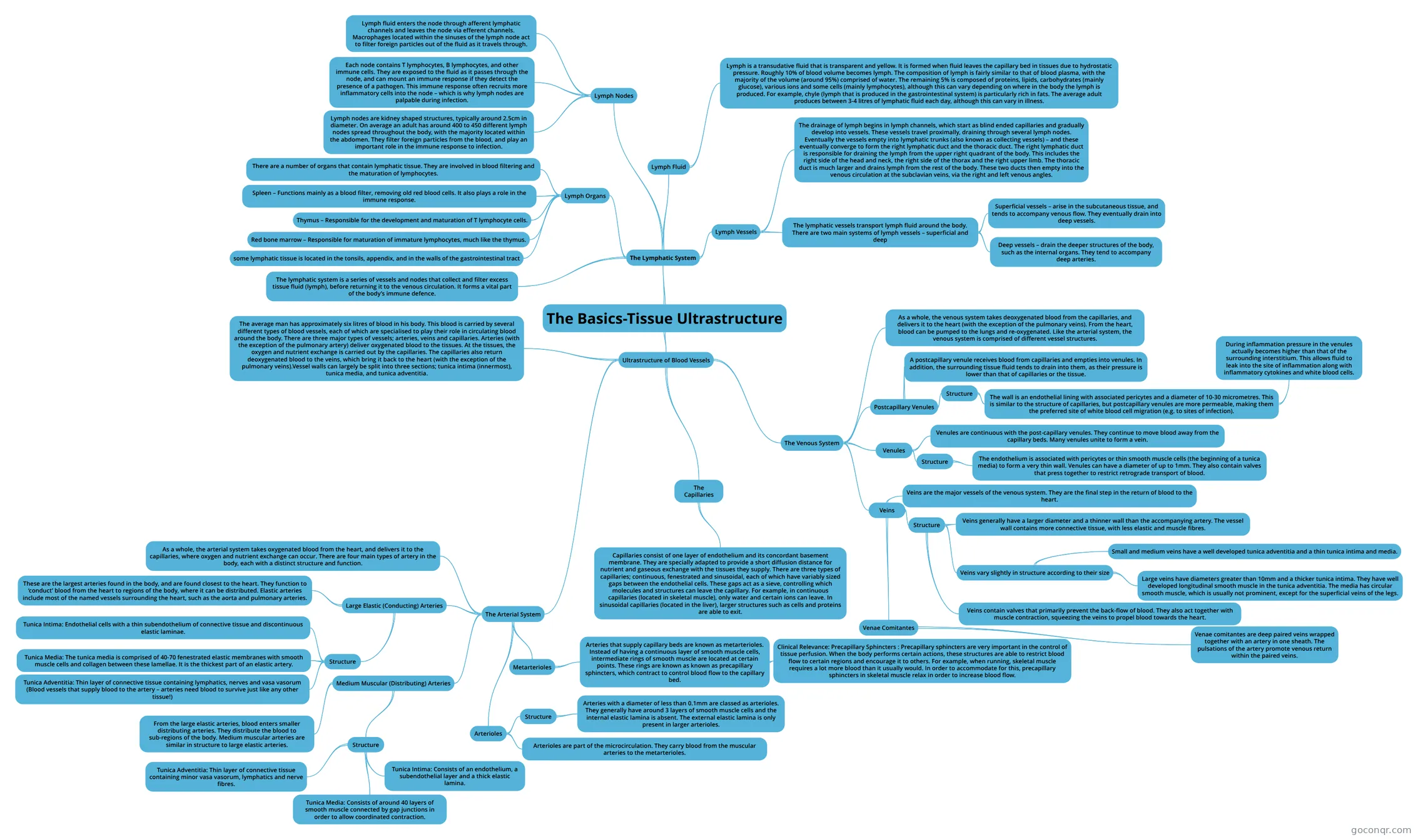
Mind map: Tissue Ultrastructure of Circulatory Systems
Contenido del mapa
The Basics-Tissue Ultrastructure
The Lymphatic System
- The lymphatic system is a series of vessels and nodes that collect and filter excess tissue fluid (lymph), before returning it to the venous circulation. It forms a vital part of the body’s immune defence.
- Lymph Organs
- There are a number of organs that contain lymphatic tissue. They are involved in blood filtering and the maturation of lymphocytes.
- Spleen – Functions mainly as a blood filter, removing old red blood cells. It also plays a role in the immune response.
- Thymus – Responsible for the development and maturation of T lymphocyte cells.
- Red bone marrow – Responsible for maturation of immature lymphocytes, much like the thymus.
- some lymphatic tissue is located in the tonsils, appendix, and in the walls of the gastrointestinal tract
- Lymph Nodes
- Lymph nodes are kidney shaped structures, typically around 2.5cm in diameter. On average an adult has around 400 to 450 different lymph nodes spread throughout the body, with the majority located within the abdomen. They filter foreign particles from the blood, and play an important role in the immune response to infection.
- Each node contains T lymphocytes, B lymphocytes, and other immune cells. They are exposed to the fluid as it passes through the node, and can mount an immune response if they detect the presence of a pathogen. This immune response often recruits more inflammatory cells into the node – which is why lymph nodes are palpable during infection.
- Lymph fluid enters the node through afferent lymphatic channels and leaves the node via efferent channels. Macrophages located within the sinuses of the lymph node act to filter foreign particles out of the fluid as it travels through.
- Lymph Vessels
- The lymphatic vessels transport lymph fluid around the body. There are two main systems of lymph vessels – superficial and deep
- Superficial vessels – arise in the subcutaneous tissue, and tends to accompany venous flow. They eventually drain into deep vessels.
- Deep vessels – drain the deeper structures of the body, such as the internal organs. They tend to accompany deep arteries.
- The drainage of lymph begins in lymph channels, which start as blind ended capillaries and gradually develop into vessels. These vessels travel proximally, draining through several lymph nodes. Eventually the vessels empty into lymphatic trunks (also known as collecting vessels) – and these eventually converge to form the right lymphatic duct and the thoracic duct. The right lymphatic duct is responsible for draining the lymph from the upper right quadrant of the body. This includes the right side of the head and neck, the right side of the thorax and the right upper limb. The thoracic duct is much larger and drains lymph from the rest of the body. These two ducts then empty into the venous circulation at the subclavian veins, via the right and left venous angles.
- The lymphatic vessels transport lymph fluid around the body. There are two main systems of lymph vessels – superficial and deep
- Lymph Fluid
- Lymph is a transudative fluid that is transparent and yellow. It is formed when fluid leaves the capillary bed in tissues due to hydrostatic pressure. Roughly 10% of blood volume becomes lymph. The composition of lymph is fairly similar to that of blood plasma, with the majority of the volume (around 95%) comprised of water. The remaining 5% is composed of proteins, lipids, carbohydrates (mainly glucose), various ions and some cells (mainly lymphocytes), although this can vary depending on where in the body the lymph is produced. For example, chyle (lymph that is produced in the gastrointestinal system) is particularly rich in fats. The average adult produces between 3-4 litres of lymphatic fluid each day, although this can vary in illness.
Ultrastructure of Blood Vessels
- The average man has approximately six litres of blood in his body. This blood is carried by several different types of blood vessels, each of which are specialised to play their role in circulating blood around the body. There are three major types of vessels; arteries, veins and capillaries. Arteries (with the exception of the pulmonary artery) deliver oxygenated blood to the tissues. At the tissues, the oxygen and nutrient exchange is carried out by the capillaries. The capillaries also return deoxygenated blood to the veins, which bring it back to the heart (with the exception of the pulmonary veins).Vessel walls can largely be split into three sections; tunica intima (innermost), tunica media, and tunica adventitia.
- The Arterial System
- As a whole, the arterial system takes oxygenated blood from the heart, and delivers it to the capillaries, where oxygen and nutrient exchange can occur. There are four main types of artery in the body, each with a distinct structure and function.
- Large Elastic (Conducting) Arteries
- These are the largest arteries found in the body, and are found closest to the heart. They function to ‘conduct’ blood from the heart to regions of the body, where it can be distributed. Elastic arteries include most of the named vessels surrounding the heart, such as the aorta and pulmonary arteries.
- Structure
- Tunica Intima: Endothelial cells with a thin subendothelium of connective tissue and discontinuous elastic laminae.
- Tunica Media: The tunica media is comprised of 40-70 fenestrated elastic membranes with smooth muscle cells and collagen between these lamellae. It is the thickest part of an elastic artery.
- Tunica Adventitia: Thin layer of connective tissue containing lymphatics, nerves and vasa vasorum (Blood vessels that supply blood to the artery – arteries need blood to survive just like any other tissue!)
- Medium Muscular (Distributing) Arteries
- From the large elastic arteries, blood enters smaller distributing arteries. They distribute the blood to sub-regions of the body. Medium muscular arteries are similar in structure to large elastic arteries.
- Structure
- Tunica Intima: Consists of an endothelium, a subendothelial layer and a thick elastic lamina.
- Tunica Media: Consists of around 40 layers of smooth muscle connected by gap junctions in order to allow coordinated contraction.
- Tunica Adventitia: Thin layer of connective tissue containing minor vasa vasorum, lymphatics and nerve fibres.
- Arterioles
- Arterioles are part of the microcirculation. They carry blood from the muscular arteries to the metarterioles.
- Structure
- Arteries with a diameter of less than 0.1mm are classed as arterioles. They generally have around 3 layers of smooth muscle cells and the internal elastic lamina is absent. The external elastic lamina is only present in larger arterioles.
- Metarterioles
- Arteries that supply capillary beds are known as metarterioles. Instead of having a continuous layer of smooth muscle cells, intermediate rings of smooth muscle are located at certain points. These rings are known as known as precapillary sphincters, which contract to control blood flow to the capillary bed.
- Clinical Relevance: Precapillary Sphincters : Precapillary sphincters are very important in the control of tissue perfusion. When the body performs certain actions, these structures are able to restrict blood flow to certain regions and encourage it to others. For example, when running, skeletal muscle requires a lot more blood than it usually would. In order to accommodate for this, precapillary sphincters in skeletal muscle relax in order to increase blood flow.
- Arteries that supply capillary beds are known as metarterioles. Instead of having a continuous layer of smooth muscle cells, intermediate rings of smooth muscle are located at certain points. These rings are known as known as precapillary sphincters, which contract to control blood flow to the capillary bed.
- The Capillaries
- Capillaries consist of one layer of endothelium and its concordant basement membrane. They are specially adapted to provide a short diffusion distance for nutrient and gaseous exchange with the tissues they supply. There are three types of capillaries; continuous, fenestrated and sinusoidal, each of which have variably sized gaps between the endothelial cells. These gaps act as a sieve, controlling which molecules and structures can leave the capillary. For example, in continuous capillaries (located in skeletal muscle), only water and certain ions can leave. In sinusoidal capillaries (located in the liver), larger structures such as cells and proteins are able to exit.
- The Venous System
- As a whole, the venous system takes deoxygenated blood from the capillaries, and delivers it to the heart (with the exception of the pulmonary veins). From the heart, blood can be pumped to the lungs and re-oxygenated. Like the arterial system, the venous system is comprised of different vessel structures.
- Postcapillary Venules
- A postcapillary venule receives blood from capillaries and empties into venules. In addition, the surrounding tissue fluid tends to drain into them, as their pressure is lower than that of capillaries or the tissue.
- Structure
- The wall is an endothelial lining with associated pericytes and a diameter of 10-30 micrometres. This is similar to the structure of capillaries, but postcapillary venules are more permeable, making them the preferred site of white blood cell migration (e.g. to sites of infection).
- During inflammation pressure in the venules actually becomes higher than that of the surrounding interstitium. This allows fluid to leak into the site of inflammation along with inflammatory cytokines and white blood cells.
- The wall is an endothelial lining with associated pericytes and a diameter of 10-30 micrometres. This is similar to the structure of capillaries, but postcapillary venules are more permeable, making them the preferred site of white blood cell migration (e.g. to sites of infection).
- Venules
- Venules are continuous with the post-capillary venules. They continue to move blood away from the capillary beds. Many venules unite to form a vein.
- Structure
- The endothelium is associated with pericytes or thin smooth muscle cells (the beginning of a tunica media) to form a very thin wall. Venules can have a diameter of up to 1mm. They also contain valves that press together to restrict retrograde transport of blood.
- Veins
- Veins are the major vessels of the venous system. They are the final step in the return of blood to the heart.
- Structure
- Veins generally have a larger diameter and a thinner wall than the accompanying artery. The vessel wall contains more connective tissue, with less elastic and muscle fibres.
- Veins vary slightly in structure according to their size
- Small and medium veins have a well developed tunica adventitia and a thin tunica intima and media.
- Large veins have diameters greater than 10mm and a thicker tunica intima. They have well developed longitudinal smooth muscle in the tunica adventitia. The media has circular smooth muscle, which is usually not prominent, except for the superficial veins of the legs.
- Veins contain valves that primarily prevent the back-flow of blood. They also act together with muscle contraction, squeezing the veins to propel blood towards the heart.
- Venae Comitantes
- Venae comitantes are deep paired veins wrapped together with an artery in one sheath. The pulsations of the artery promote venous return within the paired veins.
Información
- Descripción
- Mapa Mental sobre The Basics-Tissue Ultrastructure, creado por J Niharika el 06/09/2018.
- Creado por
- J Niharika
- Visitas
- 0
- Valoración