
Mapa mental
Physics P3 Topic 2
Contenido del mapa
Physics P3 Topic 2
X-Rays
- Electromagnetic waves with a very short wavelength
- Very ionising because they transfer more energy
- Use them to produce images of bones and organs
- Able to penetrate soft body tissue
- When X-Rays pass through body can be absorbed by molecules
- These become ionised
- X-Rays lose energy and eventually cannot ionise any more tissue
- Producing X-Rays
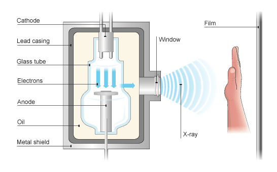
- In X-Ray machine electrons produced by cathode, which is heated by filament in an evacuated tube (a vacuum tube)
- Electrons accelerate towards an anode because high potential difference between anode and cathode
- Embedded in anode is tungsten target
- When very energetic electrons strike the target some of energy converted into x-rays
- Beam in an x-ray machine consists of large numbers of electrons travelling from the cathode to tungsten target
- Since electrons carry charge beam provides current through vacuum in x-ray tube
- Beam in an x-ray machine consists of large numbers of electrons travelling from the cathode to tungsten target
- When very energetic electrons strike the target some of energy converted into x-rays
- Embedded in anode is tungsten target
- Vacuum means no gas molecules to scatter and absorb energy of electrons striking target
- Anode positive
- Cathode negative
- Electrons accelerate towards an anode because high potential difference between anode and cathode
- In X-Ray machine electrons produced by cathode, which is heated by filament in an evacuated tube (a vacuum tube)
- Electromagnetic waves with a very short wavelength
X- Ray calculations
- Current in beams of charged particles
- Size of current in x-ray tube is given by the formula: I = N x q
- I = Current (amps, A)
- N = Number of particles per second (seconds, s)
- q = Charge carried by each electron (coulomb ,C)
- Size of current in x-ray tube is given by the formula: I = N x q
- The kinetic energy of particles in beams
- In an x-ray electron beam the electrons gain KE as they are accelerated though the vacuum by the potential difference
- The unit of potential difference is the volt
- One coulomb of electric charge gains one joule of energy when it is accelerated through a potential difference of one volt
- The unit of potential difference is the volt
- KE = e x V
- KE = Kinetic energy (joules, J)
- e = Charge on one electron (coulomb, C)
- V = Accelerating potential difference (volts, V)
- KE = 1/2 mv²
- m = Mass of electron (kg)
- v = Velocity of electron (m/s)
- e x v = 1/2 mv²
- In an x-ray electron beam the electrons gain KE as they are accelerated though the vacuum by the potential difference
- Current in beams of charged particles
Using X-rays
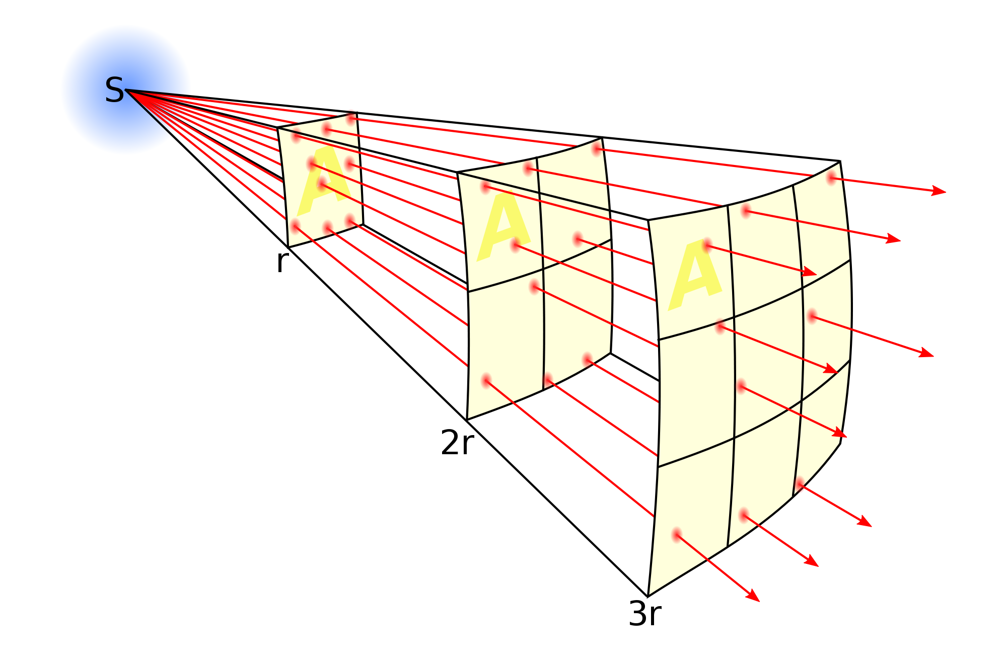
- When electromagnetic radiation (such as light) spreads from a source its energy is spread out over an area when increases with distance
- Inverse square law
- Area increases with the square of distance so intensity decreases with the square of distance
- Example: at twice the distance from the source the intensity is one quarter as much as area is four times greater
- Applies to any energy source
- Area increases with the square of distance so intensity decreases with the square of distance
- Absorption of X-Rays
- Increases the thickness of the material increases the x-rays absorbed
- CAT scans and fluoroscopes
- Conventional x-rays produce a 2-dimensional image of a patient
- In a CAT scanner an x-ray source is used to produce a 3D image of internal structure of patient
- Fluoroscopes use x-ray images on a screen to see real time images of patients internet condition
- Advantages
- Help to give a reliable diagnosis and make sure treatment is fast and accurate
- X-rays are non-invasive (no surgery)
- Disadvantages
- X-rays can damage living cells
- Risks are greater for babies and children so exposure kept to a minimum
ECGs and pacemakers
- Action potentials
- Electrical signals that control the heart muscles
- Generated by the hearts natural pacemaker to make heart beat at correct rate for body to be supplied with Oxygen
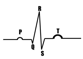
- ECGS (electricalcardiogram) measures changing electrical signals in heart
- An ECG shows possible problems with the heart as it may have an abnormal rhythm or be irregular
- Labelled PQRST = time for one complete heart beat
- Heart/pulse rate
- A normal ECG shows heart beating regularly
- Average = 72 Beats/Minute
- Pacemaker monitors heart and detects irregular heartbeats, if irregular heartbeat pacemaker produces electrical signals to correct heart rate
- f = I/T
- f = Frequency (hertz, Hz)
- T = time period (seconds, s)
- Pulse rate usually given in beats per minute so frequency given by this formula multiplied by 60 to give pulse in beats/minute
- A normal ECG shows heart beating regularly
- Heart/pulse rate
- Labelled PQRST = time for one complete heart beat
- An ECG shows possible problems with the heart as it may have an abnormal rhythm or be irregular
- Electrical signals that control the heart muscles
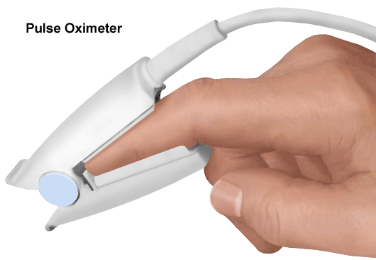
- Pulse oximetry
- Measure amount of oxygen in the blood and pulse rate
- Two light sources = one infrared and one visible red light
- Blood with a lot of oxygen absorbs move infrared light as it is a different colour from deoxgyenated blood
- The difference allows amount of oxygen in the blood to be calculated
- Blood with a lot of oxygen absorbs move infrared light as it is a different colour from deoxgyenated blood
- Action potentials
Imágenes y archivos del mapa
Información
- Descripción
- GCSE (P3) Science Mapa Mental sobre Physics P3 Topic 2, creado por Lydia C el 25/01/2015.
- Creado por
- Lydia C · ene 2015
- Actualizado
- 19 feb 2016
- Visitas
- 2
- Valoración