
Mapa mental
Studying Cells
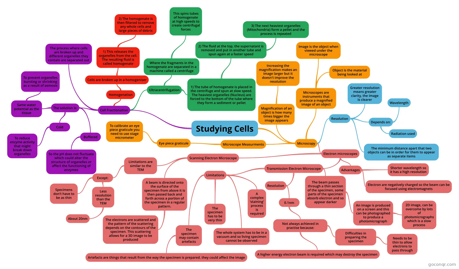
Mind map: Studying cells
Contenido del mapa
Studying Cells
Microscopy
- Microscopes are instruments that produce a magnified image of an object
- Object is the material being looked at
- Image is the object when viewed under the microscope
- Magnification of an object is how many times bigger the image appears
- Increasing the magnification makes an image larger but it doesn't improve the resolution
- Resolution
- The minimum distance apart that two objects can be in order for them to appear as separate items
- Depends on:
- Wavelength
- Radiation used
- Greater resolution means greater clarity, the image is clearer
- Electron microscopes
- Advantages
- Shorter wavelength so it has a high resolution
- Electron are negatively charged so the beam can be focused using electromagnets
- Transmission Electron Microscope
- The beam passes through a thin section of the specimen, some parts of the specimen absorb electron and so appear darker
- An image is produced on a screen and this can be photographed to produce a photomicrograph
- 2D image, can be overcome by lots of photomicrographs which is a slow process
- An image is produced on a screen and this can be photographed to produce a photomicrograph
- Resolution
- 0.1nm
- Not always achieved in practise because:
- Difficulties in preparing the specimen
- Needs to be thin to allow electrons to pass through
- A higher energy electron beam is required which may destroy the specimen
- Difficulties in preparing the specimen
- Not always achieved in practise because:
- 0.1nm
- Limitations
- The whole system has to be in a vacuum and so living specimen cannot be observed
- A complex 'staining' process is required
- The specimen has to be very thin
- The specimen may contain artefacts
- Artefacts are things that result from the way the specimen is prepared. they could affect the image
- The beam passes through a thin section of the specimen, some parts of the specimen absorb electron and so appear darker
- Scanning Electron Microscope
- Limitations are similar to the TEM
- Except:
- Specimens don't have to be as thin
- Less resolution than the TEM
- About 20nm
- Except:
- A beam is directed onto the surface of the specimen from above it is then passed back and forth across a portion of the specimen in a regular pattern.
- The electrons are scattered and the pattern of the scattering depends on the contours of the specimen. This scattering allows for a 3D image to be produced
- Limitations are similar to the TEM
- Advantages
- Microscope Measurments
- Eye piece graticule
- To calibrate an eye piece graticule you need to use stage micrometer
- Eye piece graticule
- Microscopes are instruments that produce a magnified image of an object
Cell Fractionation
- The process where cells are broken up and different organelles they contain are separated out
- The solution is:
- Cold
- To reduce enzyme activity that might break down organelles
- Same water potential as the tissue
- To prevent organelles bursting or shrinking as a result of osmosis
- Buffered
- So the pH does not fluctuate which could alter the structure of organelles or affect the functioning of enzymes
- Cold
- Homogenation
- Cells are broken up in a homogeniser
- 1) This releases the organelles from the cell. The resulting fluid is called homogenate
- 2) The homogenate is then filtered to remove any whole cells and large pieces of debris
- 1) This releases the organelles from the cell. The resulting fluid is called homogenate
- Cells are broken up in a homogeniser
- Ultracentrifugation
- Where the fragments in the homogenate are separated in a machine called a centrifuge
- This spins tubes of homogenate at high speeds to create centrifugal forces
- 1) The tube of homogenate is placed in the centrifuge and spun at slow speed. The heaviest organelles (Nucleus) are forced to the bottom of the tube where they form a sediment or pellet
- 2) The fluid at the top, the supernatant is removed and put in another tube and spun again at a faster speed
- 3) The next heaviest organelles (Mitochondria) form a pellet and the process is repeated
- 2) The fluid at the top, the supernatant is removed and put in another tube and spun again at a faster speed
- Where the fragments in the homogenate are separated in a machine called a centrifuge
Información
- Descripción
- Mapa Mental sobre Studying Cells, creado por Rhiannon Connor el 16/08/2016.
- Creado por
- Rhiannon Connor
- Visitas
- 1
- Valoración