
Mapa mental
Thorax Anatomy
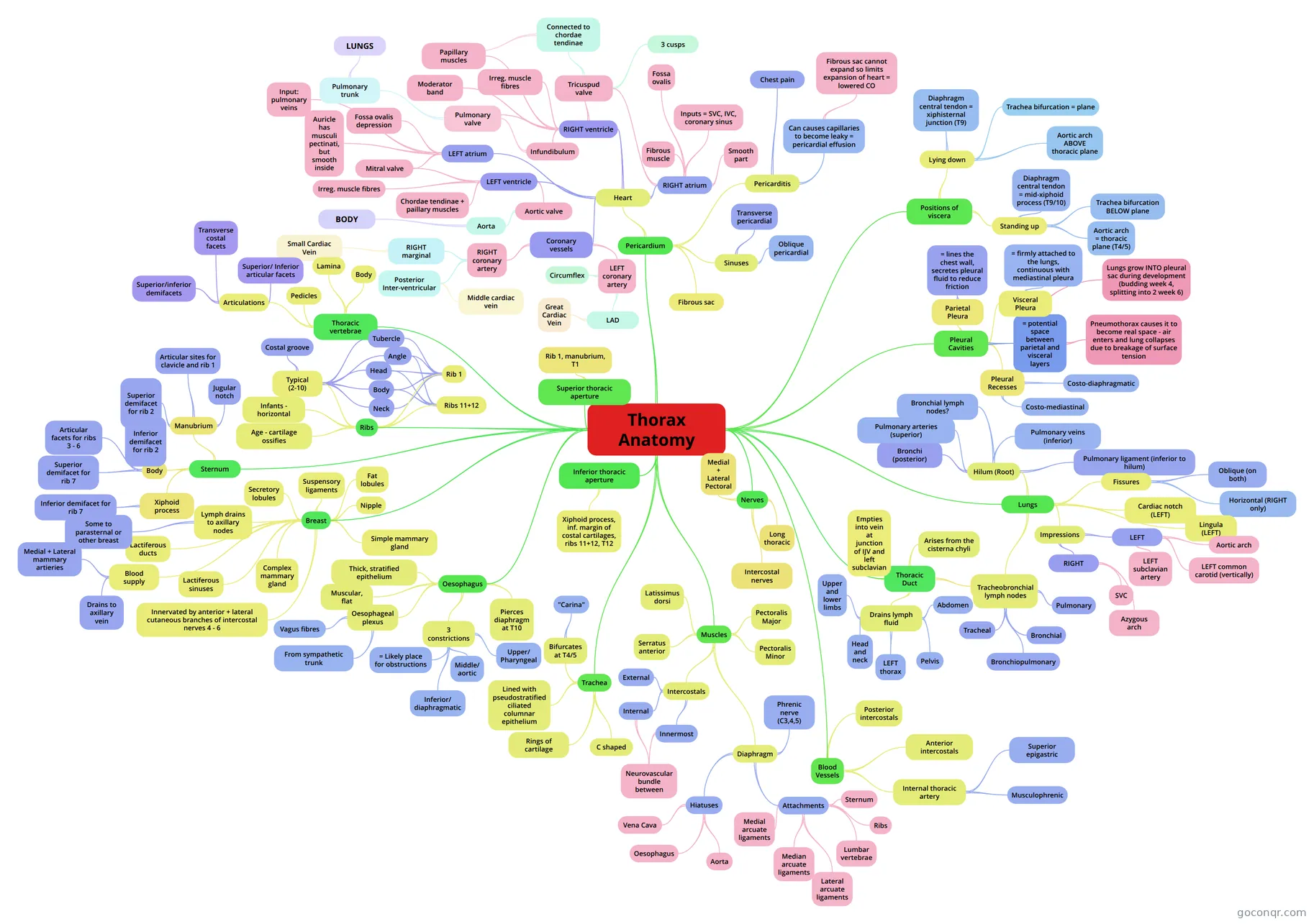
Mind map: thorax anatomy
Contenido del mapa
Thorax Anatomy
Pleural Cavities
- Parietal Pleura
- Aspects: --> Cervical --> Costal --> Mediastinal --> Diaphragmatic
- = lines the chest wall, secretes pleural fluid to reduce friction
- = potential space between parietal and visceral layers
- Pneumothorax causes it to become real space - air enters and lung collapses due to breakage of surface tension
- Lungs grow INTO pleural sac during development (budding week 4, splitting into 2 week 6)
- Visceral Pleura
- = firmly attached to the lungs, continuous with mediastinal pleura
- Pleural Recesses
- Parts of pleural cavity NOT filled during QUIET inspiration
- Costo-diaphragmatic
- Costo-mediastinal
- Parietal Pleura
Lungs
- Surfaces: --> Costal --> Vertebral --> Diaphragmatic --> Cardiac
- Hilum (Root)
- Bronchi (posterior)
- Cartilagenous
- Pulmonary arteries (superior)
- Pulmonary veins (inferior)
- Bronchial lymph nodes?
- may appear black due to pollution
- Pulmonary ligament (inferior to hilum)
- Just a fold of pleural membrane - attaches lung to mediastinum
- Bronchi (posterior)
- Fissures
- Oblique (on both)
- Horizontal (RIGHT only)
- Cardiac notch (LEFT)
- where heart sits
- Lingula (LEFT)
- Angle between diagphragmatic surface and cardiac notch
- Impressions
- LEFT
- Aortic arch
- LEFT common carotid (vertically)
- LEFT subclavian artery
- RIGHT
- SVC
- Azygous arch
- LEFT
- Tracheobronchial lymph nodes
- Tracheal
- either side of trachea
- Bronchial
- around bifurcation
- Bronchiopulmonary
- hilum of lungs
- Pulmonary
- within lungs
- Tracheal
Positions of viscera
- Standing up
- Aortic arch = thoracic plane (T4/5)
- Trachea bifurcation BELOW plane
- Diaphragm central tendon = mid-xiphoid process (T9/10)
- Lying down
- Aortic arch ABOVE thoracic plane
- Trachea bifurcation = plane
- Diaphragm central tendon = xiphisternal junction (T9)
- Standing up
Pericardium
- Fibrous sac
- Sinuses
- Transverse pericardial
- separates aorta and pulmonary trunk
- Oblique pericardial
- posterior; behind left atrium
- Transverse pericardial
- Pericarditis
- Inflammation of the pericardium
- Can causes capillaries to become leaky = pericardial effusion
- Fibrous sac cannot expand so limits expansion of heart = lowered CO
- Chest pain
- Heart
- RIGHT atrium
- Inputs = SVC, IVC, coronary sinus
- Smooth part
- Crista terminalis
- Fibrous muscle
- Musculi pectinati
- Fossa ovalis
- Should be closed - foramen ovale in embryo
- Tricuspud valve
- 3 cusps
- Anterior, posterior, septal
- Connected to chordae tendinae
- 3 cusps
- RIGHT ventricle
- Papillary muscles
- innervated by Purkinje fibres
- Irreg. muscle fibres
- = Trabeculae carnae
- Moderator band
- nerve tissue connecting anterior papillary muscle to septum
- Infundibulum
- Smooth wall leading to pulmonary valve
- Pulmonary valve
- 3 semilunar cusps
- Pulmonary trunk
- LUNGS
- Papillary muscles
- LEFT atrium
- Input: pulmonary veins
- Auricle has musculi pectinati, but smooth inside
- Fossa ovalis depression
- Mitral valve
- LEFT ventricle
- Mitral valve
- Bicuspid - anterior and posterior
- Irreg. muscle fibres
- trabeculae carnae
- Chordae tendinae + paillary muscles
- Aortic valve
- Semilunar
- Aorta
- BODY
- Mitral valve
- Coronary vessels
- RIGHT coronary artery
- in atrio-ventricular groove
- RIGHT marginal
- Small Cardiac Vein
- Posterior Inter-ventricular
- (Right posterior descending)
- Middle cardiac vein
- LEFT coronary artery
- Circumflex
- LAD
- Great Cardiac Vein
- RIGHT coronary artery
- RIGHT atrium
Thoracic Duct
- Arises from the cisterna chyli
- root of the mesentery of the small intestine
- Drains lymph fluid
- Abdomen
- Pelvis
- LEFT thorax
- Head and neck
- Upper and lower limbs
- Empties into vein at junction of IJV and left subclavian
- Arises from the cisterna chyli
Trachea
- C shaped
- Because oesophagus is behind it
- Rings of cartilage
- hold trachea open
- Bifurcates at T4/5
- (transverse thoracic plane)
- "Carina"
- mucus membrane here responsible for cough reflex
- Lined with pseudostratified ciliated columnar epithelium
- glands to secrete mucus
- C shaped
Oesophagus
- Pierces diaphragm at T10
- through muscular part to help prevent reflux
- 3 constrictions
- Upper/ Pharyngeal
- level of cricoid cartilage
- Middle/ aortic
- at bifurcation of trachea
- Inferior/ diaphragmatic
- at piercing of diaphragm (lower oesophageal sphincter)
- = Likely place for obstructions
- Upper/ Pharyngeal
- Oesophageal plexus
- Vagus fibres
- From sympathetic trunk
- Thick, stratified epithelium
- to resist damage/abrasion as food travels down throat
- Muscular, flat
- except when food is passing down it
- Pierces diaphragm at T10
Thoracic vertebrae
- Body
- Lamina
- between transverse and spinous processes
- Pedicles
- between body and transverse processes
- Articulations
- Superior/ Inferior articular facets
- articulate with inferiort/superior articular facets of vertebra above/below
- Transverse costal facets
- articulate with tubercles of ribs
- Superior/inferior demifacets
- on vertebral body - articulate with head of rib at that level and one below
- Superior/ Inferior articular facets
Superior thoracic aperture
- Rib 1, manubrium, T1
Inferior thoracic aperture
- Xiphoid process, inf. margin of costal cartilages, ribs 11+12, T12
Ribs
- Typical (2-10)
- Head
- Neck
- Tubercle
- Angle
- most likely to break (not in infants)
- Body
- Costal groove
- protects neurovascular bundle
- Rib 1
- Head
- Neck
- Tubercle
- Angle
- Body
- Ribs 11+12
- Head
- Angle
- Body
- Infants - horizontal
- Age - cartilage ossifies
- becomes harder to move ribcage to breathe
- Typical (2-10)
Sternum
- Manubrium
- Jugular notch
- Articular sites for clavicle and rib 1
- primary cartilagenous joints = no movement
- Superior demifacet for rib 2
- Body
- Inferior demifacet for rib 2
- Articular facets for ribs 3 - 6
- Superior demifacet for rib 7
- Xiphoid process
- Inferior demifacet for rib 7
- Manubrium
Breast
- Fat lobules
- Suspensory ligaments
- Secretory lobules
- produce milk
- Lactiferous ducts
- Lactiferous sinuses
- Simple mammary gland
- All lobules supplying one duct
- Complex mammary gland
- All glands serving one nipple
- Nipple
- Blood supply
- Medial + Lateral mammary artieries
- from internal thoracic
- Drains to axillary vein
- Medial + Lateral mammary artieries
- Innervated by anterior + lateral cutaneous branches of intercostal nerves 4 - 6
- Lymph drains to axillary nodes
- Anterior Posterior Infraclavicular Central Apical Lateral
- Some to parasternal or other breast
Blood Vessels
- Posterior intercostals
- 11 pairs: Upper 2 from costocervical trunk of subclavian Lower 9 from thoracic aorta
- Anterior intercostals
- 9 pairs: Upper 6 from internal thoracic artery Lower 3 from musculophrenic artery
- Internal thoracic artery
- Musculophrenic
- Superior epigastric
- Posterior intercostals
Nerves
- Intercostal nerves
- in costal grooves of ribs
- Long thoracic
- supplies serratus anterior
- Medial + Lateral Pectoral
- Medial = both Pec major and minor Lateral = ONLY Pec major
- Intercostal nerves
Muscles
- Pectoralis Major
- Origins: medial half of clavicle, sternum, costal cartilages 1-7 Insertions: Proximal part of humerus Innervation: Medial and Lateral Pectoral nerves
- Pectoralis Minor
- Origins: Surface of ribs 3-5 Insertions: Coracoid process Innervation: Medial pectoral nerves
- Diaphragm
- Phrenic nerve (C3,4,5)
- Central tendon developed at C2, then picked up myotomes from C3,4,5 during development
- Attachments
- Sternum
- Ribs
- Lumbar vertebrae
- L and R crurae
- Lateral arcuate ligaments
- over quadratus lumborum muscle
- Medial arcuate ligaments
- Over psoas major
- Median arcuate ligaments
- over aorta
- Hiatuses
- Vena Cava
- T8
- Oesophagus
- T10
- Aorta
- T12
- Vena Cava
- Phrenic nerve (C3,4,5)
- Serratus anterior
- Origins: Anterior surface of ribs 1-8/9 Insertion: Anterior surface of medial border of scapula Innervation: Long thoracic nerve (C5-C7)
- Latissimus dorsi
- Origins: spinous processes of T6-L5+sacrum, iliac crest, ribs 10-12 Insertions: intertubercular sulcus of humerus Innervation: C6-C8
- Intercostals
- External
- \\// used in inspiration
- Internal
- //\\ in forced expiration
- Neurovascular bundle between
- Innermost
- //\\ in forced expiration
- Neurovascular bundle between
- External
- Pectoralis Major
Información
- Descripción
- Degree Anatomy Mapa Mental sobre Thorax Anatomy, creado por Hannah Tribe el 24/04/2014.
- Creado por
- Hannah Tribe
- Visitas
- 462
- Etiquetas
- Valoración