
Mapa mental
Tissues Paisley Davies
Contenido del mapa
Tissues Paisley Davies
Muscular tissue
- Striated
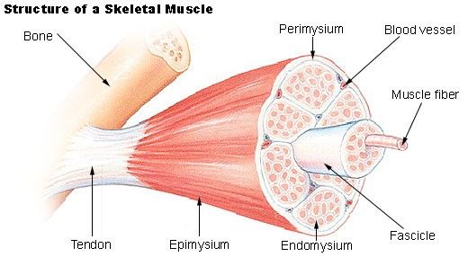
- Striated muscle is also known as skeletal muscle are muscle that is under our conscious control. some examples of striated muscle are; biceps muscle
- Structure
- Striated muscle tissue are made up of fibres and are attached to bones by stretchy tendons although they are also attached to facial muscle. Each fibre is cylinder in shape and are positioned adjacent to one another. The muscle looks like it has dark and lights bands which is because of the protein filaments that it is made of.
- Function
- Nearly all body movement is caused by striated muscle, they contract and relax to cause movement of the bone the are attached to. They are controlled by motor nerves. Also skeletal muscles are almost continuously functioning to keep posture, to keep our bodies upright. Striated muscle also generates heat as a by product which helps maintain our body temperature.
- Non striated/Smooth
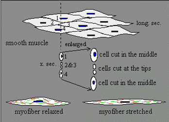
- Non striated muscle is involuntary (which means we have no conscious control over its movements). The fibres are spindle like in shape with a single, central nuclei. Smooth muscle is located in hollow organs - in the stomach, intestines, bladder and the iris (in the eye).
- Structure
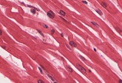
- Non striated/smooth muscle contains protein filaments, although they do not lie in any specific pattern, therefor they do not have the banding that is so distinctive about striated muscle. The fibres are spindle shaped with a single, central nuclei and each fibre intertwines with one another. This fibre mostly forms sheets of tissue and requires stimulation from nerves. Smooth muscle fibres are small and tapered - with the ends reducing in size. Every non striated muscle fibre has a single central located nucleus
- Function
- More often than not smooth muscle occurs in two sheets, going in different directions (this is known as antagonistic muscles) Smooth muscle contracts and stretches to help muscles move, these contractions and stretches are involuntary but are controlled by the autonomic nervouse system. For example in the digestion tract - one sheet will run in a circular motion around the intestine and the other will run against it, down the length. This helps push the contents down the intestines. Another example is of smooth muscle is the wall of the bladder will contract to expel urine from the body.
- Cardiac
- Cardiac muscle is only found in the four chambers of the heart. It is not a muscle under our conscious control and can contract without receiving any stimuli from the nervous system.
- Function
- The cardiac muscle role is to contract to expel blood and then relax to refill with blood, so the requirements of blood needed around the body when exercising or resting are met. Cardiac muscle cannot contract myogenically (which means to contract independently from any signals from the brain) because the upper chamber muscle has a different contraction rate to the lower ventricular muscle and this would cause deficient heart action. The nervous system controls how often the contraction occur, as they change when resting or exercising.
- Structure
- The cells spread repeatedly so they form a network, which the contraction quickly runs through. Every cell contains a central nuclei. The cell is both horizontally and vertically striped. The space between cells are specifically so, so that impulses can be transmitted sufficiently - this is known as intercalated discs.
- Striated
Nervous tissue
- Nervous tissue is only found in the brain, spinal cord and nerves - these as a collective make up the nervous system. Its main purpose is to co-ordinate and to communicate between different parts of the body by receiving information from sources outside and inside the body. For example, the nervous system will receive and read signals from the sense organs so that vision, hearing and smell can take place.
- Neurons
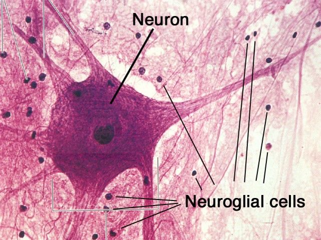
- Neurons are a type of nervous tissue (nervous cell) There are many types of neurons, Some of these are; Motor nerves sensory neurons relay neurons Each neurons are sensitive to a specific type of stimuli, such as cold/head and light/dark .
- Function
- The role of a neuron is to send electrical impulses which contain information around the body. For example when its light these neurons will send a signal to our brain to tell our pupils to contract.
- Structure
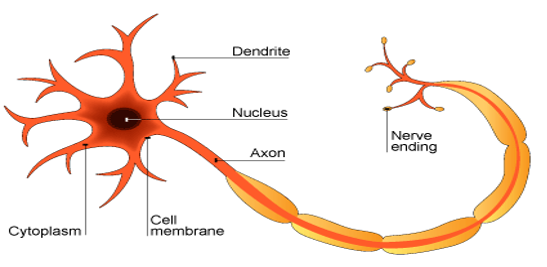
- Each neuron has a nerve cell body, which contains a central nucleus and organelles (such as mitochondria, ER and Golgi apparatus) It appearance is almost like a tree, and the ‘branches’ that branch off it are called dendrites which act like antennae’s picking up information. The ‘trunk’ of the neuron called the axon contains a Myelin sheath (which also contains a nucleus). At the very end of a neuron (which looks like roots) is the nerve ending.
- Neuroglia
- Neuroglia cells (also known as glail cells) are support cells for the nervous system.
- Function
- Neuroglia cells have no function in relaying information like neurons but are to support the neurons by securing their position so they aren’t damaged by other neurons. Another function of a neuroglia cell is to source useful substances such as nutrients and oxygen to neurons.They also remove waste such as damaged or dead neurons. Its almost like a connective tissue.
- Structure
- Neuroglia come in different shapes and sizes, however they all have long processes (nerve fibre) which form the nerves. They are intermingled with neurons and are found in the brain and spinal cord.
Epithelial
- Epithelial tissue is composed cells laid together in sheets with the cells connected tightly together.
- Simple
- Simple epithelial tissue is a single layer of cells.
- Squamous epithelial tissue
- Function
- Squamous epithelial tissue allows substances to pass through it via diffusion. For example in the alveoli it allows oxygen to pass into the blood stream. In the capillary walls it allows oxygen, carbon dioxide and glucose travel through it.
- Structure
- Squamous epithelial tissue is a single layer of cells. One side of the cell is orientated towards the surface of tissue and the then is joined to a basement membrane. They can be found lots of places such as : lungs heart kidneys capillaries
- Function
- Cuboidal
- Structure
- Like squamous tissue, cuboidal tissue is a single layer. However cuboidal tissue is thicker and can withstand more trauma. They can be found in many places in the body for example in the ovaries and kidneys.
- function
- Like the squamous tissue, cuboidal also allows substances to pass through by diffusion (e.g. water in the kidney tubules) They also specialise in secreting substances such as sweat, mucus, enzymes and other products that are delivered by ducts.
- Structure
- columnar epithelial and ciliated columnar epithelia tissue
- function
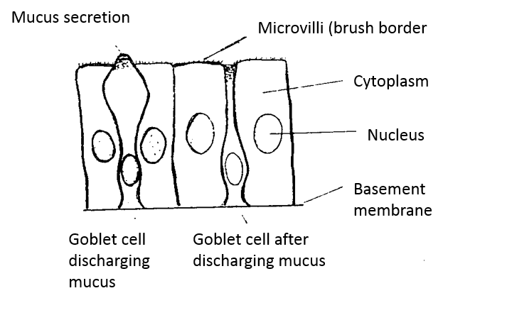
- To trap bacteria and dust that could be harmful to the body and then move it away from the organs via cilia. For example in the trachea the mucus traps bacteria and dust and then moves it up into the throat until its coughed out of the mouth, therefor eliminated any possible causes of infection.
- structure
- Again they are a single layer of tissue with goblets inbetween them which secretes mucus. You can find it in the trachea and intestines.
- function
- Compound
- Stratified squamous epithelial tissue - keratinised
- structure
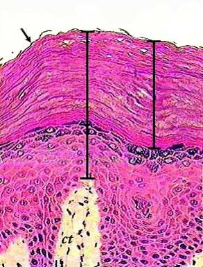
- Unlike simple epithelial tissue, Stratified squamous epithelial tissue is layered. The top layers are dead cells, filled with a protein called keratin. The cells beneath the top dead cells are cuboidal in shape. The skin is a type of Stratified squamous epithelial tissue.
- function
- The primary function is to protect underlying tissues. It also continually divides to replace the dead cells
- structure
- non- keratinised
- Function
- Like the keratinised tissue the cell surface falls off through friction and then is replaced by the cells underneath, which are cuboidal cells.
- Structure
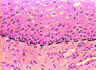
- Non-kerantinised tissue is used to line cavaties and tubes in our body. For example it is the lining in the mouth, the oesophagus and the cornea.
- Function
- Stratified squamous epithelial tissue - keratinised
connective tissue
- These tissues hold everything together in the body.
- Blood
- Functiion
- Blood is used to transport materials around the body. For example is carries C02 and 02 to and from the lungs. It transports nutrients and waste products around the body.
- Structure
- Blood consists of cells and liquids , it contains plasma (55%), White (<1%), Red blood cells (45%)
- Functiion
- Bone
- Function
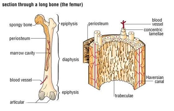
- the bone aids the bodies movement and helps keep the bodies posture. The spongy bone marrow in the middle of the bone contains blood cells and stem cells that creat blood cells or white blood cells.
- Structure
- Bone is hard live tissue that is made of calcium salts that are deposited around the protein fibres. In the middle of the bone is a spongy bone tissue (bone marrow)
- Function
- Areolar tissue
- Structure
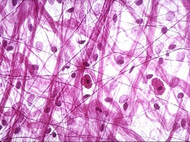
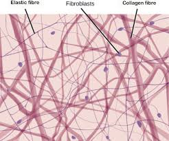
- A stretchy white material that's sticky. It contains elastin fibres and collagen.
- Function
- It holds muscle groups, blood vessels and nerves together.
- Structure
- Adipose tissue
- Function
- Its function is to protect tissue and to insulate. Its also used to store energy.
- Structure
- Like the areolar tissue, its a sticky white material that contains collagen and elastin but it contains more fat cells
- Function
Imágenes y archivos del mapa
Información
- Descripción
- BTEC Health and Social Care Mapa Mental sobre Tissues Paisley Davies, creado por Paisley Olivia Davies el 28/09/2017.
- Creado por
- Paisley Olivia Davies
- Visitas
- 22
- Valoración