
Mapa mental
Anatomy
Contenido del mapa
Anatomy
Anatomy
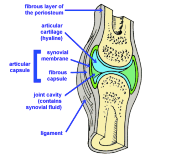
- Synovial Joints
- Synovial Membrane(synovium)
- Produces a synovial fluid
- lubrication, free movement, and nutrition of the cartilage since it doesn’t have a blood supply
- viscous, non-Newtonian fluid found in the cavities of synovial joints.
- hyaluronic acid
- lubricin
- collagenases
- proteinases
- Produces a synovial fluid
- They allow free movement
- Articular surfaces of the bone
- Hyaline cartilage(articular cartilage)
- Intra-capsular (intra-articular) structures can be found; meniscus, labrum, tendons, and disc.
- A synovial sheath .
- Synovial Membrane(synovium)
- Bones
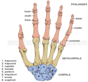
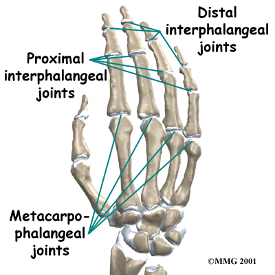
- 14 phalanges
- 8 Carpals
- Trapezium
- Trapezoid
- Capitate
- Hamate
- Scaphoid
- Lunate
- Triquetrium
- Pisiform
- 5 metacarpals
- Arteries
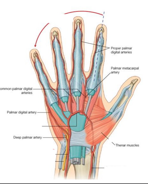
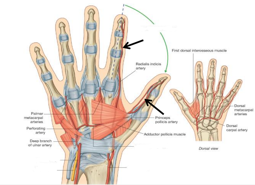
- Superficial Palmar Arch
- mainly by ulnar a.)
- is superficial to the long flexor tendons just deep to the palmar aponeurosis. On the lateral side of the palm, the arch communicates with the superficial br. of the radial artery.
- Deep Palmar Arch
- mainly by radial a
- penetrates the origin of the hypothenar muscles; access the deep plane of the palm to anastomose with the deep palmar arch derived from the radial artery.
- Superficial Palmar Arch
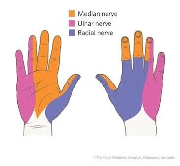
- Nerves
- Ulnar N.: medial 2/3 of the palm & dorsum + medial 11/2 fingers
- Median N.: Lateral 2/3 of the palm + lateral 3 1/2
- Radial n.: lateral 2/3 of the dorsum of hand + lateral 3 1/2 fingers
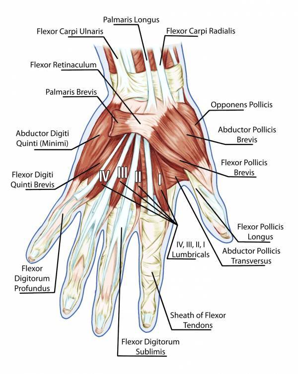
- Muscles
- Joints
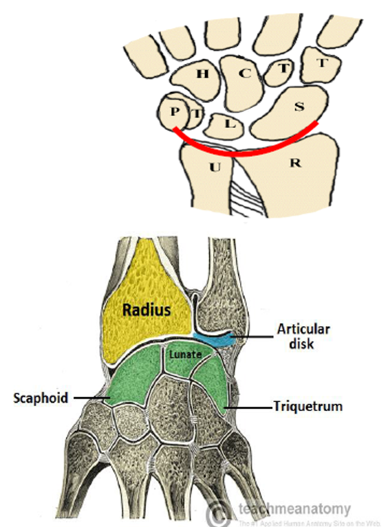
- Wrist Joint
- Synovial , Ellipsoid , biaxial
- Articular disc that separates the head of ulna from the triquetral .
- Synovial , Ellipsoid , biaxial
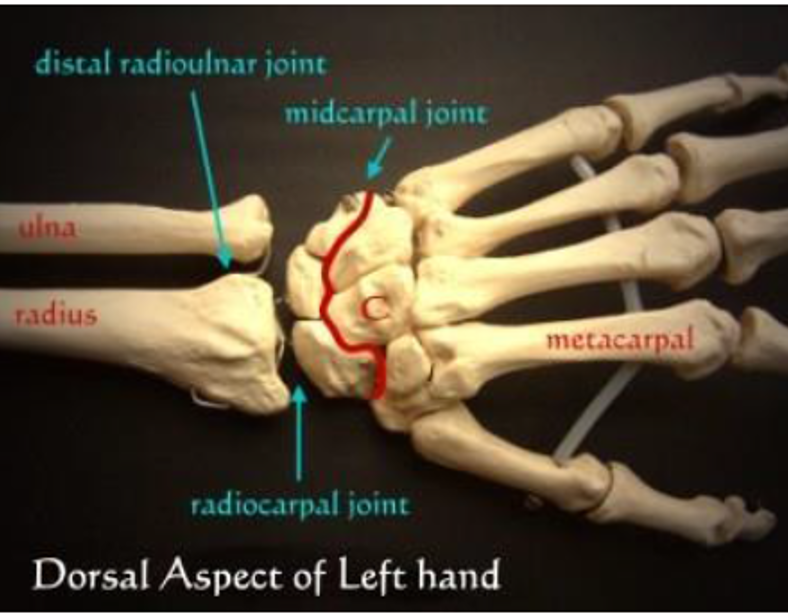
- Mid-Carpal Joint
- Synovial joints
- Between the two rows of carpal bones
- Positioning of the hand in abduction , adduction , flexion and particularly extension
- Between the two rows of carpal bones
- Synovial joints
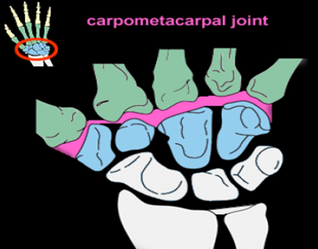
- Carpometacarpal Joint
- Between the distal row of carpal bones and proximal end of the metacarpal bones .
- more stability than mobility.
- Between the distal row of carpal bones and proximal end of the metacarpal bones .
- Metacarporhalangeal Joints
- Between the distal heads of the metacarpals and the proximal phalanges of the digits .
- Interphalangeal Joints
- Hinge Joints
- Between the phalanges of the hand .
- Hinge Joints
- Wrist Joint
- Synovial Joints
Diseases that affect 1 or > joints
- Infectious diseases
- acute and chronic septic arthritis
- Inflammatory diseases-
- autoimmune disorders such as rheumatoid arthritis
- Degenerative diseases
- osteoarthritis
- Infectious diseases
Joint Arthritis
- inflammation of the joints
- the term is used to describe many rheumatic diseases that affect the joints.
- Injury
- Degenerative arthritis
- inflammation, breakdown, and eventual loss of the cartilage of the joints.
- hands, feet, spine, and large weight-bearing joints, such as the hips and knees.
- Osteoarthritis
- hands, feet, spine, and large weight-bearing joints, such as the hips and knees.
- inflammation, breakdown, and eventual loss of the cartilage of the joints.
- Degenerative arthritis
- Genetic
- Rheumatoid Arthritis
- signs and symptoms
- fever, malaise, arthralgias, and weakness
- progresses to joint inflammation and swelling
- Persistent symmetric polyarthritis (synovitis) of hands and feet
- hallmark feature)
- Progressive articular deterioration
- Extra-articular involvement
- Difficulty performing activities of daily living (ADLs)
- fever, malaise, arthralgias, and weakness
- onset is insidious
- Investigations
- Blood Investigations
- Anti-CCP(cyclic citrullinated peptide)
- identifying autoantibodies
- normocytic anemia :anemia of chronic disease(ACD)
- The most common cause is a long-term (chronic) disease.
- High Erythrocyte sedimentation rate and C-reactive protein (CRP) level
- markers of inflammation
- Rheumatoid factor (RF)
- RF antibody can attach to normal body tissue, resulting in damage.
- Anti-CCP(cyclic citrullinated peptide)
- Joint imaging
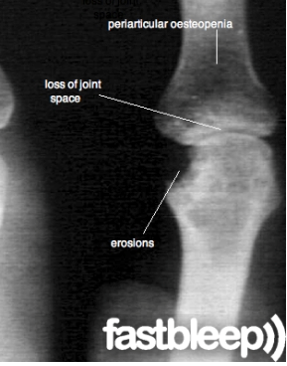
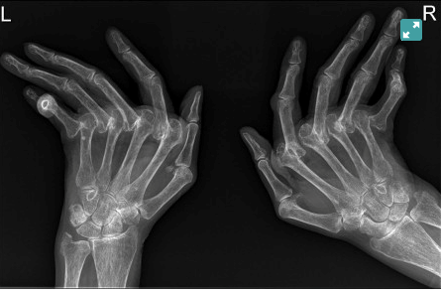
- Plain x-ray
- symmetric joint
- soft tissue swelling
- space loss
- subchondral erosions.
- Plain x-ray
- Blood Investigations
- Clinical Findings
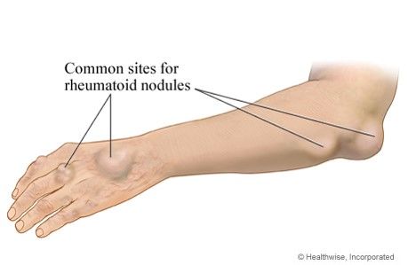
- Rheumatoid nodules
- firm, noticeable lumps that form underneath the skin
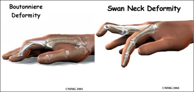
- Characteristic deformities
- Z deformity of the thumb
- Rheumatoid nodules
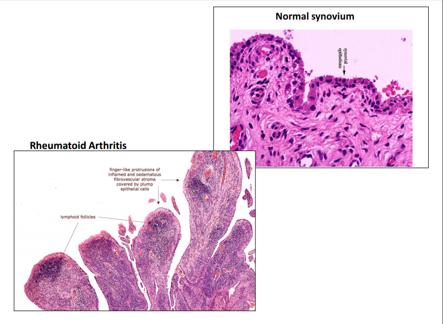
- Pathohistology
- synovial cell hyperplasia and proliferation
- dense perivascular inflammatory cell infiltrates (frequently forming lymphoid follicles)
- RA joint is hypoxic
- angiogenesis
- Activation of osteoclasts and chondrocytes
- Investigations
- History Taking
- basic patient details and statement of presenting problem.
- Where
- Progression
- Quality
- Alleviating factors
- Beliefs
- basic patient details and statement of presenting problem.
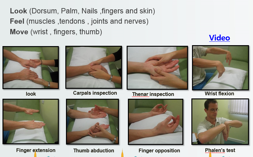
- Physical Examination of the hand
- Causes
- not fully understood
- Complications
- Haematological
- Anemia of chronic disease
- Iron deficiency anemia
- Bone marrow hypoplasia with anemia
- Vasculitis
- Digital arteritis
- Ulcers
- Cardiac
- Pericarditis
- Myocarditis
- Endocarditis
- Pulmonary
- Pleural effusions
- Bronchiolitis
- Neurological
- Cervical cord compression
- Peripheral neuropathy
- Systemic
- Weight loss
- Susceptibility to infection
- Lymphatic
- Splenomegaly
- Musculoskeletal
- Osteoporosis
- Bursitis
- Haematological
- Pathophysiology
- Autoreactive T cells are activated by antigen-presenting cells presenting self-peptide to CD4 cells.
- Macrophages exacerbate the disease by secreting TNF usually found in rheumatoid synovial tissues, resulting in cartilage destruction via effects on chondrocytes
- Rheumatoid factors are released as a consequence to the activation of B cells.
- Macrophages also release the chemokine IL-8 which recruit neutrophils that cause more damages to the tissues
- Inflammation
- Cartilage damage and joint space narrowing
- Bone erosion
- During the process of the disease, cells are damaged releasing anti-DNA. RF then binds the DNA-anti-DNA forming a complex that binds and damages basement membranes.
- Macrophages also release the chemokine IL-8 which recruit neutrophils that cause more damages to the tissues
- Rheumatoid factors are released as a consequence to the activation of B cells.
- Macrophages exacerbate the disease by secreting TNF usually found in rheumatoid synovial tissues, resulting in cartilage destruction via effects on chondrocytes
- Autoreactive T cells are activated by antigen-presenting cells presenting self-peptide to CD4 cells.
- Management
- Pharmacotherapy
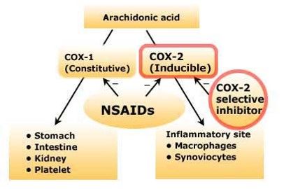
- NSAIDs
- Ibuprofen Ketoprofen Acetyl salicylic acid Naproxen Diclofenac Indomethacin Piroxicam
- Disease Modifying Anti-Rheumatic Drugs (DMARDs).
- Methotrexate
- Hydroxychloroquine
- Sulfasalazine
- Leflunomide
- Sulfasalazine
- Hydroxychloroquine
- Minocycline
- Control current inflammatory features
- Reduce joint damage and deformity
- Reduce long-term disability
- Reduce joint damage and deformity
- Methotrexate
- Non-TNF biologic therapy
- preventing full T-cell activation
- B-Cell Depletion
- Interleukin-1 Receptor Antagonists
- B-Cell Depletion
- preventing full T-cell activation
- NSAIDs
- Physiotherapy
- Cold/Hot Applications
- cold in acute
- where intra-articular heat increase is undesired
- cold-pack, ice, nitrogen spray, and cryotherapy
- where intra-articular heat increase is undesired
- hot in chronic
- Analgesia
- Relieve muscle spasms
- elasticity of periarticular structures obtained
- cold in acute
- Electrical Stimulation
- Transcutaneous electrical nerve stimulation (TENS) therapy
- Hydrotherapy
- exercises in a pool
- Rehabilitative Treatment
- Joint Protection Strategies
- Therapeutic Exercise
- swimming, walking, and cycling with adequate resting periods are recommended
- Cold/Hot Applications
- Intermittent splinting
- external supports applied to body parts
- decrease inflammation
- relieve pain
- protect a joint
- improve function
- correct deformity
- external supports applied to body parts
- Pharmacotherapy
- Prognosis
- 40% of RA
- disabled within 3 years
- 80% are moderately to severely disabled
- within20 years
- life expectancy could be shortened
- 40% of RA
- signs and symptoms
- Rheumatoid Arthritis
- Autoimmunity
- RA and Systemic lupus erythematous.
- SLE: It can affect the skin, joints, kidneys, brain, and other organs.
- RA and Systemic lupus erythematous.
- Injury
- Infection
- Septic Arthritis
- most common in the knees and hips
- may lead to permanent joint damage
- life threatening
- may lead to permanent joint damage
- most common in the knees and hips
- Reactive Arthritis
- triggered by an infection in another part of the body
- the knees and the joints of your ankles and feet
- triggered by an infection in another part of the body
- Septic Arthritis
- the term is used to describe many rheumatic diseases that affect the joints.
- inflammation of the joints
Imágenes y archivos del mapa
Información
- Descripción
- Mapa Mental sobre Anatomy, creado por kholod haj hussain el 23/02/2020.
- Creado por
- kholod haj hussain
- Visitas
- 0
- Historial
-
- Creado por Fatma Shwaylia · 11 mar 2017
- Copiado por Raghad Abbas · 23 feb 2020
- Copiado por kholod haj hussain · 23 feb 2020
- Valoración