
Mapa mental
Structure of the Eye and Retina
Contenido del mapa
Structure of the Eye and Retina
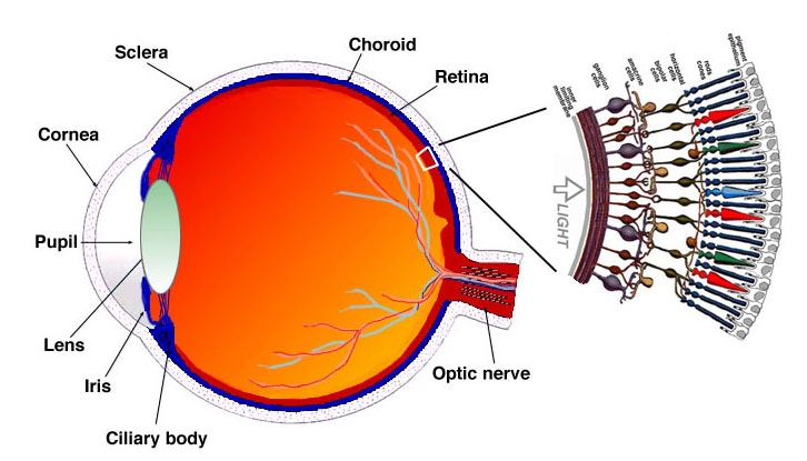
Pupil- center of the iris (coloured area) in which light enters through and opening in the centre and is projected on the back of the eye.
- Lens- focuses the pupil, is adjustable
- Cornea- focus the pupil, is not adjustable
Retina- where the light is projected at the rear surface of the eye. Is lined with visual receptors.
- Light always strikes the opposing side
Fovea- the pit at the back of the eye that is specialised for acute, detailed vision.
- Almost absent of blood vessels and ganglion cells creating unimpeded vision.
Rods- respond to faint light and are not useful in daylight. Bright light bleaches them. Abundant in the periphery of the human retina.
Cones- less active in dim light and are essential in colour vision. Abundant in and near the fovea.
- Red- longwave 700nm
- Green- medium wave 500nm
- Blue- shortwave 350nm
Ganglion cells- recieves messages from bipolar cells (rods and cones) and the ganglion cells join together (close to the center of the eye) and travel to the brain.
- Blind spot- the ganglion cells form the optic nerve. The point in which it leaves the eye is a blind spot as there are no receptors.
Imágenes y archivos del mapa
Información
- Descripción
- Behavioural Neuroscience Mapa Mental sobre Structure of the Eye and Retina, creado por lynda_ashford_25 el 22/10/2013.
- Creado por
- lynda_ashford_25
- Visitas
- 830
- Etiquetas
- Valoración