
Mapa mental
Spinal Cord (CNS)
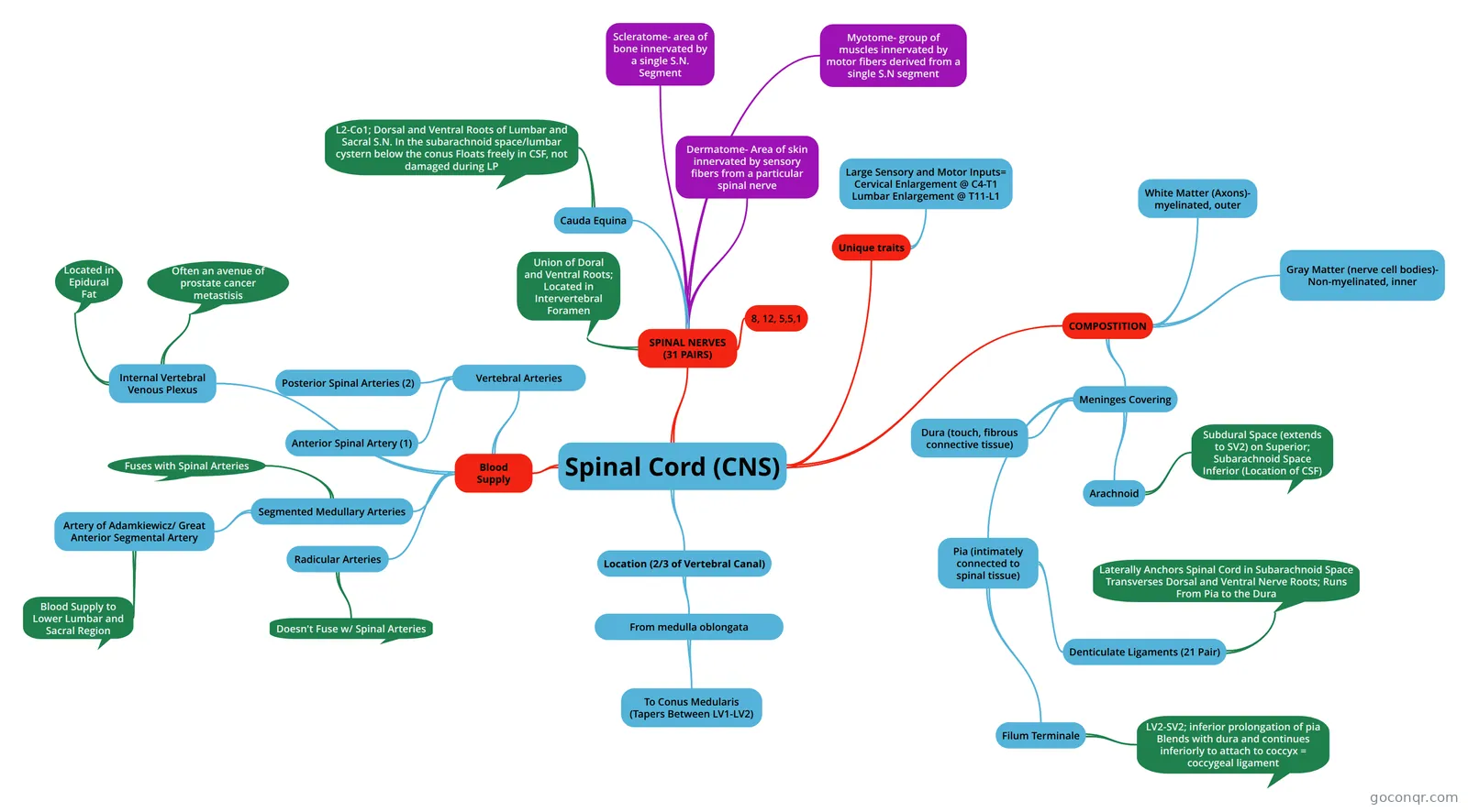
Mind map: spinal cord CNS anatomy
Contenido del mapa
Spinal Cord (CNS)
Blood Supply
- Vertebral Arteries
- Anterior Spinal Artery (1)
- Posterior Spinal Arteries (2)
- Segmented Medullary Arteries
- Fuses with Spinal Arteries
- Artery of Adamkiewicz/ Great Anterior Segmental Artery
- Blood Supply to Lower Lumbar and Sacral Region
- Radicular Arteries
- Doesn't Fuse w/ Spinal Arteries
- Internal Vertebral Venous Plexus
- Often an avenue of prostate cancer metastisis
- Located in Epidural Fat
- Vertebral Arteries
Location (2/3 of Vertebral Canal)
- From medulla oblongata
- To Conus Medularis (Tapers Between LV1-LV2)
- From medulla oblongata
Unique traits
- Large Sensory and Motor Inputs= Cervical Enlargement @ C4-T1 Lumbar Enlargement @ T11-L1
COMPOSTITION
- Gray Matter (nerve cell bodies)- Non-myelinated, inner
- Meninges Covering
- Dura (touch, fibrous connective tissue)
- Arachnoid
- Subdural Space (extends to SV2) on Superior; Subarachnoid Space Inferior (Location of CSF)
- Pia (intimately connected to spinal tissue)
- Denticulate Ligaments (21 Pair)
- Laterally Anchors Spinal Cord in Subarachnoid Space Transverses Dorsal and Ventral Nerve Roots; Runs From Pia to the Dura
- Filum Terminale
- LV2-SV2; inferior prolongation of pia Blends with dura and continues inferiorly to attach to coccyx = coccygeal ligament
- Denticulate Ligaments (21 Pair)
- White Matter (Axons)- myelinated, outer
SPINAL NERVES (31 PAIRS)
- Union of Doral and Ventral Roots; Located in Intervertebral Foramen
- 8, 12, 5,5,1
- Cauda Equina
- L2-Co1; Dorsal and Ventral Roots of Lumbar and Sacral S.N. In the subarachnoid space/lumbar cystern below the conus Floats freely in CSF, not damaged during LP
- Dermatome- Area of skin innervated by sensory fibers from a particular spinal nerve
- Scleratome- area of bone innervated by a single S.N. Segment
- Myotome- group of muscles innervated by motor fibers derived from a single S.N segment