
Mapa mental
CT
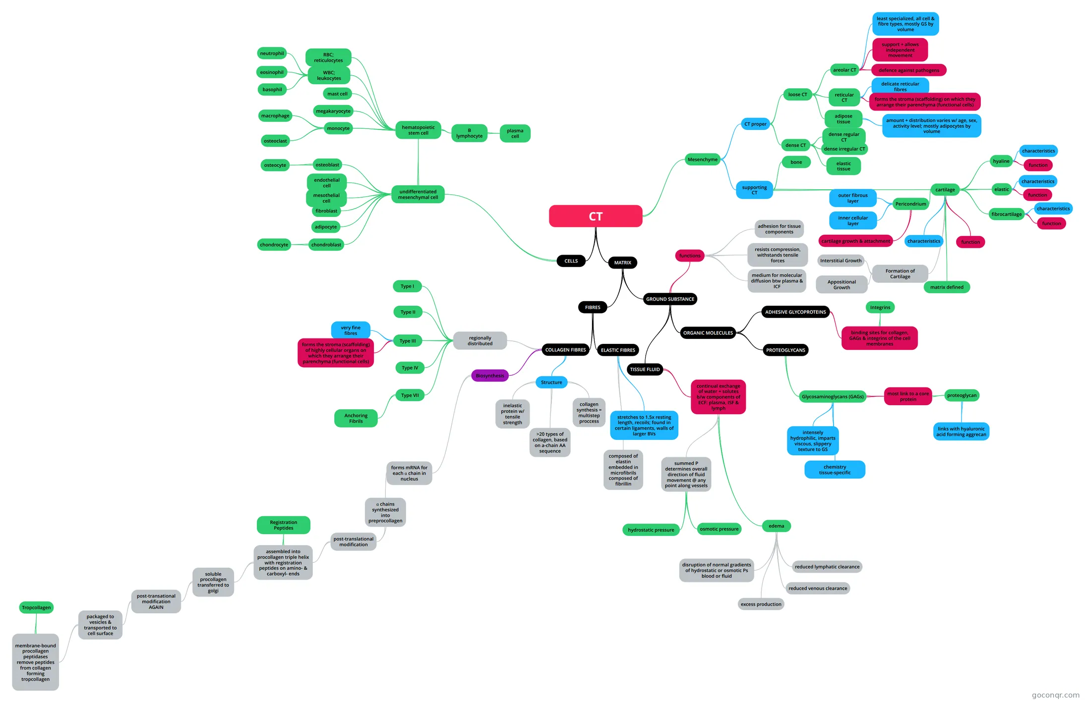
Mind map: connective tissue
Contenido del mapa
CT
- N.B. tissues = cells + cell products CT = mostly cell products
CELLS
- fixed cells primarily function locally i.e. fibroblasts, fixed macrophages, adipocytes, mast cells wandering cells increase w/ # of tissue damage or infection i.e. monocytes, lymphocytes, microphages (neutrophils & eosionphils)
- undifferentiated mesenchymal cell
- multipotential stem cells
- osteoblast
- osteocyte
- endothelial cell
- mesothelial cell
- fibroblast
- - produces fibres, proteoglycans & glycoproteins of the matrix - abundant RER, well-developed Golgi - oval nucleus - pale in active fibroblasts - darker in quiescent "fibrocytes"
- adipocyte
- - lipid storing cells - "signet ring" shape w/ thin rim of cytoplasm & flattened nucleus surrounding large droplet of stored lipid - lipid extraction during preparation makes the cell appear empty - when adipocytes predominate in certain areas, tissue is referred to as adipose
- chondroblast
- chondrocyte
- hematopoietic stem cell
- WBC; leukocytes
- - carried in the circulation but function in CT - an increased number in connective sisue is indicative of inflammation
- neutrophil
- drawn by chemotactic signals
- eosinophil
- drawn by chemotactic signals
- basophil
- mast cell
- - store chemical mediators of inflammation & hypersensitivity rxns - oval cells w/ granular cytoplasm + round, central nucleus - granules are metachromatic i.e. certain stains that bind with the granules change their colour e.g. toluidine blue stains the granules purple/magenta - activated by trauma or by presence of antigen - become sensitized to specific antigens when antibodies made against that antigen become bound to receptors on the mast cell membranes activated mast cells immediately release: 1. Histamine: vasodilation increases blood flow, increased vascular permeability (esp. post-capillary venules) increases production of protein-rich tissue fluid and slows blood flow 2. NCF attracts neutrophils 3. ECF attracts eosinophils
- megakaryocyte
- monocyte
- macrophage
- - derived from blood monocytes - mature in tissue spaces - can proliferate locally - avidly phagocytic cells - antigen presenting cells - relatively small & inconspicious unless they have been active in phagocytosis - are the Kuppfer cells in the liver, microglial cells of fthe CNS, Langerhands cells of the skin, osteoclasts of bone tissue
- osteoclast
- macrophage
- RBC; reticulocytes
- B lymphocyte
- become antibody-producing plasma cells with antigen exposure
- plasma cell
- - derived from B-lymphocytes - antibody-producing - relatively small, round cells with basophilic cytoplasm (filled with RER) - round, eccentric nucleus, like a "clock face" i.e. prominent nucleolus and clumps of peripheral heterochromatin - juxtanuclear Golgi forms pale "halo" around nucleus
- WBC; leukocytes
MATRIX
- FIBRES
- COLLAGEN FIBRES
- Structure
- inelastic protein w/ tensile strength
- >20 types of collagen, based on a-chain AA sequence
- collagen synthesis = multistep proccess
- - prepcollagen - procollagen - tropocollagen - fibrils - fibres
- Biosynthesis
- forms mRNA for each ɑ chain in nucleus
- ɑ chains synthesized into preprocollagen
- post-translational modification
- assembled into procollagen triple helix with registration peptides on amino- & carboxyl- ends
- Registration Peptides
- maintain solubility of procollagen
- soluble procollagen transferred to golgi
- post-transational modification AGAIN
- packaged to vesicles & transported to cell surface
- membrane-bound procollagen peptidases remove peptides from collagen forming tropcollagen
- Tropcollagen
- tropocollagen molecules aggregate to form collagen fibrils, covalent cross-links b/w tropocollagen molecules reinforce fibrilar structure
- Tropcollagen
- membrane-bound procollagen peptidases remove peptides from collagen forming tropcollagen
- packaged to vesicles & transported to cell surface
- post-transational modification AGAIN
- Registration Peptides
- assembled into procollagen triple helix with registration peptides on amino- & carboxyl- ends
- post-translational modification
- ɑ chains synthesized into preprocollagen
- forms mRNA for each ɑ chain in nucleus
- regionally distributed
- Type I
- most common, CT proper, bone
- Type II
- cartilages
- Type III
- forms "reticular fibres"
- very fine fibres
- forms the stroma (scaffolding) of highly cellular organs on which they arrange their parenchyma (functional cells)
- Type IV
- forms the meshwork in the BM
- Type VII
- forms anchoring fibrils
- Anchoring Fibrils
- bind the BM to underlying type 1 & type III collagen
- Type I
- Structure
- ELASTIC FIBRES
- stretches to 1.5x resting length, recoils; found in certain ligaments, walls of larger BVs
- composed of elastin embedded in microfibrils composed of fibrillin
- COLLAGEN FIBRES
- GROUND SUBSTANCE
- consistency varies from liquid to solid; gel-like substance in which cells & fibres are embedded
- TISSUE FLUID
- continual exchange of water + solutes b/w components of ECF: plasma, ISF & lymph
- summed P determines overall direction of fluid movement @ any point along vessels
- hydrostatic pressure
- synonymous with blood pressure, pushes fluid out of BVs N.B. lowest hydrostatic pressure is found within lymphatic vessels so excess interstitial fluid becomes lymph
- osmotic pressure
- fluid pulled into blood vessels b/c of plasma proteins
- hydrostatic pressure
- edema
- excess tissue fluid
- reduced lymphatic clearance
- reduced venous clearance
- excess production
- disruption of normal gradients of hydrostatic or osmotic Ps blood or fluid
- summed P determines overall direction of fluid movement @ any point along vessels
- continual exchange of water + solutes b/w components of ECF: plasma, ISF & lymph
- ORGANIC MOLECULES
- PROTEOGLYCANS
- Glycosaminoglycans (GAGs)
- inflexible unbranched polysaccharides composed of repeating disaccharide units
- intensely hydrophilic, imparts viscous, slippery texture to GS
- chemistry tissue-specific
- most link to a core protein
- proteoglycan
- responsible for.. gel-like consistency of matrix; turgidity & ability of CT to resist compression; molecular filter of varying pore size, charge distribution; slow diffusion of substances, cells, pathogens through ISF.
- links with hyaluronic acid forming aggrecan
- proteoglycan
- Glycosaminoglycans (GAGs)
- ADHESIVE GLYCOPROTEINS
- inc, fibronectin, laminin
- binding sites for collagen, GAGs & integrins of the cell membranes
- Integrins
- transmembrane link glycoproteins (recall hemidesmosomes)
- Integrins
- PROTEOGLYCANS
- functions
- resists compression, withstands tensile forces
- medium for molecular diffusion btw plasma & ICF
- adhesion for tissue components
- FIBRES
Mesenchyme
- CT proper
- loose CT
- reticular CT
- e.g. liver, kidneys, lymph nodes, smooth muscle & bone marrow
- delicate reticular fibres
- forms the stroma (scaffolding) on which they arrange their parenchyma (functional cells)
- adipose tissue
- abundant deep to skin, behind eyes, around kidneys
- amount + distribution varies w/ age, sex, activity level; mostly adipocytes by volume
- areolar CT
- e.g. superficial fascia, b/w muscles, BVs, Ns, around joints
- defence against pathogens
- support + allows independent movement
- least specialized, all cell & fibre types, mostly GS by volume
- reticular CT
- dense CT
- dense regular CT
- largely collagen type I fibres arranged in parallel arrays thus imparts tensile strength in 1 direction e.g. tendons, aponeuroses, ligaments
- dense irregular CT
- largely collagen type I fibres arranged in a meshwork sheet thus imparts tensile strength in >1 direction e.g. reticular layer of dermis, periosteum, perichondrium, organ capsules, joint capsules, submucosa of GI tract
- elastic tissue
- high concentration of elastic fibres = high resiliency (stretch + recoil); certain ligaments e.g. ligamentum flavum, supporting transitional epithelium, blood vessels
- dense regular CT
- loose CT
- supporting CT
- bone
- cartilage
- hyaline
- e.g. articular cartilage, tracheal rings, costal cartilage, immature skeleton, nasal cartilages
- characteristics
- most common & weakest cartilage, closely packed collagen type II fibrils in a firm gel-like GS
- function
- resists compression, provides touch, flexible support; reduces friction b/w bony surfaces in articulations
- elastic
- e.g. pinna of external ear, epiglottis, pharyngotympanic tube
- characteristics
- like hyaline but w/ a higher proportion of elastic fibres
- function
- provides greater flexibility and resiliency i.e. "bounces back"
- fibrocartilage
- anulus fibrosis of intervertebral disks, pubic symphysis, intra-articular disks, tendon & ligament insertions
- characteristics
- little ground substance, high proportion of density interwoven collagen type I fibres arranged in perpendicular arrays, intermediate in strength b/w hyaline caartilage & densse regular CT; chondrocytes arranged in rows b/w bundles of collagen type I fibres
- function
- resists both compression + tension
- matrix defined
- characteristics
- EC matrix abundant, firm, gel-like, resilient, smooth all types are avascular, no lymphatic vessels, no nerves thus chondrocytes metabolize glucose mainly by anaerobic glycolysis; nourished by diffusion through the GS from vessels in the surrounding perichondrium and/or ordinary CT
- function
- gives structure to soft tissues in development & growth of long bones (covered under bone)
- Pericondrium
- most cartilages surrounded by perichondrium (dense & irregular CT which has BVs, Ns, Ls)
- inner cellular layer
- contains chondroblasts: synthesize the organic components of the matrix; contributes to cartilage growth and thus less prominence with maturity
- outer fibrous layer
- contains mainly collagen fibres, fibroblasts
- cartilage growth & attachment
- Formation of Cartilage
- A. cartilage develops from condensations of embryonic mesenchyme B. mesenchymal cells diifferentiate into chondroblasts which secrete the organic components of the matrix C. chondroblasts ultimately surround themselves with matrix thus becoming isolated in their lacunae D. these chondroblasts may divide thus causing the cartilage to grow from within in a process called intersittial growth N.B. isolated chondroblasts become quiescent, maintain matrix as chondrocytes.
- Interstitial Growth
- the cartilage expands from within as chrondrocytes in the matrix divide, grow, and produce new matrix; chrondrocyte undergoes division within a lacuna surrounded by cartilage matrix, as daughter cells secrete additional matrix, they move apart, expanding the cartilage from within
- Appositional Growth
- - mesenchymal cells at the surface of the newly formed cartilage differentiate into fibroblasts which form the fibrous layer of the perichondrium; - mesenchymal cells deep in the fibrous layer differentiate into chondroblasts forming the cellular layer of the perichondrium; - these cells secrete matrix onto the surface of the newly formed cartilage in a process called appositional growth
- hyaline
- CT proper
Información
- Descripción
- Flow Chart for CT Lectures
- Creado por
- thashali.anthony
- Visitas
- 110
- Etiquetas
- Valoración