
Mapa mental
Physiology
Contenido del mapa
Physiology
Structure and function of exercising muscle
- Skeletal muscle
- Structural adaptations in skeletal muscle
- Happen withing 6 weeks of concentric and eccentric training
- Eccentric training: ↑ muscle fascicle length; ↓ pennation* angle (i.e. the fascicles align better with the direction of the muscle
- Concentric training: ↓ muscle fascicle length; ↑ pennation angle (i.e. fascicles angled more away from the full muscle’s direction)
- 4 weeks detraining: o Eccentric training induced adaptations are reversed. oConcentric adaptations are maintained.
- Thus, structural adaptations to strength training is highly specific to the mode of training.
- Has effect on injury prevention and development of rehabilitation programs
- Function of skeletal muscle
- Force production for locomotion and breathing
- Force production for postural support
- Heat production during cold stress
- Anatomy of skeletal muscle
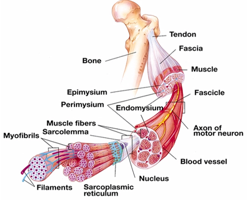
- Entire muscle surrounded by epimysium and consists of many bundles (Fasciculi)
- Fasciculi surrounded by perimysium and consists of individual muscle cell (muscle fiber)
- Each muscle fiber is a muscle cell with multiple nuclei, surrounded by endomysium, consists of myofibrils divided into sarcomers
- Structure of muscle fiber
- Plasmalemma (cell membrane) fuses with tendon, conducts AP from motor neuron to muscle fiber, maintains pH, transports nutrients into muscle fiber
- Sarcolemma consists of plasmalemma and the basement membrane
- Satellite cells are between the plasmalemma and the basement membrane, there for muscle growth and development, assists with injury, immobilization, training.
- Sarcoplasm is cytoplasm of muscle cell, glycogen storage, myoglobin
- Transverse tubules are extensions of plasmalemma, carry action potential and other substances deep into muscle fiber, waste products out of cell.
- Sarcoplasmic Reticulum (SR) is longitudinal network of tubules and Ca2+ storage
- Sarcomeres
- • Skeletal muscle – striated muscle o A-bands: dark stripes o I-bands: light stripes o H-zone: middle of A-band o M-line: middle of H-zone • Common boundary structure: Z-disk
- Used for muscle contraction. Made up of actin and myosin
- Myosin two intertwined filaments with myosin heads. Myosin heads composed of compactly folded polypeptide chains. Stabilized by titin
- Titin is a regulatory protein. Located in space between two filaments, attached at various points to the myosin and runs all the way to the Z-disk. Acts as spring mechanism that keeps myosin filaments centred.
- Actin composed of actin, tropomyosin and troponin. Anchored at Z-disk.
- Myosin two intertwined filaments with myosin heads. Myosin heads composed of compactly folded polypeptide chains. Stabilized by titin
- Structure of muscle fiber
- Structural adaptations in skeletal muscle
- Skeletal muscle
Imágenes y archivos del mapa
Información
- Descripción
- Mapa Mental sobre Physiology, creado por Strauss Viljoen el 18/05/2023.
- Creado por
- Strauss Viljoen
- Visitas
- 1
- Valoración