
Apuntes
Muscle Tissue
Contenido del apunte (10)
Página 1
Muscle is specialised for contraction and transmission of impulses that aid contraction
Characterised by ability to contract
3 types based on:
Organisation/appearance of cells
Nervous control
Location in the body
Muscles have very specific and limited functions - highly specialised
Associated with CT and nervous tissue
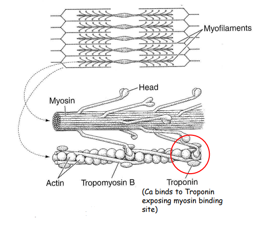
Contraction due to interaction of actin and myosin as microfilaments in the sarcoplasm
Transmission of force goes through connective tissue of collagen fibres
Outermost covering (fascia) is connected to origin/insertion/tendons/ligaments
No connective tissue means no force transmitted
Página 2
Types:
Striated Muscle:
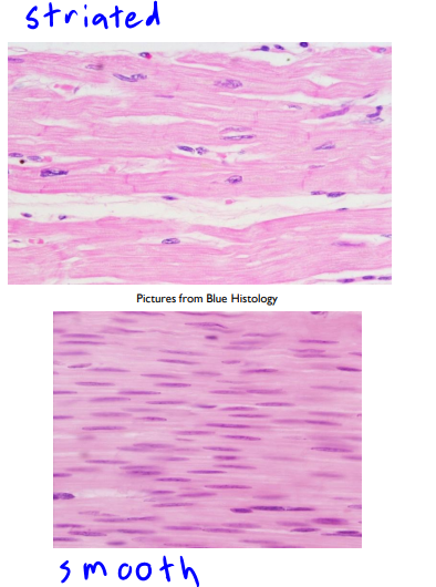
Characteristic banding pattern visible on light microscope
Skeletal muscle: voluntary muscle
Cardiac muscle: involuntary muscle
Smooth muscle:
Visceral or "involuntary" muscle
Found in internal organs and other locations
Página 3
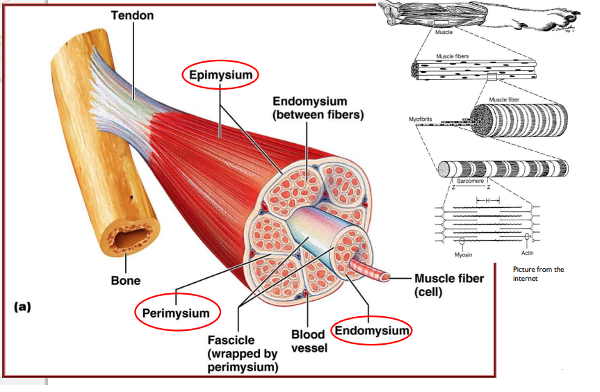
CT forms basework:
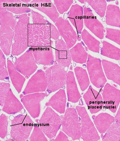
Endomysium surrounds individual cells
Perimysium surrounds muscle bundles
Epimysium surrounds entire muscle
Specialisation has caused loss of other capabilities such as ability to divide. Repair is by scarring.
Página 4
Muscle repair:
Highly differentiated muscle cells cannot divide
Injury will cause a loss of material. Scarring will fill in the space with collagen, but collagen is not contactible, which causes loss of function
Skeletal muscle has a reserve of myoblasts, meaning they may regenerate but not much.
No regeneration in cardiac and smooth muscles.
Página 5
Neural relationship in muscle:
Varies with the muscle type:
Skeletal muscle most elaborate
Smooth muscle simple but direct
Cardiac muscle controlled by non neuronal network and internally
Control is monitored closely by the central nervous system (brain & spinal cord)
Página 6
Skeletal Muscle
Appearance:
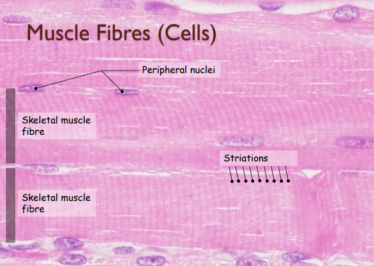
Striation
Long fibres
Multinucleated (peripheral ie. sits on the side of cells.)
Very large cells 100-150 microns diameter and several centimeters to a meter of length
Blood vessels between
Invested by intercellular collagen
Nervous control:
Somatic (voluntary)
Location:
Usually attached to bones of skeleton
Definitions:
Muscles are bundles of individual cells, MYOFIBERS
MYOFIBERS contain contractile MYOFIBRILS (1000)
MYOFIBRILS are composed of MYOFILAMENTS (1000)
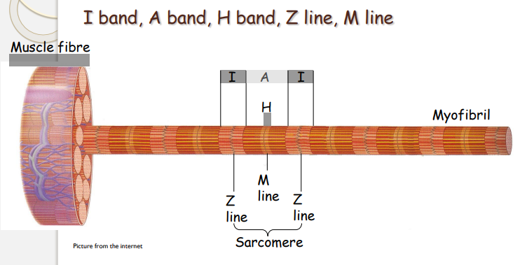
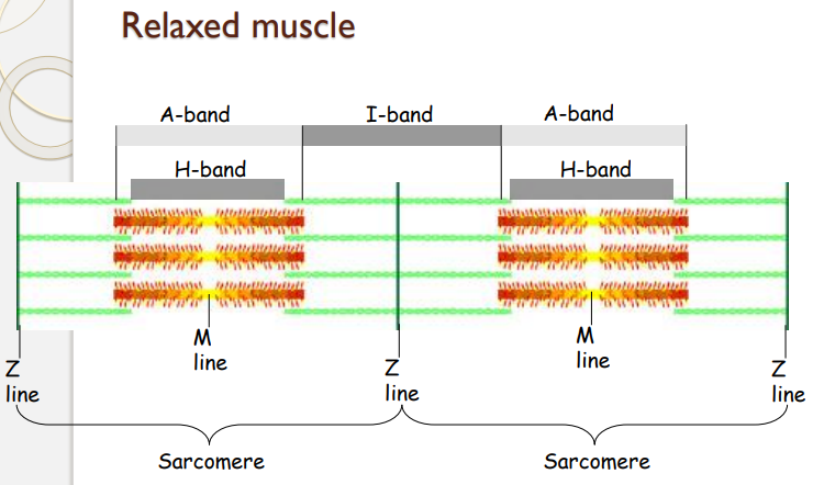
MYOFILAMENTS are organized in SARCOMERES (1000)
SARCOMERE is the basic unit of contraction and contains Actin and Myosin
Myosin: Multiple heads at each end of a long molecule - filament. Heads change angle.
Actin: Receptor sites for heads in long filament
Página 7
Bands:
I-band: Only Actin
A-band: Actin and myosin
H-Band: Only myosin
Página 8
Cardiac Musles
Cells:
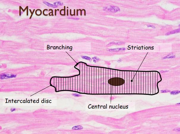
Striation (not easy to see under Light Miscroscope)
Branched
Mononucleated
Nervous Control:
Visceral (involuntary)
Contraction not an inherent property
No direct neuronal stimulation needed
Location:
Heart
Página 9
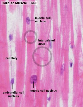
Myocytes of cardiac muscle are individual cells that are much smaller that myofiber of skeletal muscle. MONONUCLEATED with the nucleus in the center
Myocytes butted end-to-end to form anastomosing network (cross connection)
Extremely well vascularised
Capillaries lies among and between cells
Cardiac muscle can not sustain any oxygen deficit (need O2 )
unique intercalated disk - Diagnostic feature of cardiac muscle
Contain myofibrils and sarcomeres
Cardiac fibres surrounded by loose connective tissue
No perimysium
Connective tissue surrounding heart is called epicardium
Página 10
Smooth Muscle
Cells:
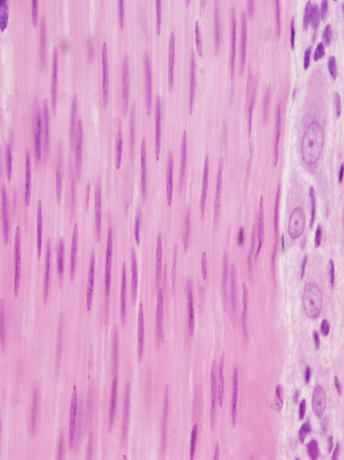
Fusiform cells
No striations
Each cell only has one oval nucleus in the centre
Not branched
No sarcomeres but have actin and myosin so can still contract. But float inside muscle cell rather than align.
Nervous cells:
Involuntary
Location:
Walls of GIT, airways and urogenital tracts
Información
- Descripción
- TV1003 Apunte sobre Muscle Tissue, creado por Erika Cobb el 19/08/2019.
- Creado por
- Erika Cobb
- Visitas
- 2
- Valoración